Radiologic–pathologic association of tumor‐like lesions with inflammation in cerebral white matter: Comparison of two cases with distinct clinical outcomes
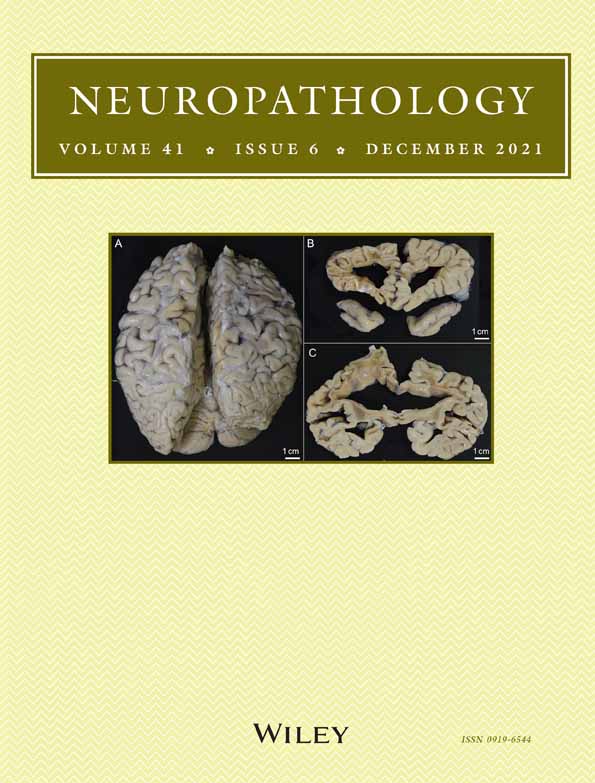
Here, we report two cases showing tumor-like white matter lesions; one case was diagnosed as having inflammatory disease, and the other was diagnosed as having astrocytoma. Their outcomes were completely distinct despite similar pathology. Prior to biopsy, perfusion computed tomography (CT) and magnetic resonance imaging (MRI) were conducted. The two mass-forming lesions were distinct in edema level and vascularity patterns on CT and MRI. However, pathological examination of brain biopsy specimens revealed commonalities, including (1) proliferation of glial cells, (2) perivascular lymphocytic infiltration, and (3) appearance of numerous macrophages. Although atypical astrocytes proliferated in both cases, nuclear atypia was more distinct in case 2 than in case 1. The immunohistochemical results were the same for both cases: isocitrate dehydrogenase 1 (IDH1) R132H mutation was negative, and alpha thalassaemia mental retardation X-linked (ATRX) was retained. Faint immunoreactivity for p53 was observed in a few glial cells, and Ki-67 immunoreactive cells were markedly reduced in numbers (< 1%). Inflammatory reactions were evident in both cases: T cells dominantly infiltrated over B cells in the perivascular area in case 1, whereas both T and B cells infiltrated in case 2. Molecular analysis revealed wild-type IDH1 and IDH2 in both cases. However, a telomerase reverse transcriptase (TERT) sequence mutation was detected in case 2 but not in case 1. Eventually, case 1 was diagnosed as having inflammatory lesions, whereas case 2 was diagnosed as having diffuse astrocytoma associated with inflammatory reactions. The prognosis was favorable for case 1, whereas case 2 died 10 months following biopsy. These data indicated the diagnostic value of molecular analysis, for example, a TERT mutation, in association with the radiological findings. Although in case 2, histopathological evidence did not suggest high-grade glioma, the case met the new diagnostic criteria: “diffuse astrocytic glioma, IDH wild-type, with molecular features of glioblastoma, World Health Organization (WHO) grade IV,” according to cIMPACT-NOW, update 3. Thus, interdisciplinary approaches are essential for accurate diagnosis of newly categorized white matter diseases.


留言 (0)