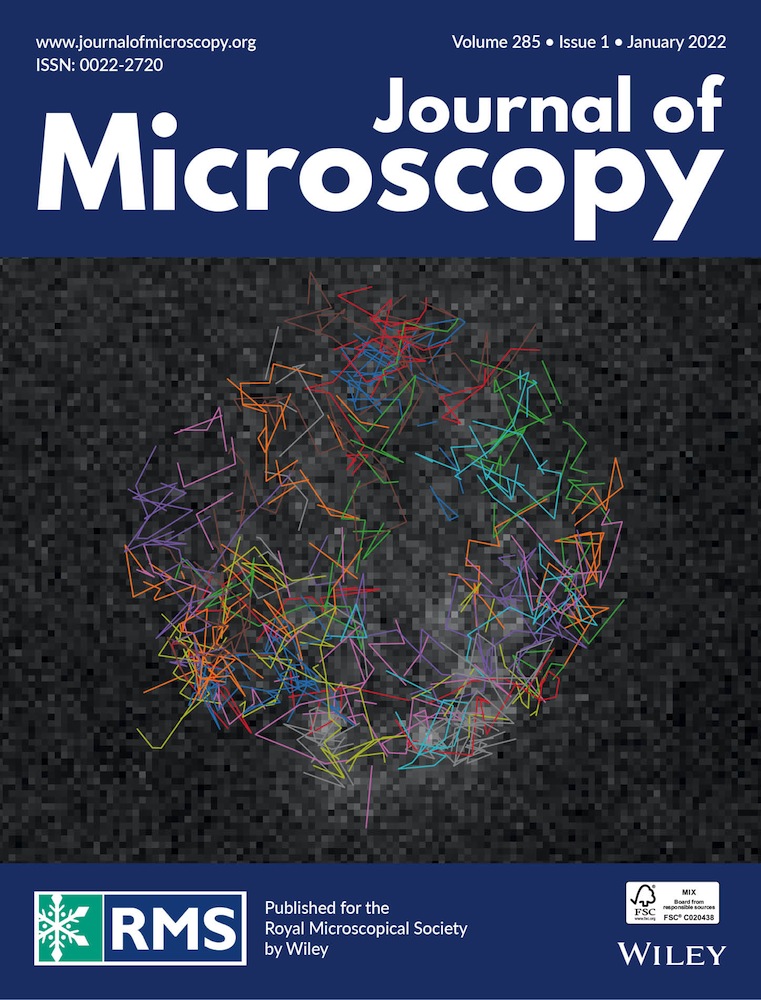
Fast 3D imaging of giant unilamellar vesicles using reflected light‐sheet microscopy with single molecule sensitivity
Observation of highly dynamic processes inside living cells at the single molecule level is key for a better understanding of biological systems. However, imaging of single molecules in living cells is usually limited by the spatial and temporal resolution, photobleaching and the signal-to-background ratio. To overcome these limitations, light-sheet microscopes with thin selective plane illumination, for example, in a reflected geometry with a high numerical aperture imaging objective, have been developed. Here, we developed a reflected light-sheet microscope with active optics for fast, high contrast, two-color acquisition of z-stacks. We demonstrate fast volume scanning by imaging a two-color giant unilamellar vesicle (GUV) hemisphere. In addition, the high contrast enabled the imaging and tracking of single lipids in the GUV cap. The enhanced reflected scanning light sheet microscope enables fast 3D scanning of artificial membrane systems and potentially live cells with single-molecule sensitivity and thereby could provide quantitative and molecular insight into the operation of cells.
This article is protected by copyright. All rights reserved


留言 (0)