"A picture is worth a thousand words": the use of microscopy for imaging neuroinflammation
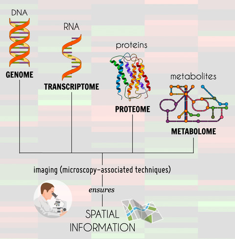
Since the first studies of the nervous system by the Nobel laureates Camillo Golgi and Santiago Ramon y Cajal using simple dyes and conventional light microscopes, microscopy has come a long way – to the most recent techniques that make it possible to perform images in live cells and animals, in health and disease. Many pathological conditions of the central nervous system have already been linked to inflammatory responses. In this scenario, several available markers and techniques can help imaging and unveil the neuroinflammatory process. Moreover, microscopy imaging techniques have become even more necessary to validate the large quantity of data generated in the era of "omics". This review aims to highlight how to assess neuroinflammation by using microscopy as a tool to provide specific details about the cell’s architecture during neuroinflammatory conditions. First, we describe specific markers that have been used in light microscopy studies and that are widely applied to unravel and describe neuroinflammatory mechanisms in distinct conditions. Then, we discuss some important methodologies that facilitate the imaging of these markers, such as immunohistochemistry and immunofluorescence techniques. Emphasis will be given to studies using two-photon microscopy, an approach that revolutionized the real-time assessment of neuroinflammatory processes. Finally, some studies integrating omics with microscopy will be presented. The fusion of these techniques is developing, but the high amount of data generated from these applications will certainly improve the comprehension of the molecular mechanisms involved in neuroinflammation.


留言 (0)