
記住我


DrReposER predicted numerous potential 3D-drug binding motifs of both left (L) and right (R) superpositions for 7K3N, 6WEY, 6M03, 7JLT, 6W4B, 6ZCT, 6M71, 7NIO, 5C8S, 6VWW and 7BQ7 (Additional file 1: S1, Additional file 2: S2, Additional file 3: S4, Additional file 4: S4, Additional file 5: S5, Additional file 6: S6, Additional file 7: S7, Additional file 8: S8, Additional file 9: S9, Additional file 10: S10 and Additional file 11: S11). Known drugs that bind these motifs bind either human, bacterial or viral proteins. Results after analyzing the 3D structures of the target molecules and complexes were further filtered for anti-viral drugs. From the hit results, 14 anti-viral drugs i.e., Amphetamine (Drug bank ID-DB00182), Amprenavir (Drug bank ID-DB00701), Atazanavir (Drug bank ID-DB01072), Darunavir (Drug bank ID-DB01264), Grazoprevir (Drug bank ID-DB11575), Indinavir (Drug bank ID-DB00224), Lopinavir (Drug bank ID-DB01601), Nelfinavir (Drug bank ID-DB00220), Nevirapine (Drug bank ID-DB00238), Ribavirin (Drug bank ID-DB00811), Rimantadine (Drug bank ID-DB00478), Ritonavir (Drug bank ID-DB00503), Saquinavir (Drug bank ID-DB01232), and Tipranavir (Drug bank ID-DB00932) were selected for having unique 3D-drug binding motifs (Tables 1, 2, 3, 4, 5, 6, 7, 8, 9, 10 and 11). The findings showed that several anti-viral drugs had binding interfaces on a single protein or protein complexes and moreover, each anti-viral drug had one to several binding motifs (Tables 12 and 13).
Table 1 Possible binding sites of NSP1 against known anti-viral drugsTable 2 Possible binding sites of NSP3 against known anti-viral drugsTable 3 Possible binding sites of NSP5 against known anti-viral drugs.Table 4 Possible binding sites of NSP7-NSP8 against known anti-viral drugsTable 5 Possible binding sites of NSP9 against known anti-viral drugsTable 6 Possible binding sites of NSP10 against known anti-viral drugsTable 7 Possible binding sites of NSP7-NSP8-NSP12 complex against known anti-viral drugsTable 8 Possible binding sites of NSP13 against known anti-viral drugsTable 9 Possible binding sites of NSP14 against known anti-viral drugsTable 10 Possible binding sites of NSP15 against known anti-viral drugsTable 11 Possible binding sites of NSP16-NSP10 complex against known anti-viral drugsTable 12 Comparison of drug binding motifs of analyzed NSPs for antiviral drugs Table 13 Comparison of NSPs binding of the drugs analyzedAmphetamine (DB00182) targeted only a single binding interface on Nsp5 (6M03) (Tables 3, 12, 13). Amprenavir (DB00701) targeted four binding motifs on Nsp3 (6WEY), three motifs onNsp1 (7K3N), Nsp7-8-12 complex (6M71), Nsp13 (7NIO) and Nsp14 (5C8S), and two binding motifs on Nsp7-8 complex (7JLT), Nsp15 (6VWW) and Nsp16-10 complex (7BQ7) (Tables 2, 1, 7, 8, 9, 4, 10, 11, 12, Figs. 1, 23, 4, 5, 6, 7, 8, 9, 10 and 11). Atazanavir (DB01072) targeted three motifs on Nsp16-10 complex (7BQ7), two motifs on Nsp10 (6ZCT) and single motif each on Nsp1, Nsp7-8-12, Nsp13, Nsp14 and Nsp15 (Tables 11, 6, 12). Darunavir (DB01264) is the most promising drug as it targeted the greatest number of binding motifs and targeted every molecule except Nsp9. It targeted ten motifs on Nsp1 (7K3N), seven motifs on Nsp14 (5C8S), six motifs on Nsp3 (6WEY), five motifs on Nsp15 (6VWW) and Nsp16-10 complex (7BQ7), four motifs on Nsp7-8-12 complex (6M71), three motifs on Nsp10 (6ZCT), two motifs each on Nsp5 (6M03) and Nsp13 (7NIO), respectively and a single motif on Nsp7-8 complex (Tables 1, 9, 2, 10, 11, 7, 6, 3, 8, 4, 12, Figs. 1, 2, 3, 4, 5, 6, 7, 8, 9, 10 and 11). Grazoprevir (DB11575) targeted two motifs, on Nsp10 (6ZCT) and two on Nsp16-10 complex (7BQ7) and single motif each on Nsp9 and Nsp14 (Tables 6, 11, 5, 9, 12). Indinavir (DB00224) significantly targeted three motifs, each on Nsp13 (7NIO) and Nsp15 (6VWW) (Tables 8, 10, 12). Lopinavir significantly targeted three motifs on Nsp15 and 2 motifs each on Nsp13 and Nsp14 (Tables 10, 8, 9). Nelfinavir targeted two interfaces on Nsp1 and Nsp7-8–12 complexes (Tables 1, 7). On the other hand, Nevirapine targeted only a single motif on Nsp5 (Table 3). Rimantadine (DB00478) significantly targeted five binding interfaces on Nsp14 (5C8S), three binding motifs each on Nsp5 (6M03) and Nsp9 (6W4B), and two motifs on Nsp3 (6WEY), Nsp13 (7NIO), Nsp16-10 (7BQ7) and a single motif on Nsp1, Nsp7-8 and Nsp7-8-12 complex (Tables 9, 3, 5, 2, 8, 11, 1, 4, 7, 12, Figs. 1, 2, 3, 4, 5, 6, 7, 8, 9, 10 and 11). Ritonavir targeted two motifs on Nsp16-10 complex (Table 11). Saquinavir (DB01232) targeted four motifs on Nsp16-10 complex (7BQ7), three interfaces each on Nsp7-8–12 (6M71) and Nsp15 (6VWW), two motifs on Nsp1 and Nsp14 (5C8S) and a single motif on Nsp3, Nsp7-8, Nsp10 and Nsp13 (Tables 11, 7, 10, 1, 9, 3, 4, 6, 8, Figs. 1, 2, 3, 4, 5, 6, 7, 8, 9, 10 and 11). Finally, Tipranavir (DB00932) targeted two binding motifs; each on Nsp3, Nsp7-8–12 complex and Nsp14 (Tables 3, 7, 9), whereas single binding interface each on Nsp1, Nsp5, Nsp9, Nsp13, Nsp15 and Nsp16-10 (Table 12).
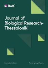
Fig. 1
3D-binding interfaces of NSP1with Amprenavir, Darunavir, Rimantadine &Saquinavir. a–c Binding motifs of Amprenavir. d All the binding motifs of Amprenavir. e–n Binding interfaces of Darunavir. o All the binding motifs of Darunavir. p, q Rimantadine binding motif. r, s Saquinavir binding motifs and t All the motifs on NSP1. Numbers indicate the motif forming amino acids. Three letter codes of amino acids have been mentioned
Fig. 2
3D-binding interfaces of NSP3 with Amprenavir, Darunavir, Rimantadine &Saquinavir. a–d Binding motifs of Amprenavir. e All the binding motifs of Amprenavir together. f–k Binding interfaces of Darunavir. L Combined binding motifs of Darunavir. m, n Rimantadine binding motifs. o All motifs of RIM. p, q Saquinavir binding motif. Numbers indicate the motif forming amino acids. Three letter codes of amino acids have been mentioned
Fig. 3
3D-binding interfaces of NSP5 with Darunavir&Rimantadine. a–c Binding motifs of Rimantadine. d All the binding motifs of RIM on NSP5. e, f Binding interfaces of Darunavir. g All the binding motifs of Darunavir. Numbers indicate the motif forming amino acids. Three letter codes of amino acids have been mentioned
Fig. 4
3D-binding interfaces of NSP7-8 complex with Amprenavir, Darunavir, Rimantadine &Saquinavir. a, b. Binding motifs of Amprenavir. c All the binding motifs of Amprenavir together. d, e Binding interfaces of Darunavir. f, g Rimantadine binding motifs. h, i Saquinavir binding motif. Numbers indicate the motif forming amino acids. Three letter codes of amino acids have been mentioned
Fig. 5
3D-binding interfaces of NSP9 with Rimantadine. a–c Three binding motifs of Rimantadine. d All the binding motifs of RIM together. Numbers indicate the motif forming amino acids. Three letter codes of amino acids have been mentioned
Fig. 6
3D-binding interfaces of NSP10with Darunavir&Saquinavir. a–c. Binding motifs of Darunavir. d Combined binding motifs of Darunavir. e, f Saquinavir binding motif. Numbers indicate the motif forming amino acids. Three letter codes of amino acids have been mentioned
Fig. 7
3D-binding interfaces of NSP7-8–12 complex with Amprenavir, Darunavir, Rimantadine &Saquinavir. a–c Binding motifs of Amprenavir. d All the binding motifs of Amprenavir together. e–h Binding interfaces of Darunavir. i Combined binding motifs of Darunavir. j, k Rimantadine binding motifs. l–n Saquinavir binding motifs. o Combined motifs of ROC. Numbers indicate the motif forming amino acids. Three letter codes of amino acids have been mentioned
Fig. 8
3D-binding interfaces of NSP13 with Amprenavir, Darunavir, Rimantadine &Saquinavir. a–c Binding motifs of Amprenavir. d All the binding motifs of Amprenavir together. e, f. Binding interfaces of Darunavir. g Combined binding motifs of Darunavir. h, i. Rimantadine binding motifs. j All motifs of RIM. k, l Saquinavir binding motif. Numbers indicate the motif forming amino acids. Three letter codes of amino acids have been mentioned
Fig. 9
3D-binding interfaces of NSP14 with Amprenavir, Darunavir, Rimantadine &Saquinavir. a–c Binding motifs of Amprenavir. d All the binding motifs of Amprenavir together. e–k.Binding interfaces of Darunavir. l Combined binding motifs of Darunavir. m–q Rimantadine binding motifs. r All motifs of RIM. s, t Saquinavir binding motifs. u All the Saquinavir motifs together. Numbers indicate the motif forming amino acids. Three letter codes of amino acids have been mentioned
Fig. 10
3D-binding interfaces of NSP15 with Amprenavir, Darunavir&Saquinavir. a, b Binding motifs of Amprenavir. c All the binding motifs of Amprenavir together. d–h Binding interfaces of Darunavir. i Combined binding motifs of Darunavir. j–l Saquinavir binding motifs. m All the ROC binding interfaces. Numbers indicate the motif forming amino acids. Three letter codes of amino acids have been mentioned
Fig. 11
3D-binding interfaces of NSP16-10 complex with Amprenavir, Darunavir, Rimantadine &Saquinavir. a, b Binding motifs of Amprenavir. c All the binding motifs of Amprenavir together. d–h Binding interfaces of Darunavir. i Combined binding motifs of Darunavir. j, k Rimantadine binding motifs. l All motifs of RIM. m–p Saquinavir binding motifs. q All the Saquinavir binding interfaces. Numbers indicate the motif forming amino acids. Three letter codes of amino acids have been mentioned
All the binding results were further compiled and analyzed. Results revealed that Darunavir (DB01264) had 45 unique binding sites and targeted 10 SARS-CoV-2 PDB entries or 10 NSPs (Tables 12, 13). The Lowest Root Mean Square Deviation (RMSD) value of Darunavir among all the target molecules was 0.54 Å for Nsp16-10 complex and maximum number of residues involved in interaction was 27 (Tables 1, 2, 3, 4, 5, 6, 7, 8, 9, 10 and 11). Significant binding interfaces were again targeted by Amprenavir (DB00701) and Saquinavir (DB01232) with 22 and 18 (Tables 12, 13), respectively. The two drugs had eight and nine binding partners, respectively (Tables 12, 13). The lowest RMSDs for them were 0.54 Å and 0.52 Å and maximum residues involved in drug-target binding were 28 and 31, respectively (Tables 1, 2, 3, 4, 5, 6, 7, 8, 9, 10 and 11). Additionally, Rimantadine (DB00478) had 20 drug binding motifs that targeted nine binding partners (Tables 12, 13) with the lowest RMSD value of 0.67 Å and maximum number of residues involved in binding were 10 (Tables 1, 2, 3, 4, 5, 6, 7, 8, 9, 10 and 11). Again, Tipranavir (DB00932) and Indinavir (DB00224) both showed 12 binding motifs for nine and eight binding partners, respectively (Tables 12, 13). Lowest RMSD values for these two drugs were 0.53 Å and 0.72 Å and maximum number of residues involved in binding were 27 and 24, respectively (Tables 1, 2, 3, 4, 5, 6, 7, 8, 9, 10 and 11).
Results showed that Darunavir, Amprenavir, Rimantadine, Saquinavir, Tipranavir and Indinavir were more effective in targeting the twelve SARS-CoV-2 proteins and their complexes (Tables 12, 13). Darunavir is a nonpeptidic benzenesulfonamide inhibitor that targets active site of HIV-1 protease [38, 39]. Amprenavir is a hydroxyethylamine sulfonamide derivative that inhibits HIV-1 protease [40, 41]. Rimantadine is an alkylamine that specifically targets Influenza A virus M2 protein [42,43,44]. Saquinavir is a L-asparagine derivative that acts as HIV-1 protease inhibitor [45, 46]. Tipranavir is a sulfonamide that acts as HIV-1 protease inhibitor [47]. Moreover, Indinavir is a piperazinecarboxamide having HIV-1 protease inhibitory activity [
留言 (0)