Bacterial isolates and susceptibility testing
Nine XDR P. aeruginosa clinical isolates were obtained from different Spanish hospitals in the context of two multicentric studies performed in 2017 and 2019 [21, 22]. These isolates were previously characterized by whole genome sequencing, and the main mechanisms of antimicrobial resistance were analysed. They were preserved in Muller-Hinton broth supplemented with 20% glycerol and stored at -80ºC.
Antimicrobial susceptibility testing was performed and interpreted according to EUCAST breakpoints (V13.1) and guidelines for broth microdilution using cation-adjusted Muller-Hinton broth (CAMHB, Becton-Dickinson and Co) for CZA, aztreonam and aztreonam/avibactam, and iron-depleted cation-adjusted Muller-Hinton broth (ID-CAMHB) for cefiderocol [23]. The reference strain PAO1 was included in the susceptibility testing experiments as quality control.
Culture media
CAMHB was prepared according to the manufacturer’s instructions. Tryptic Soy Agar II (TSA II, Becton-Dickinson and Co.) used for colony quantification, was prepared following the manufacturer’s instructions. Columbia blood agar plates (COS) (bioMérieux) were used to reculture the isolates.
For cefiderocol testing, iron-depleted liquid media was required. ID-CAMHB was prepared following the CLSI recommendations, finally adjusted to pH 7.3 using hydrochloric acid and kept at 4ºC until usage [24]. To ensured proper preparation of the ID-CAMHB medium, a minimum inhibitory concentration (MIC) of cefiderocol was tested on the reference control strain (PAO1).
Antibiotics
Aztreonam and ceftazidime were from Merck & Co., Inc (Kenilworth, NJ) and avibactam was provided by Pfizer (Ringaskiddy, County Cork, Ireland). Stocks were prepared according to the European Committee on Antimicrobial Susceptibility Testing (EUCAST) recommendations [23].
Clinical vials of cefiderocol (Fetcroja®, Shionogi, Inc) were used. The powder (1 g) was reconstituted with 10 mL of water for injection.
Concentrations for time-kill experiments were based on area under the curve (AUC) serum levels for 24 h (h): for aztreonam 2 g every 6 h, AUC24 of 1050 µg · h/ml [25]; for ceftazidime 2 g every 8 h, AUC24 of 800 µg · h/ml [26]; for avibactam 2 g every 8 h, AUC24 of 147 µg · h/ml [26]; and for cefiderocol 2 g every 8 h, AUC24 of 1050 µg · h/ml [27].
In the chemostat model, antibiotics were administered to simulate free plasma concentrations in critically ill patients under treatment for several infections. The simulated CZA dosing regimen was 2/0.5 g every 8 h by intravenous infusion over 2 h (current standard) to achieve a free maximum concentration of 74 mg/L (avibactam fixed at 4 mg/L), with a simulated elimination half-life of 2 h [17]. The simulated aztreonam dosing regimen was 2 g every 6 h by intravenous infusion over 1 h to achieve a free maximum concentration of 110 mg/L, with a simulated elimination half-life of 2 h [17]. For cefiderocol, the simulated dosing regimen was 2 g every 8 h by intravenous infusion over 3 h, to achieve a free maximum concentration of 142 mg/L and with a simulated half-life of 2 h [27].
Time-kill curves
Time-kill curves were conducted in duplicate for each isolate and each experimental condition. Time-kill experiments were performed with each antibiotic alone and in combination at clinically achievable free drug concentrations.
Bacterial isolates were streaked onto Sheep Blood Agar plates (bioMérieux) and further incubated aerobically at 37 °C for 18–24 h. The overnight culture of each isolate was diluted in 30 mL of CA-MHB and incubated at 37 °C in a water batch shaker for 1–2 h to achieve early-log-phase growth. The inoculum used to start the time-kill assay was calculated by determining the optical density (OD) at 630 nm using a spectrophotometer. Erlenmeyer flasks were used for each isolate and time-kill experiment, each containing 30 mL of CAMHB supplemented with the corresponding antibiotics. The final bacterial inoculum was approximately 6 to 7 log10 CFU/mL per flask. Flasks were incubated at 37ºC in a shaker water bath for 24 h. Bacterial growth was measured at different time points (0, 4, 8, and 24 h). A 1-mL aliquot was obtained from each flask at each time point and centrifuged at 13,000 rpm for 3 min. Supernatants were discarded, and bacterial pellets were washed with sterile saline solution to minimize drug carryover. Serial decimal dilutions of samples were performed in CAMHB. 50 µL of each dilution were streaked onto TSA agar plates. Plates were incubated at 37ºC for 18 to 24 h. Colony-forming units (CFU) were quantified after overnight incubation; the bacterial density from the original sample was obtained considering the dilution factor. The limit of quantification was stablished at 400 CFU/mL (equivalent to 20 colonies per plate). All data were converted to a log10 scale.
Pharmacodynamic parameters
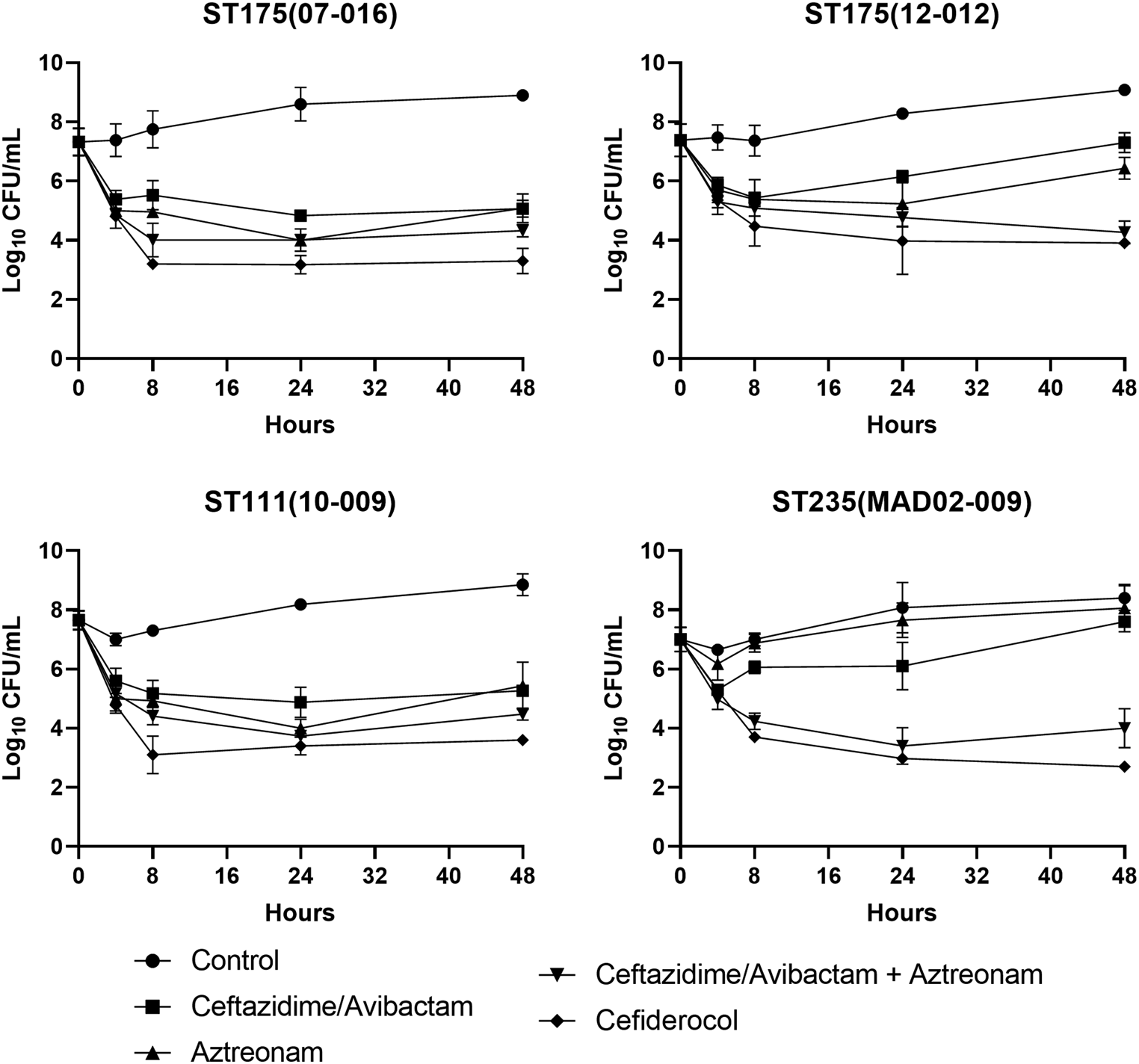
Bactericidal effect was defined as a decrease of ≥ 3log10 CFU/mL in the colony count after 24 h, starting from the initial bacterial density. Synergy was defined as a reduction of at least ≥ 2log10 CFU/mL in the colony count after 24 h when comparing the antibiotic combination with the most potent individual drug. Additive effect was defined as a reduction of ≥ 1-<2log10 CFU/mL in the colony count after 24 h when comparing the combination with the most active antibiotic alone. In the present time-kill studies, a ΔCFU/mL ≥ 2.80log10 was considered bactericidal. The same pharmacodynamic parameters were used for the chemostat results [28].
Chemostat model
Four XDR P. aeruginosa isolates belonging to the most common high-risk clones were selected due to their high prevalence in Spain [5]. A one-compartment in vitro chemostat model was performed to validate the activity of the antibiotics studied by time-kill curves. Chemostat experiments were conducted in duplicate for each isolate and each experimental condition. The chemostat model consisted of four independent glassware reactors. Isolates were cultured on COS plates (Columbia Agar with 5% Sheep Blood) and incubated at 37ºC aerobically overnight. Colonies were inoculated into an Erlenmeyer flask containing 35 mL of CAMHB and incubated at 37ºC overnight in a water bath shaker. Four Reactors were filled with 100 mL of CAMHB and one was filled with 100 mL of ID-CAMHB. Reactors were filled with 100 mL of CAMHB and were supplemented with the corresponding concentration of the selected antibiotic. One reactor was filled with 100 mL of ID-CAMHB and supplemented with cefiderocol. All antibiotics were infused via antibiotic pumps (CADD-Legacy® PLUS; Smiths Medical ASD, Inc.). A reactor without antibiotic was included as a control. The chemostat experiment was placed in an incubator at 37ºC for 48 h. Fresh broth was supplied via a peristaltic pump (Masterflex L/S model 7524-40; Cole-Parmer Instrument Company, Vernon Hills, IL) programmed to achieve the human-simulated half-life of the antimicrobial being tested. Samples were obtained from each of the reactors at specific time points (0, 2, 4, 8 and 24 h) and were centrifuged at 13,000 rpm for 3 min. Serial dilutions in CAMHB were performed with each bacterial suspension. 50 µL of these diluted samples were plated onto TSA II plates and incubated at 37 °C overnight. Colonies were counted after the incubation, and CFU/mL were determined for each condition.
Pharmacokinetic studies
Antibiotic concentrations were collected from the chemostat studies at predetermined time points until the end of the study, and immediately stored at -80ºC until analysis. These samples were taken to validate the antibiotic concentrations previously modelled. Pharmacokinetic samples were determined by high-performance liquid chromatography (HPLC) and validated by a simple linear regression model (r2).
Statistical analysis
In the chemostat model, differences in bacterial concentrations (in logarithmic phase) among antibiotic regimens for each included isolate were assessed using analysis of variance (ANOVA). To ensure the appropriateness of ANOVA application, data normality was verified via the Shapiro-Wilk test, and variance homogeneity was evaluated using Levene’s test. Upon demonstration of statistically significant differences between antibiotic regimens within an isolate by ANOVA, a post-hoc analysis employing Tukey’s test was conducted to delineate these differences. The means of differences (x ?), their respective 95% confidence intervals (CI) and the adjusted p-values for multiple comparisons were reported. A p-value < 0.05 was considered statistically significant. The analysis was performed using the R programming language in the RStudio software version 2023.12.1.


留言 (0)