
記住我


This retrospective case series was approved by the Ethical Review Board of Nihon University School of Medicine (approval no: RK-230411-8) and adhered to the tenets of the Declaration of Helsinki. Written informed consent was obtained from all participants.
Patients who underwent DMEK using this technique between August 2022 and July 2023 were enrolled in this study. To evaluate the effectiveness of this technique, we measured the best spectacle-corrected visual acuity (BSCVA), corneal endothelial cell density (ECD), and central corneal thickness (CCT) before and 6 months after the surgery. After measuring the BSCVA, its decimal values were converted to logarithmic values for statistical analysis. Additionally, the CCT and postoperative ECD were measured using corneal tomography (SS1000; Tomey, Aichi, Japan) and a specular microscope (FA3509; Konan Medical, Nishinomiya, Japan), respectively.
Surgical procedureProcedures before graft repositioningThree experienced surgeons performed the DMEK procedures, with all surgeries performed under local anesthesia. The pre-stripped donor tissue was prepared to an estimated size (approximately 8.0 mm) using a vacuum punch (Moria Japan, Tokyo, Japan) and subsequently stained with 0.06% trypan blue or 0.1% brilliant blue G dye. Descemet’s membrane was stripped from the posterior stroma using a reverse Sinsky hook under air. A 25-G infusion cannula (Kobayashi 25 g DSAEK Chamber Maintainer, Catalog #AE-7802, ASICO, Westmont, IL) was used to preserve the anterior chamber depth by way of a paracentesis. Additionally, peripheral iridectomy was performed at the 6 o’clock position, and a DMEK shooter (G-3863; Geuder, Heidelberg, Germany) or intraocular lens inserter (WJ-60 M; Santen, Osaka, Japan) was used to insert the graft into the anterior chamber through the corneal incision.
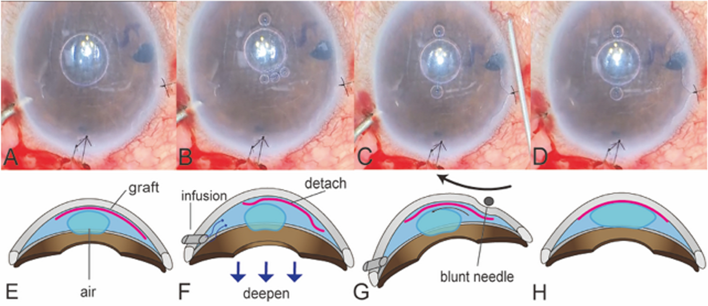
Graft unfolding using the infusion and non-touch techniqueIn cases where the graft was deployed off-center, the volume of air was first reduced to an amount smaller than the graft (Fig. 1A, E). The infusion flow was then turned on and off several times to detach the graft from the posterior surface of the cornea (Fig. 1B, F). At this time, the graft was only detached but remained unrolled. Subsequently, the infusion cannula was grasped, and the eyeball was tilted toward which the graft was intended to be moved. Once the graft was detached, a blunt 27-G needle was stroked from the scleral side toward the center of the cornea (Fig. 1C, G), with slow, deliberate strokes guiding the graft toward the corneal center (Fig. 1D, H). After the graft was moved to the target position, sulfur hexafluoride gas was injected into the anterior chamber, and the graft was allowed to attach (see Video, Supplemental Digital Content 1).
Fig. 1
Illustrations of the graft-repositioning technique using an infusion cannula (A, E) Before repositioning, the DMEK graft is dislocated to the upper right side. B, F The infusion cannula is inserted into the anterior chamber, and the infusion switch is turned on and off several times. The anterior chamber deepens, and the graft detaches from the posterior surface of the cornea through aqueous flow, with the air bubble smaller than the graft. C, G The eye is tilted in the displaced direction, and a blunt needle is stroked from the sclera to the center of the cornea. D, H After performing the repositioning technique, the graft moves to the center. As with conventional DMEK, the anterior chamber is replaced with gas or air, and the grafts are attached DMEK, Descemet’s membrane endothelial keratoplasty
Postoperative medicationsPostoperatively, 1.5% levofloxacin (Cravit; Santen, Osaka, Japan) was administered four times daily for 2 weeks. Additionally, betamethasone (Sanbetason; Santen, Osaka, Japan) and 2% rebamipide ophthalmic solution (Mucosta; Otsuka, Tokyo, Japan) were prescribed four times daily for 3 months and were gradually tapered.
Statistical analysisThe Wilcoxon rank-sum test was used to compare the preoperative and postoperative values of the BSCVA, CCT, and ECD measurements. All analyses were performed using GraphPad Prism version 10.2.3 for MacOS Software (GraphPad Software, Boston, MA, USA). Statistical significance was set at a p-value of < 0.05.
留言 (0)