
記住我


The institutional review board of Seoul National University Hospital approved this study, and the requirement for informed consent was waived because of the retrospective nature of the study. ChatGPT was used for English grammar and expression corrections, but it did not affect the overall content of the paper.
Data sourcesTwo CT scan datasets were used for model development: 105 abdominal CT scans for the abdominal CT model and 60 low-dose chest CT scans for the low-dose chest CT model. Additionally, external test datasets comprising 55 abdominal CT and 10 low-dose chest CT scans were used to assess the performance of these models. The CT acquisition and reconstruction parameters for non-enhanced abdominal CT and low-dose chest CT used in the development and external test sets are presented in Supplementary tables 1 and 2.
Non-enhanced abdominal CT scansThe model development included 105 abdominal CT scans that incorporated non-enhanced phase images obtained from 105 patients. These scans were conducted in the emergency department of our institution between June and November 2020 using a multidetector CT machine (Aquilion ONE, Canon Medical Systems). The inclusion criteria comprised patients aged ≥ 18 years who had not undergone surgical resection or interventional treatment on any of the target organs (the liver, spleen, right kidney, and left kidney) and had no mass-like lesions that altered the normal organ contours. Patients with large or extensive cysts, such as those with autosomal dominant polycystic kidney disease, visible on non-enhanced CT, were excluded. However, organ size was not applied as a separate inclusion or exclusion criterion. The external test dataset consisted of 55 non-enhanced abdominal CT scans acquired from 55 patients at our institution using CT machines different from those used in the development set.
Low-dose chest CT scansModel development included a dataset of 60 low-dose chest CT scans from 60 patients obtained during health check-ups at our institution between January and February 2021. The inclusion criteria for selecting the low-dose chest CT scans matched those used for the abdominal CT dataset, with a focus on the liver and spleen as target organs. While the scan range of low-dose chest CT typically includes a significant portion of the liver and spleen, it does not always capture their lower edges. Therefore, patients with incomplete scans of these organs were included in the study, and only the portions of the liver and spleen within the scan range were evaluated.. The low-dose chest CT scans for the development dataset were obtained using three different CT machines, including Somatom Force (Siemens Healthineers, n = 25), Somatom Definition (Siemens Healthineers, n = 20), and iCT 256 (Philips Healthcare, n = 15). For the external test dataset, 10 low-dose chest CT scans from 10 patients acquired at our institution using a CT machine (Discovery CT750 HD, GE Healthcare) different from those used for the development set.
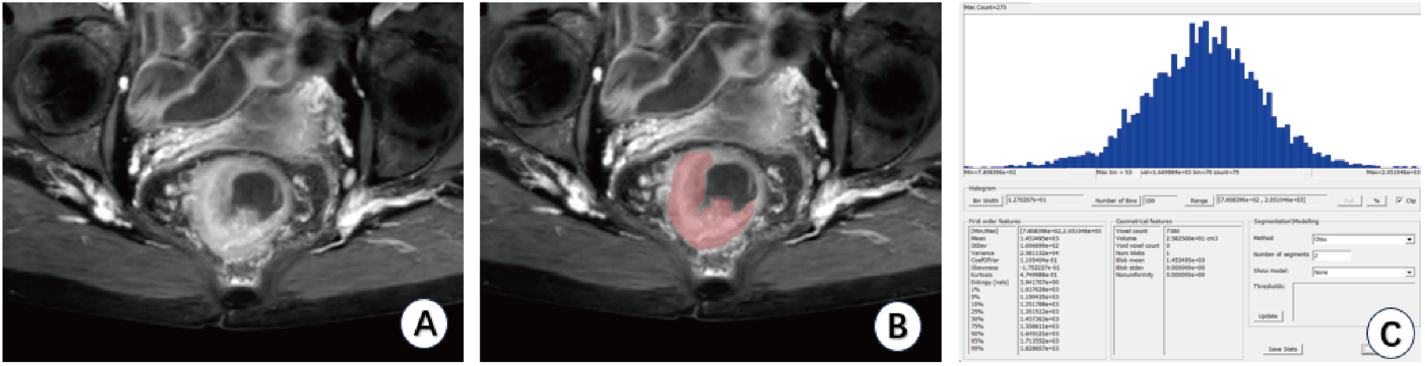
Data preparationAll CT data in the development and external test sets were initially processed using a commercially available software program (MEDIP PRO, v2.0.0, MEDICAL IP Co., Ltd., Seoul, Korea) to efficiently obtain the 3D organ label for each target organ in CT images. The target organs included the liver, spleen, right kidney, and left kidney for abdominal CT, whereas only the liver and spleen were targeted for low-dose chest CT. A board-certified radiologist (J.P., with 6 years of experience in body CT interpretation) manually adjusted the preliminary labels slice-by-slice to create the ground truth (GT). Several rules were applied during the process: (1) the hepatic fissures, intrahepatic portion of the inferior vena cava, and large portal veins (such as the main and lobar portal veins) were excluded from the liver mask as much as possible; (2) only the renal cortex and medulla, excluding the renal sinus, were included in the kidney mask; and (3) all focal lesions within the organs were included in the organ mask, considering the limitations of non-enhanced CT in identifying and delineating focal lesions.
Development of 3D nnU-Net modelsTwo separate 3D nnU-Net models were developed for multi-organ segmentation in the abdominal CT and low-dose chest CT scans (Fig. 1). The development datasets of each model were randomly categorized into training, tuning, and internal test sets with 85, 10, and 10 scans, respectively, for abdominal CT, and 40, 10, and 10 scans, respectively, for low-dose chest CT. Each model was designed to consider internal organ areas, predicted using a body composition segmentation algorithm [24] as input. Subsequently, the model output segmented areas of the liver, spleen, right kidney, and left kidney in abdominal CT and the liver and spleen in low-dose chest CT. The 3D nnU-Net network determined the preprocessing methods and network hyperparameters based on the dataset (including patch size, number of pooling layers, and convolutional kernel size). The patch size was initialized to the median image shape and iteratively reduced to adapt the network topology until the network could be trained with a batch size of at least two, considering the memory constraints of the graphics processing unit. The final patch size configurations were 56 × 192 × 160 and 96 × 160 × 160 for the abdominal CT and low-dose chest CT models, respectively. The last activation function was softmax, and the loss function was the sum of the Dice and cross-entropy losses. Stochastic gradient descent (Nesterov momentum = 0.99) was the optimization algorithm. The polynomial learning rate scheduler was initialized at 0.01. The models were trained for 1000 epochs.
Fig. 1
Architecture of our 3D nnU-Net-based multi-organ segmentation model. InstNorm = instance normalization, LReLU = leaky rectified linear unit
Organ volumetry and 3D radiomics feature extractionThe clinical applicability of the developed models was evaluated by testing their accuracy in measuring organ volume and extracting radiomics features of the target organ. Organ volume and volume-based radiomics features were assessed for each target organ. These radiomics features included three shape-based parameters (sphericity, elongation, and flatness) and seven first-order statistics (mean Hounsfield unit [HU], median HU, standard deviation, skewness, kurtosis, uniformity, and entropy). These features were extracted from the automatically segmented masks generated by the developed models and the GT masks.
Statistical analysisThree widely used metrics for evaluating medical imaging segmentation—including Dice similarity coefficients (DSCs), sensitivity, and precision—were calculated to evaluate the accuracy of the automated segmentation performed by the developed model. These metrics were separately computed into the internal and external test sets by comparing the model-derived masks with the corresponding GT masks [25].
In the external test set, the agreements in organ volumes and radiomics features between the model-derived and the GT masks were evaluated using the intraclass correlation coefficient (ICC) and Bland–Altman analysis. ICC values below 0.5, 0.5–0.75, 0.75–0.9, and above 0.9 were categorized as poor, moderate, good, and excellent agreements, respectively [26]. Furthermore, the volume differences between the model-derived and GT masks were examined. A volume difference below 5% was considered an accurate estimation. Conversely, differences exceeding 5% were categorized as an overestimation or underestimation. Additionally, we determined whether there were significant differences in organ volume and radiomic features measured from model-derived and GT masks using paired t-tests. The significance level was set at p < 0.05.
All statistical analyses were performed using the R statistical software (version 4.2.0; R Foundation for Statistical Computing, Vienna, Austria).
留言 (0)