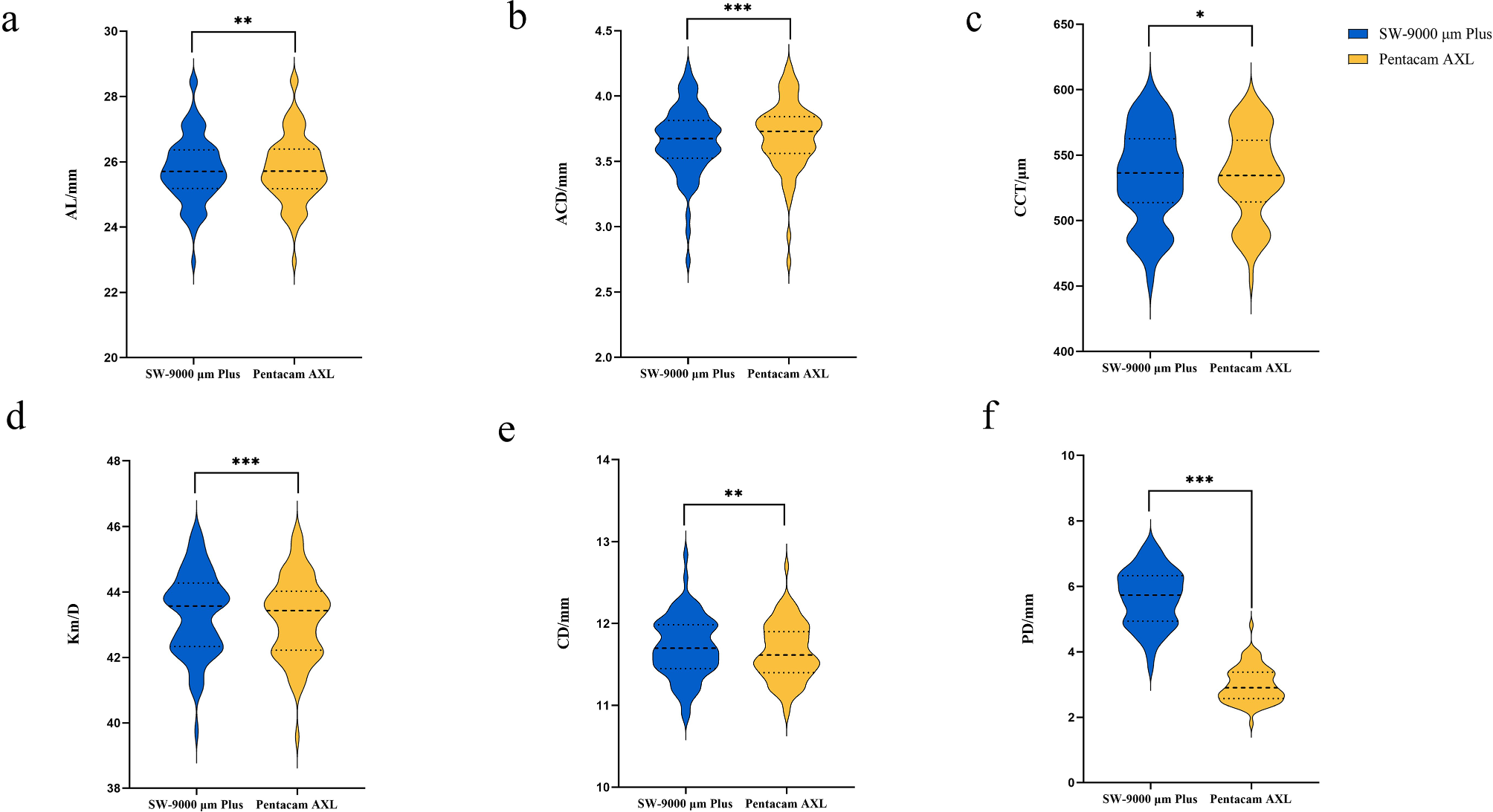
In this study, we found that TNFα was significantly superior in the group treated with insulin, and IL-8 was slightly higher in the group treated with hypoglycemic agents, but without statistical difference. Furthermore, the concentrations of both cytokines were higher in cases where vitreous hemorrhage occurred concomitantly with DR, regardless of the treatment used, although the inverse effect of IL-8 and TNFα was notable, possibly related to the mechanisms of action of each drug; nonetheless, these differences were no significant either. By the immunological technique employed, interleukins 1β, 6, and 10 could not be detected.
Some studies have described that the increase in IL-1β can cause neurodegeneration, apoptosis of retinal capillary cells, and dysfunction of the blood-retinal barrier, [24,25,26,27] which would partly explain the visual dysfunction present in patients with retinal damage. However, IL-1β is not the only one that increases its concentration when DR occurs; Capitão et al. mention that IL-1β, IL-8, and TNFα increase as the disease progresses [25]. In contrast, Lee et al [28]. reported that although there may be alterations in the cytokine profile in cases where proliferative DR coexists with vitreous hemorrhage, the role of IL-1β could be weak in cases of retinal capillary nonperfusion, given that the concentration of this interleukin is low (0.39 ± 0.08 pg/mL), which is similar to the result reported in the study by Koskela et al. (0.3 ± 0.9 pg/mL) [29].
Moreover, Gustavsson and his group could not find detectable values of IL-1β in vitreous samples from patients with proliferative DR by a chemiluminescence assay equivalent to ELISA tests, whose lower limit of detection was 5 pg/mL for this interleukin [30]. Likewise, we could not detect IL-1β concentrations by the CBA technique, which uses antigen-antibody binding as a principle similar to other immunoenzymatic assays. Retinal microglia also synthesize other cytokines such as TNFα, found in early stages of DR, [31] and as we mentioned above, can increase during the disease development. A Korean group reported concentrations of 0.49 ± 0.28 pg/mL,28 much lower than the values of the meta-analysis of Yao et al., [32] where the means of TNFα range from 1.08 ± 0.22 to 973.2 ± 115.4 pg/mL, while in a Brazilian study, the concentrations of this factor were undetectable; [33] these heterogeneous values may be due to the different detection techniques used. Our result exceeded 50 pg/mL in each group evaluated and was higher in the insulin treatment group.
TNFα has also been correlated with IL-6, [34] an interleukin with pro- and anti-inflammatory responses whose concentrations in DR cases have varied from 1.08 ± 0.22 to 276.1 ± 13.9 pg/mL; [35] values within this range were also reported by Mallman et al. (69.37 pg/mL), in patients with proliferative DR who had mean glucose values of 165.1 ± 59.4 mg/dL, and who had associated complications such as vitreous hemorrhage or retinal detachment [33]. Furthermore, it is known that the regulatory capacity of IL-6 changes over time and that its concentration tends to decrease as the duration of DM increases [36, 37] During the development of T2DM there is also the suppression of some genes that encode proteins that regulate the immune response, such as IL-10, an anti-inflammatory cytokine that inhibits the translocation and infiltration of pro-inflammatory cells from damaged capillaries due to oxidative stress; the reduction of this cytokine and other dual inflammatory response proteins, as well as some transcription factors that are precursors of antioxidant enzymes, could have a repercussion in the progression of retinal damage [36].
On the other hand, previous research such as those conducted by Pessoa et al. or Yang et al. suggests that the presence of IL-8 in the retina is the result of the degranulation of immune regulatory cells in the eye, [38, 39] as a consequence of the presence of TNFα that intervenes in the mechanism of the enzyme nitric oxide synthase (NOS), increases the production of nitric oxide (NO) and promotes oxidative stress, a precursor to the activation of these cells [40]. IL-8 is a cytokine that stimulates neovascularization; [41] in patients with proliferative DR, vitreous humor concentrations of 79.6 ± 9.7 mg/dL have been found, which exceed those found in cadaveric controls (19.0 ± 3.9 pg/mL) without a history of ocular or systemic disease [42]. This increase would be related to the proliferation of vessels in DR, but the detectable concentrations of IL-8 have also been contradictory.
In a study from Slovenia that evaluated 68 patients with T2DM and proliferative DR, where more than 80% were insulin-dependent, a mean of 358 ± 698 pg/mL of IL-8 was found, [43]which was similar to our mean result (346.18 ± 174.57), although the first had a higher statistical dispersion; furthermore, the duration of diabetes was similar to the reported in our study (19.02 ± 6.86 versus 17.26 ± 7.37 years), and subjects who had vitreous hemorrhage or macular detachment were included in the analysis. However, none of these reports evaluated the cytokine profile of antidiabetic treatments or other pharmacological therapies included in their studies.
In this sense, pharmacotherapy has demonstrated its effect on the progression of DR independently of glycemic control. In the review by Saw et al. (2019), [44] different antidiabetic therapies were addressed, such as metformin, whose cardioprotective effect has been widely described, so it is considered that its antiangiogenic and anti-inflammatory effects could also limit the microvascular alterations typical of DR [45]. Glibenclamide has also shown that it can prevent the progression of DR, although its effect is inferior to other sulfonylureas such as glycoside; [46] even when this drug is not considered among the first-line therapies in the recent years, it is commonly found in the therapeutic scheme of T2DM patients in our country. As part of the most common antidiabetic therapies, insulin, and its analogs are key to maintaining the normal functioning of the retinal microvasculature. Since a patient with T2DM usually has defects in the action of this hormone, the secretion of insulin-like growth factor 1 (IGF-1) decreases in the systemic blood circulation, affecting vascular homeostasis (including retinal microvasculature); therefore, it is considered that insulin and its analogs may have direct and indirect effects involved in the development of DR [47].
A 2023 study identified that both insulin and its analogs could be associated with an increased risk of developing clinically significant DR; however, these results should be taken with caution due to differences in the mechanisms of action of the drugs, and a possible “worsening effect” arising from the rapid reduction of serum glucose levels and the transient increase in inflammatory mediators and growth factors in response to changes in the environment [48].
In our study, the cytokine profile in the vitreous was analyzed and compared between the antidiabetic treatments commonly administered for T2DM, to evaluate the indirect therapeutic effect that some drug combinations can have on diseases for which they are not directed in the first place, which could serve to guide therapeutic indications in patients with T2DM who present microvascular complications in the retina. Still, as potential limitations, we did not consider body weight, body mass index (BMI), or other metabolic variables, and the specific effect of the types of insulin, or other therapies such as those administered in patients with SAH, which could modify the inflammatory response through additional signaling pathways that indirectly participate in the proliferative DR development. Even though the presence of molecules such as IL-8 and TNFα in both treatment groups suggests an ongoing pro-inflammatory response, it would be necessary to take these results with caution, because the sample sizes in the stratified groups (vitreous hemorrhage vs. retinal detachment) were small enough to give us a full outlook of the possible indirect effects of the treatments.
Also, we did not include other immunoassays for the detection of cytokines that could have added value to this study; however, multiplex studies such as the one used, have demonstrated their reproducibility and usefulness for the simultaneous measurement of proteins even at low concentrations, with a broad cost-benefit advantage. The fact that some of the cytokines of interest, such as IL-1β, IL-6, and IL-10, have not been detected, could be due to the duration of T2DM or the activity of each cytokine, but this will require further analysis, such as molecular biology techniques, to evaluate the gene expression that coded those proteins.
In summary, the concentration of TNFα in the vitreous is superior in patients receiving insulin treatment, and IL-8 is higher in the oral hypoglycemic drugs group of metformin + glibenclamide; however, in this interleukin, the concentrations did not reach a significant difference between the groups. Yet, this response is more pronounced in patients with vitreous hemorrhage, possibly due to the migration of cytokines from the retina. Until the last review of the literature, no similar reports were found that compared the cytokine profile in vitreous between different antidiabetic therapies, so these findings emphasize the impact that treatments can have on vascular and microvascular complications, and could expand the outlook for interdisciplinary management of preventive therapies in patients with retinal complications associated with T2DM.


留言 (0)