
記住我
Knee osteoarthritis (KOA) is a degenerative disease characterized by clinical symptoms and joint tissue deformations. KOA is associated with damage to the articular cartilage and related pain, swelling, and joint stiffness, which are the main causes of disability (1). As a chronic degenerative disease, the pathological damage caused by KOA involves inflammation, injury, genetics, and other factors (2–4). Due to the depth of research, various treatment methods, including nondrugs, drugs, and surgery, play certain advantages in the clinical treatment of KOA. However, owing to the complexity of its pathogenesis, there is currently no single treatment for the complete eradication of KOA (5–8). The incidence of KOA is high and is expected to increase (9). Studies have shown that the overall prevalence of hip and knee OA worldwide is approximately 300 million (10). The prevalence of mild, moderate, and severe KOA in China is 1.5%, 3.3%, and 3.9%, respectively, and the incidence in women is much higher than that in men (11). According to a study by American scholars, about 9.29% of people over the age of 60 in the United States have been diagnosed with symptomatic KOA (12). Diagnosis of KOA is critical given its high incidence. Imaging plays an important role in KOA diagnosis.
The imaging methods used to detect KOA include ultrasonography, radiography, computed tomography, and magnetic resonance imaging (MRI), all of which have advantages (13, 14). Tissue damage, including cartilage and meniscal damage, can affect the onset of KOA. However, MRI is more sensitive to changes such as cartilage injury, soft tissue lesions, and chondroedema than radiographs, which can only evaluate the knee joint space. This helps to assess the occurrence of KOA early and prevent it as soon as possible (15). The value for the diagnosis and treatment of KOA has gained increasing attention. For example, an imaging omics-clinical nomogram model based on MRI-bone marrow edema showed good performance in the diagnosis of early osteoarthritis (16). Meanwhile, using semi-quantitative methods, MRI can classify KOA into meniscus, cartilage, inflammation, and other structural phenotypes. The Whole Organ Magnetic Resonance Imaging Score (WORMS) and other MRI-based scoring systems have also been developed for epidemiological studies and risk factor analysis of KOA (17, 18). Quantitative morphological cartilage assessment is occasionally performed using MRI, including the segmentation of bone, cartilage, bone surface, cartilage surface, and cartilage morphology measurements. Component MRI analysis and MRI pulse sequence analysis have also been applied in imaging research for KOA, and each has certain advantages (19–22). MRI provides a more detailed observation of cartilage and bone marrow edema; therefore, it can be used for the diagnosis and clinical treatment of KOA (23, 24). For example, in an imaging omics-clinical nomogram model based on MRI-bone marrow, edema showed good performance in the diagnosis of early osteoarthritis (16). Studies by Salikhov et al. have also shown that in patients with type 1–2 KOA cartilage defects, MRI can accurately assess cartilage damage and guide the treatment of stromal vascular parts. MRI is helpful for evaluating the repair effect of the Stromal vascular fraction (SVF) on cartilage injury and provides a clearer basis for clinical treatment (25). Due to the advantages and extensive application of medical knee MRI in the diagnosis, evaluation, and treatment of KOA, it is of practical significance to clarify the application status of MRI in KOA to analyze future research hotspots and guide further research. Bibliometrics provides the possibility of conducting research as a scientific method for literature analysis.
Bibliometrics is a popular and rigorous method for exploring and analyzing large amounts of scientific data. Bibliometrics enabled us to reveal the current research situation and differences in a specific field, as well as the emerging direction in this field (26). Bibliometrics analysis can use the information of the network literature database to quantitatively and qualitatively evaluate the main content and hot spots of a certain field (27). We employ bibliometrics to provide technical support for this study to deeply investigate the current global application of medical knee MRI in patients with KOA and analyze future research trends.
2 Materials and methods 2.1 Data sourceOn January 7, 2024, 6,050 articles were identified in the Web of Science (WoS) Core Collection: the Science Citation Index Expanded (SCIE) and the Social Science Citation Index (SSCI). The bibliometric analysis was based on WoS, which is considered the best bibliometric database.
2.2 Search strategy and data collectionThe search strategies are summarized in Table 1. One author conducted a preliminary screening (J. C.) and then provided the results to the other two authors for rescreening (H. Z., Z. W.). Patients with serious knee diseases such as cancer, tuberculosis, hip osteoarthritis, acute knee injury, rheumatoid arthritis, gouty arthritis, psoriatic arthritis, and other non-KOA studies were excluded. A total of 2,996 articles remained for potential inclusion. CiteSpace (version 6.2. r7) was used to analyze literature. A total of 92 duplicate articles were excluded and 2,904 were ultimately included.
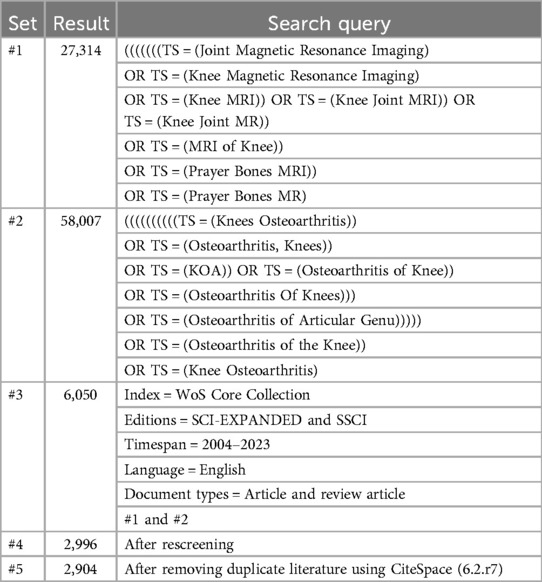
Table 1. Literature retrieval methods.
2.3 Bibliometric analysisCiteSpace and VOSviewer are the main data visualization tools used in bibliometric studies. They present the structure, laws, and distribution of knowledge on a certain subject in the form of scientific knowledge graphs to discover advances and research frontiers in specific fields. We used CiteSpace and VOSviewer to complete relevant analyses of countries, institutions, journals, authors, references, and keywords. ScimagoGraphica is a grammar-based digital imaging tool that was used to complete national geographic distribution, publication trends, references, and keyword network connection analyses.
3 Results 3.1 Annual number and trend of publicationsA literature review was conducted from January 1, 2004, to December 31, 2023. A total of 2,904 articles were published on the application of medical knee MRI in patients with KOA, with an average of 145 articles published annually. After 2009, the number of documents issued annually exceeded 100; since 2020, the number has exceeded 200. With an in-depth understanding of KOA pathogenesis and further development of MRI technology, the number of publications will also increase (Figure 1). According to the results, the total number of research studies and annual average number of medical knee MRI applications in KOA are increasing, which has certain research prospect.
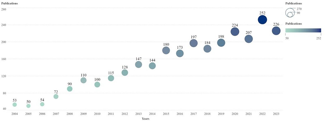
Figure 1. Annual publication trend chart of research on the application of MRI in patients with KOA. The horizontal axis is the year and the vertical axis is the number of published articles. The node size and color represent the number of publications. Smaller nodes and lighter colors represent fewer publications. The number of posts per year is indicated above the node. MRI, magnetic resonance imaging.
3.2 National analysisA national analysis revealed that from 2004 to 2023, 76 countries participated in MRI-related research on KOA. The United States had the largest number of publications (1,260), followed by Australia (618) and Germany (506). The United States had the highest intermediary centrality (0.35), followed by Germany (0.32), Australia, and the United Kingdom (0.21). The United States had the highest total citation frequency of 57,311, followed by Australia (24,608) and Germany (20,498) (Table 2).
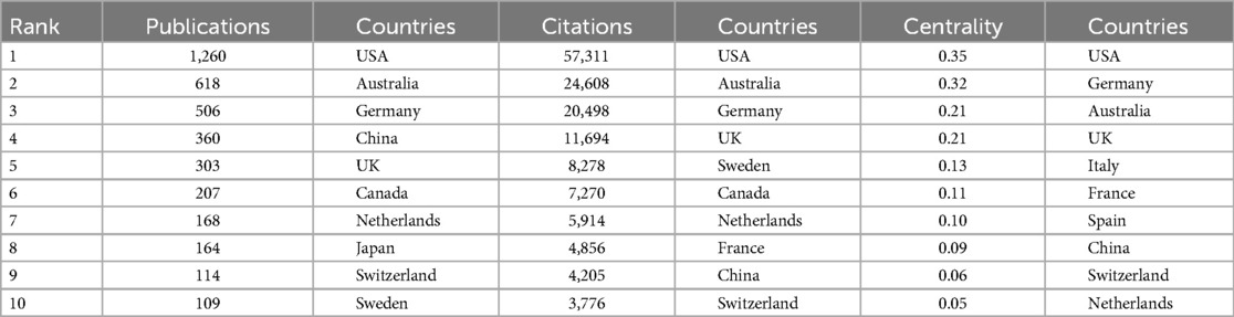
Table 2. Top ten countries in the field of publications, total citation frequency, and mediation centrality in the application of MRI in KOA.
The total citation frequencies of the United States, Australia, and Germany were consistent with the rankings of their published papers, proving that these three countries have a strong influence in this field. The national geographic distribution map of countries shows that the United States and Australia have formed close ties with European countries such as Germany and France. China and Japan have numerous publications and are closely linked (Figure 2A). As can be seen from Figure 2B, the literature published in China, India, Saudi Arabia, and other countries is relatively recent, indicating that these countries are strengthening their research on the application of MRI in KOA. In general, the United States, Australia, and some European countries are key countries in this field, and both the number of publications and the total number of citations are among the top worldwide. Among Asian countries, China and Japan are the major publishing countries.
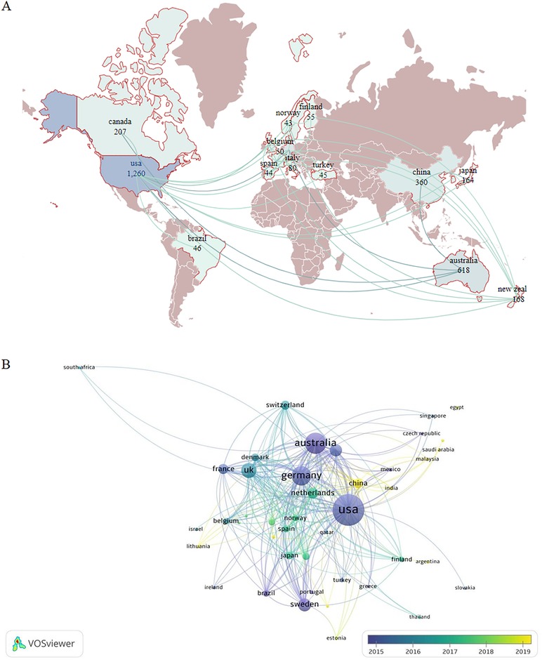
Figure 2. (A) Geographic map of the national document distribution. The figure shows countries with more than 40 publications. Dark brown color represents countries with fewer than 40 publications, whereas the light blue color represents those with a low number of publications. The more connections between the two countries in the picture, the closer the country is to other countries. The numbers below each country represent the number of publications in the field. (B) National network connectivity map. Larger node indicates more total literature citations in the country. The color of the node represents the publication time of the country. Darker color indicates earlier publishing time.
3.3 Institutional analysisAccording to data analysis, 2,873 institutions have published literature on the application of medical knee MRI in KOA. We analyzed 2,873 institutions involved in MRI research on patients with KOA over the past 20 years, of which 151 institutions published >10 articles. Boston University ranked first, with 400 publications, followed by the University of California, San Francisco (314) and Monash University (213). The University of A Coruña INSERM had the highest intermediary centrality of 0.7, followed by the Leeds Biomedical Research Centre (0.67) and Feinberg School of Medicine (0.67). Boston University had the highest citation frequency of 22,308, followed by the University of California San Francisco (17,376) and Monash University (9,354) (Table 3).
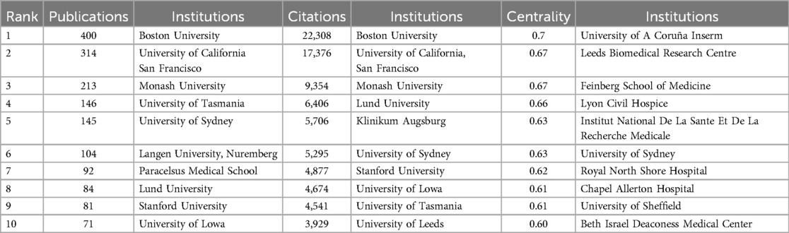
Table 3. Top 10 institutions in the field of publications, total citation frequency, and mediation centrality.
Boston University had the most citations, proving that it has considerable academic influence and research achievements in this field. In contrast, the University of A Coruña INSERM and the Leeds Biomedical Research Centre had the highest intermediary centrality, which may have been affected by the research content of the articles. The University of A Coruña INSERM has focused on the genetics of OA and the standardization of clinical symptoms and treatment. Research from the Leeds Biomedical Research Centre has focused on MRI studies of rheumatic immune system diseases and the manifestations of several knee diseases (28–30). Few studies in the literature are from the Feinberg School of Medicine, and most have focused on analyzing the risk factors for KOA (31, 32). As shown in Figure 3, institutions such as Southern Medical University and Anhui Medical University later faced issues regarding this topic. These institutions have invested considerable research into the application of mechanical knee MRI in KOA and have achieved certain results. In short, institutions in the United States and Europe have a large number of publications, and the content of different institutions in this field is inconsistent. In recent years, institutions in China have paid close attention to the content of this field and have published a certain number of posts.
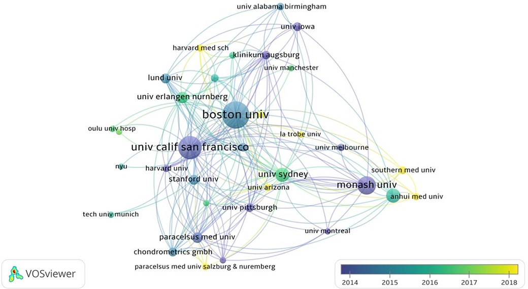
Figure 3. Institutional network connectivity diagram. The size of the nodes represents the number of posts by the agency. Larger nodes indicates more the number of posts. The color of the node represents the publishing time. Darker color indicates earlier the publishing time.
3.4 Journal analysisA total of 470 journals published articles on the application of medical knee MRI in KOA, of which 29 published >20 articles. The journal with the greatest number of articles was Osteoarthritis and Cartilage (445), followed by BMC Musculoskeletal Disorders (97) and Skeletal Radiology (84). Osteoarthritis and Cartilage ranked first in the number of citations with 20,320, followed by Annals of the Rheumatic Diseases (7,494) and Arthritis and Rheumatism (6,411). Osteoarthritis and Cartilage had the greatest total citation frequency (16,155), followed by Annals of Rheumatic Diseases (7,697) and Arthritis and Rheumatism (7,380) (Table 4). Osteoarthritis and Cartilage had the highest number of publications and total citations and was a core journal in this field (Figure 4A).
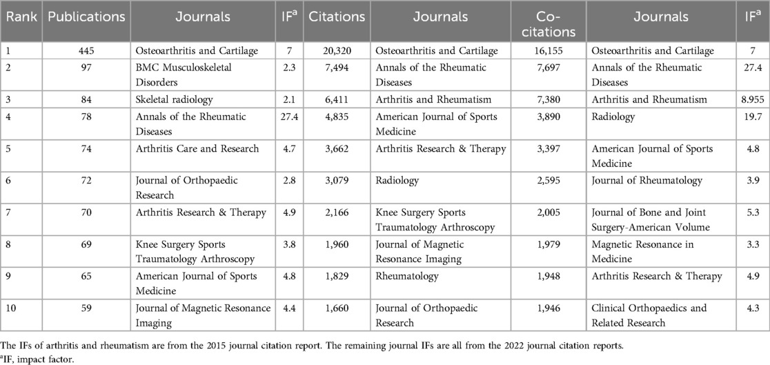
Table 4. Top 10 journals in the field of publications, total citation frequency, and total citation frequency of application of MRI in KOA.
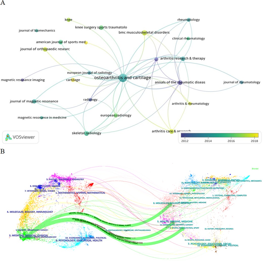
Figure 4. (A) Journal network connectivity map. The size of the nodes represents the number of articles published, where the larger the nodes, the more research articles the journal publishes in this field. The color of the node represents the time of the publication, where the darker the color, the earlier the magazine publishes the article. (B) Double image overlay of the journal. The left side is the cited journal, and the right side is the back cited journal. The colors of the connecting lines represent the citation journals derived from the same category. Thickness of the connection represents the number of references (i.e., as the number of references increases, so does the thickness of the connection).
According to the results of the journal double-image overlay analysis, medical knee MRI and KOA studies were published in orthopedic journals, but were also relevant to molecular science, nursing, sports, rehabilitation, and other fields (Figure 4B). The results show that journals published in this field have a wide range and high quality, and articles published in orthopedic journals are more likely to receive the attention of scholars.
3.5 Author analysisIn total, 10,884 authors participated in research on the application of medical knee MRI in KOA. Nineteen authors have published more than 50 articles. Guermazi A was identified as the author with the largest number of publications (309), followed by Roemer FW (183), Eckstein F (148), and Hunter DJ (148). Guermazi (0.22) had the highest intermediary centrality, followed by Cicuttini (0.21) and Hunter (0.18). Guermazi also had the highest number of citations (16,680), followed by Hunter DJ (8,908) and Felson DT (8,882) (Table 5).
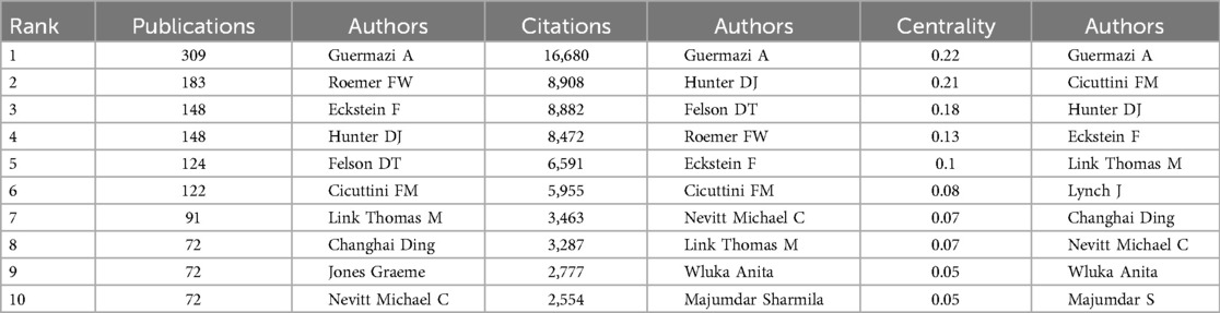
Table 5. The authors of the top 10 publications, total citation frequency, and mediation centrality in research on the application of MRI in KOA.
The author analysis showed that Guermazi A ranks first among all indicators. Guermazi et al. extensively studied not only the pathological mechanism of KOA but also the factors influencing KOA and the effect of different treatments on MRI results in patients with KOA (33–35). The number of publications and total citations for Cicuttini FM were low, but the central medium ranked second. Cicuttini et al. focused on the standardization of MRI scans in patients with OA (36–38). Although the rankings of Hunter DJ were lower than those of Guermazi A, they were stable, proving that Hunter DJ has a certain influence in this field. Hunter focused on the analysis of risk factors affecting the clinical symptoms of patients with KOA and disease prediction and judgment using MRI technology (39–41). The authors' network connections demonstrate the strength of cooperation between the authors. Figure 5 shows that the total connection strength between Guermazi and Roemer, Eckstein, and other authors is high, indicating that they not only have a high number of publications in this field, but also have willingness and strong ability to collaborate. The results show that many authors are involved in this field, and that the research content affects the degree of the authors' attention. Guermazi A is a leading author in this field.
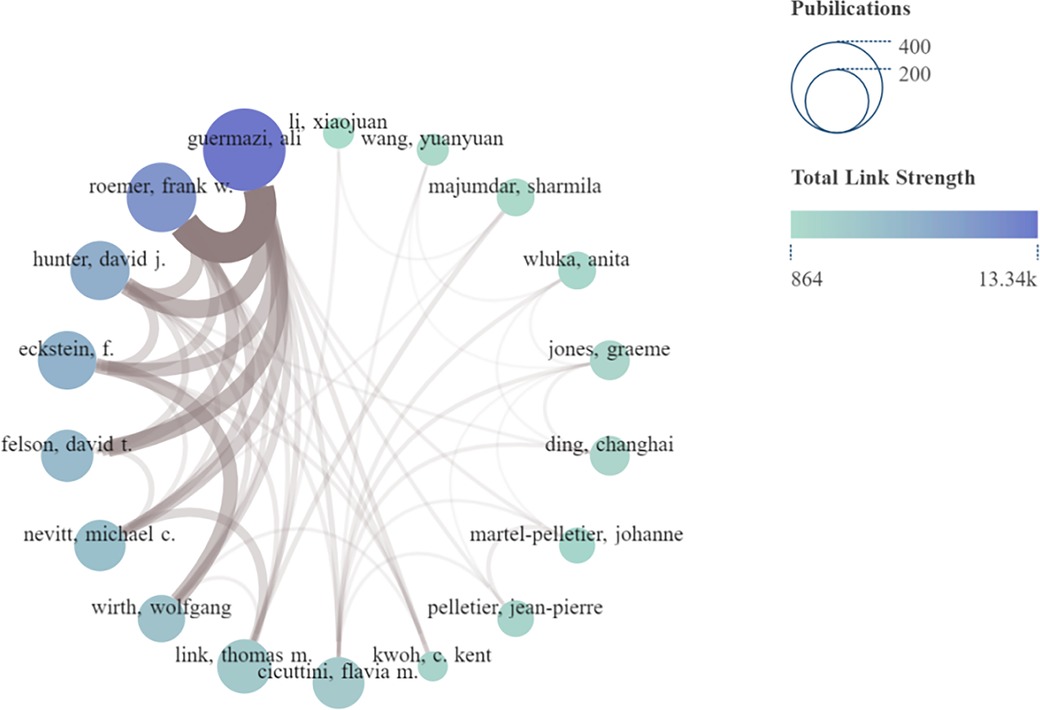
Figure 5. Network connection diagram of authors. The size of the node indicates the number of articles published (larger nodes equate to more articles), and color shade of the node indicates the total connection strength (bluer shade equates with a greater total connection strength). The thickness of the brown connector between authors indicates the strength of the connection (thicker translates to a closer connection).
3.6 Reference analysisAccording to the citation analysis, 43,288 references were cited in this study, and the top 10 were cited more than 177 times. The top three references with the highest total number of citations were the Radiological Assessment of Osteoarthrosis (42) (732), Whole-organ Magnetic Resonance Imaging Score (WORMS) of the knee in Osteoarthritis (43) (634), and Evolution of Semi-quantitative Whole-joint Assessment of Knee OA: MOAKS (MRI Osteoarthritis Knee Score) (44) (378) (Table 6). Figure 6A shows the total cited network connection strength of the references.
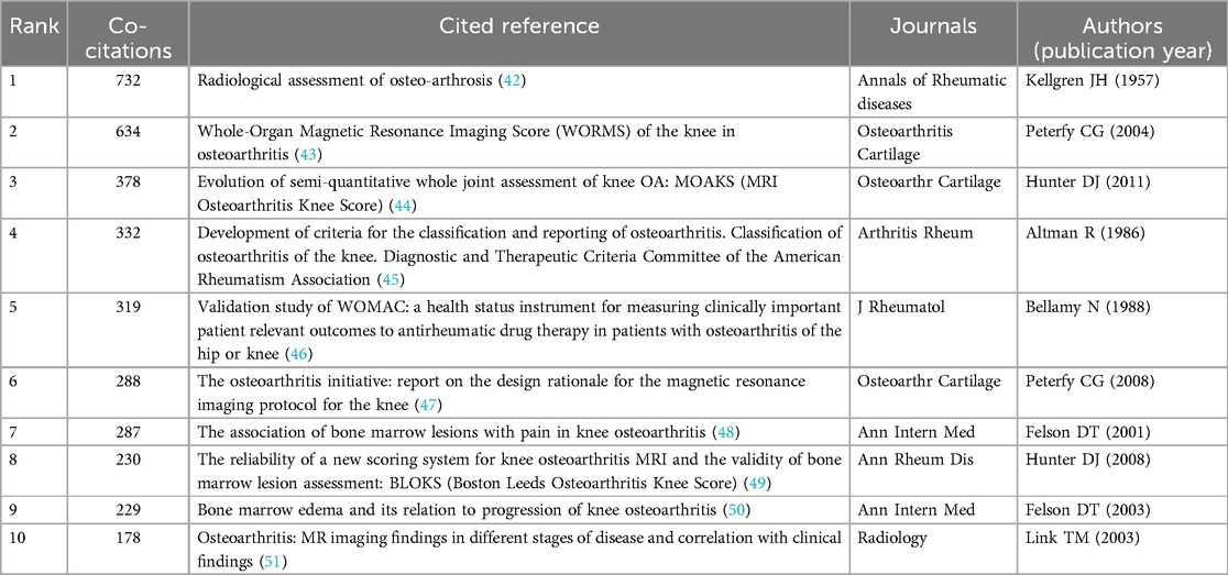
Table 6. Top 10 references of research on the application of MRI in KOA.
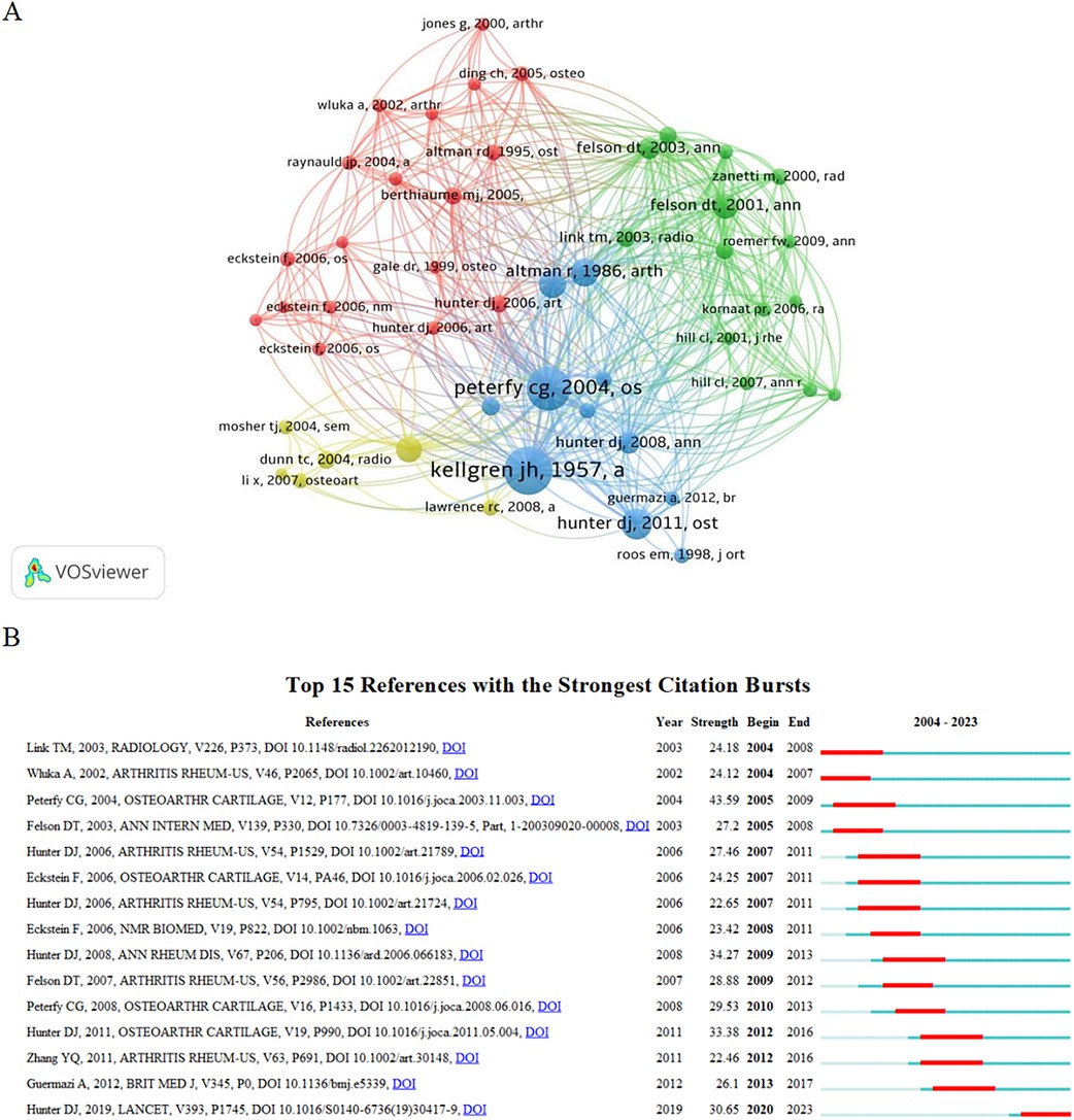
Figure 6. (A) Reference cocitation analysis diagram. The node size represents the number of cocitations, meaning that the larger the node, the more times the literature is cited. Different colors represent different species. The references of the same color have similar research content and are closely related. (B) Reference outbreak analysis diagram. “Strength” represents the burst intensity of the literature. The red line represents the concentrated outbreak time for this reference. The light blue line in the figure shows that although the literature is not a reference at this stage, it also obtains some citations.
Reference burst analysis showed that 15 references had a high burst intensity. Since 2011, literature focusing on the relationship between MRI and fluctuations in knee pain, bone marrow lesions, and changes in synovitis, as well as the diagnostic advantage of MRI for KOA, has received considerable attention (Figure 6B). According to the results, references with a large number of cocitations also had a high outbreak intensity. Their research content involved the diagnosis of KOA, research and development of evaluation tools, KOA risk factors, clinical stage, and other aspects that have attracted considerable attention.
3.7 Keyword analysis 3.7.1 Keyword co-occurrence analysisIn total, 6,178 keywords appeared in this study, 93 of which appeared >50 times. “Knee osteoarthritis” (2,189) had the highest frequency, followed by “MRI” (1,463) and “articular cartilage” (1,180) (Table 7). The results of the keyword co-occurrence analysis showed that “KOA” and “MRI” were ranked as the top two keywords. “Articular and cartilage” was the third most common keyword, with 1,180 co-occurrences, indicating that the application of MRI in patients with KOA has focused more on the articular cartilage. The keyword connection strength network shows that MRI is applied to many aspects of KOA; however, it focuses on the study of knee cartilage and symptoms of KOA (Figure 7A). The evaluation and application of medical knee MRI in KOA cartilage, such as the volume and thickness of the cartilage, is the most important aspect in this field.
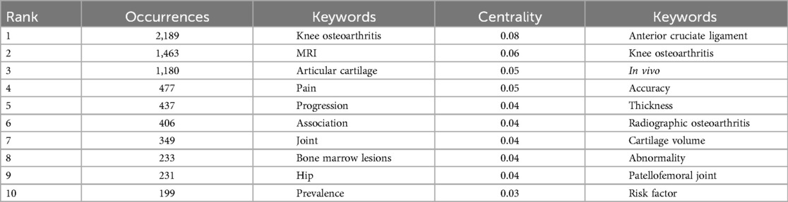
Table 7. Top 10 keywords in terms of co-occurrence frequency and mediation centrality in the field.
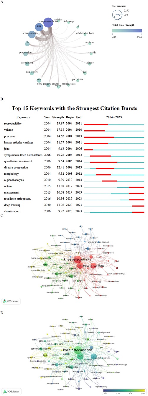
Figure 7. (A) Top 20 keyword network connection graph. The size of the node indicates the number of published articles related to the topic of the keyword (the larger the node, the higher the frequency of keyword co-occurrence, the more literature). The color shadow of the node indicates the total connection strength (the bluer the color, the greater the total connection strength, indicating that the more important the keyword is in the field). The brown joint thickness varies between keywords; the thicker the connector, the tighter the connection. The size of the node represents the number of articles published related to the topic of the keyword (the larger the node, the higher the keyword co-occurrence frequency, the more literature), and the shade of the node represents the total connection strength (the bluer the color, the greater the total connection strength, indicating the more important the keyword in the field). The thickness of the brown joint between the key words is different; The thicker the connector, the tighter the connection. (B) Explosive word analysis diagram. “Strength” represents the burst intensity of the literature. The red line represents the concentrated outbreak time of the keyword. The light blue line in the figure shows that although this keyword is not a reference at this stage, it is also cited. (C) Keywords cluster analysis. Different colors represent different clusters. The size of the node is related to occurrence; the larger nodes equate with greater occurrence. The color of the node represents the category to which the keywords belong, and the same color means that these keywords belong to the same cluster. (D) Key words network connection diagram. The node size represents the number of occurrences of the keyword. Larger node indicates more frequent occurrence of the keyword. The color of the node represents the keyword. Darker color indicates earlier appearance of keywords.
3.7.2 Analysis of outbreak-related wordsThrough the discovery of intermediary centrality, the “anterior cruciate ligament”, “cartilage thickness”, “cartilage volume”, and “patellofemoral joint” have attracted the attention of researchers. However, as the overall intermediary centrality was low, the reliability of the conclusions required additional data.
Combined with an analysis of explosive words, it was possible to further clarify the existing research hotspots. “Reproducibility” and “volume” were keywords with the highest outbreak intensities. It has been proven that before and after 2010, MRI of KOA focused on image reproduction and volume measurement, especially human articular cartilage. MRI of the knee joint also showed a high outbreak intensity in 2006–2012. As an artificial intelligence (AI) analysis method, “deep learning” experienced a high outbreak intensity in 2020. Therefore, in the next few years, the number of articles combining AI with KOA imaging is expected to increase (Figure 7B). Additionally, MRI may be used as a standardized method in clinical research on KOA and may promote the formulation of standards for imaging and symptomatology. From the analysis of the results, different research stages have different research hotspots, but compared to “management” and “classification,” “deep learning” has a high outbreak intensity after 2020, which will be a research hotspot in the next few years.
3.7.3 Keyword cluster analysisThe keyword cluster analysis clearly indicated the widespread application of medical knee MRI in KOA risk factors, clinical treatment, and pathological injury research. Four cluster groups were formed by cluster analysis, and “KOA”, “pain”, “bone marrow lesions”, and “injury” were the main keywords in the four clusters (Figure 7C). According to the cluster analysis results, the relationship between the characteristics of pathological lesions, such as cartilage and subchondral bone injury, bone marrow defects, and inflammation, and KOA is the main component of MRI research. As shown in Figure 7D, the application of MR technology in KOA is extensive and involves risk factor analysis, pathological changes, diagnosis, and treatment.
4 Discussion 4.1 Methodological analysis of the bibliometricsAs the main software for this bibliometric analysis VOSviewer, CiteSpace, and SCImago Graphica played significant roles as the main software packages for this bibliometric analysis. VOS viewer is a free computer program, which was expanded by van Eck et al. (52), who provided a viewer with a variety of bibliometric maps, including density views and cluster density views. The VOSviewer view feature has a significant advantage for map presentation, with at least a moderate number of items (e.g., at least 100 items), while also containing the cocitation and co-occurrence analysis of various elements, including countries, institutions, and authors. The CiteSpace treatment of a bibliometric atlas is more complex. However, this approach also has unique advantages. For instance, it can not only delete duplicate literature in the data but also analyze the outbreak word for each element and then show the research hotspot in a certain field in different time periods. At the same time, unlike VOSviewer, CiteSpace calculates the mediation centrality of content in a certain element. Mediator centrality is the main indicator of bibliometrics, and nodes with high mediation centrality may lie between two large clusters or subnetworks. High mediator centrality makes it easier to gain attention in the field. In addition, CiteSpace can conduct an analysis of the double-graph superposition of journals and show the relationship between different categories of journals with color lines, which allows readers to understand the intermediary centrality of relevant content in a certain element. As a tool for visual operating systems that has been developed in recent years, SCImago Graphica does not require code and can transform various data sources (53). SCImago Graphica aims to combine high levels of expressive force with an easy-to-use interface. Moreover, the design of SCImago Graphica is highly flexible for the image presentation of the data and it also can realize visual communication of the data.
In addition, VOSviewer was employed to complete the data analysis of countries, institutions, journals, authors, references, publications, and keywords; obtain specific data of different elements; and display them in the form of an analytic knowledge graph. However, CiteSpace was used to conduct the mediation centrality of each element, superposition of journal dual graphs, and correlation analysis of outbreak words to compensate for the deficiencies of VOSviewer. SCImago Graphica was used to create a national geographic distribution map and a network connection map between the author and keywords so that the total connection strength between the author and keywords was more directly displayed.
Some previous bibliometric analyses have combined VOSviewer, CiteSpace, and SCImago Graphica. For example, Wang et al. analyzed the top ten0 articles in the field of chronic fatigue syndrome by combining the three packages, and all three were used to map knowledge. These results appear to be more intuitive (54). A similar bibliometric analysis of Titis obliterans was performed by Liu et al., who used VOSviewer to analyze academic collaborations between different countries, institutions, and authors, while simultaneously drawing a visual map of the cocited authors, journals, references, and co-occurrence keywords. CiteSpace was also used to supplement the periodic double-image superposition and outburst word analysis. The Scimago Graphica application was used to map academic cooperation between different countries/regions, similar to the method used in the present study (55). However, in their study, the mediation center results were not displayed, and the in-depth discussion of the results in the Discussion was reduced, with only a brief explanation of the results. The filterometric analysis of the rough set was similar to that presented by Yu et al., where SCIE and SSCI were selected as literature sources and article types (articles or comments). Moreover, a network analysis of countries, regions, and authors was introduced, and research hotspots and trends were explored through the cocitation of keywords and analysis of outbreak-related words (56). However, in contrast to their research, our research focuses on the cocitation analysis of different elements and the top ten contents of the elements, which helps readers grasp the main content and hot spots in the analysis of each element and then helps scholars find the direction of in-depth development. Later, they conducted a quantitative bibliometric analysis of the publications of Applied Intelligent Magazines, where they divided content into different time phases, focusing on the time trend and proportion of factors in the number of applied intelligence articles and citations (57). However, relatively few studies have analyzed these results.
Compared to similar bibliometric analyses, the present study focuses on the number of research elements, cocitation or cocitation number of times and mediation centrality in the process of analysis. The display of the knowledge graph does not pay attention to the comparison of the results of the three drawing software programs but focuses on the three software programs to complement each other and show the research results in a more comprehensive way. A very large space is used to conduct in-depth discussions of the results and analyze future research trends and hotspots.
4.2 Global publication status and quality analysis of research in the application of medical knee MRI in KOAThis study included 2,904 studies. Based on the annual publication trend, research on medical knee MRI in KOA is increasing. The United States has published many studies in this field. Moreover, its intermediary centrality and total citation volume are at the forefront, have a certain output in this field, and have great influence. Several factors influenced the outcome. First, the progress in MRI technology in the United States cannot be ignored. In the functional evaluation of real-time MRI, American scholars have analyzed the morphological characteristics of the cartilage by studying its volume and thickness. Simultaneously, the automatic segmentation of the cartilage and meniscus morphology was analyzed based on a convolutional neural network (CNN), which has a significant influence in this field (58). In studies on the integrated management of KOA, studies from the United States had high numbers of citations (59). European countries, such as Germany and France, also have a large output in this field and strong connections with the United States. In authors from these countries co-operation with the United States, they not only elaborated on the current situation of the application of medical knee joint MRI in KOA but also discussed the related MRI technology and analyzed the application characteristics of MRI technology in KOA (60). At the same time, they established a magnetic resonance observation cartilage repair tissue score using the MRI semi-quantitative scoring method, which can evaluate the extent of joint surface damage by scoring the volume of the cartilage defect, degree of integration with adjacent cartilage, surface and structure of the repaired tissue, and signal strength of the repaired tissue on PD-weighted images (61). These studies further promotes the progress of medical knee joint MRI technology and helps to improve the accuracy of diagnosis. These advances in MRI technology are evident in these influential countries.
China has a large number of publications and citations, which is related to its increasing medical investments in recent years. Indeed, owing to the convenience of MRI examinations and the increased incidence of KOA in patients, Chinese scholars have been paying more attention to the application of MRI technology in KOA in recent years. By integrating meniscal and femorotibial radiomic features to predict radiographic KOA incidence using neural networks, Li et al. has also suggested that the predictive value of JS-RM, integrating meniscus and cartilage features, improves the incidence of imaging KOA, which contributes to the early diagnosis of KOA (62). YeQ et al. used MRI to analyze the subpatellar fat pad in patients with KOA, which further supports that imaging omics changes of the subpatellar fat pad were associated with the severity of KOA and knee structural abnormalities in the elderly (63). Analysis of KOA risk factors using MRI can help to evaluate and screen patients with early KOA and provide early intervention in patients with microscopic injury to improve patient outcomes. However, Chinese scholars have focused more on analyzing the risk factors of KOA using MRI, while little research has been conducted on the technological progress of MRI applications in KOA, which may explain the lower total number of citations of Chinese articles. Therefore, while studying MRI detection of KOA, strengthening technical cooperation with scholars in the United States and European countries may further improve the output.
In the analysis of issuing institutions and journals, the results demonstrate the value of MRI in interdisciplinary applications for patients with KOA. Boston University ranked first in terms of the number of published articles and citations, suggesting that it has an important influence in this field. The University of Tasmania and University of Iowa have a large number of published papers, but their total citations and intermediary centralities are low, suggesting that they must work closely with other core institutions to improve their research quality. Many indicators of the University of Sydney are in the top ten, proving that the institution has certain achievements in this field, with a focus on biomechanical and clinical research related to KOA, demonstrating MRI is valuable for the development of interdisciplinary KOA research (64–66).
Journal analysis showed that Osteoarthritis and Cartilage was the most important journal for MRI applications in KOA. Combined with a high IF, it can serve as a core journal in a field that scholars can follow to advance their research. As early as 1994, Osteoarthritis and Cartilage published articles on the use of MRI techniques to assess the extent of cartilage damage in patients with KOA and the clinical advantages of related interventions (67, 68). Later, the “Whole-organ magnetic resonance imaging score (WORMS) of the knee in osteoarthritis” (43), published in the journal, received very high citations. This further promotes the application of MRI in the clinical diagnosis of KOA. In recent years, studies on semiquantitative techniques for medical knee MRI in KOA, MRI characteristics of KOA biochemical components, and risk factors for KOA have been published in this journal (69–71). As a journal in this field, research published in KOA has made up for the lack of research on MRI image characteristics, risk factors, and formulation of imaging diagnostic criteria, providing a certain reference for scholars.
Although Annals of Rheumatic Diseases has a small number of publications, the total number of citations and co-cited are both high, indicating that the quality of the articles in this field is high, and the content has attracted the attention of many scholars. The “radiological assessment of osteo-arthrosis”, published in 1957, remains the main radiological grading standard for KOA. Arthritis & Rheumatology, Arthritis Care & Research, BMC Musculoskeletal Disorders, and other journals had relatively late publication times, suggesting that MRI in KOA has applications in the fields of nursing and rheumatology (Figure 4A).
Based on an analysis of the authors and references, MRI has certain applications in exploring the pathogenesis of KOA and standardizing research. Although the number of publications involving research on medical knee MRI in KOA has increased annually, references with strong outbreak intensities were mostly published before 2010 (Figure 6B). Therefore, scholars need to thoroughly investigate the academic research space based on previous studies of MRI in KOA to improve their influence.
The number of publications, total citations, and intermediary centrality of Guermazi A in this field are relatively high, which is sufficient to demonstrate the significant influence of the author in this field. Indeed, his 2023 review further highlights the transition of KOA from cartilage “wear” to whole-organ disease from an MRI technology perspective. Simultaneously, the advantages of semi-quantitative, quantitative, and component MRI in patellofemoral joint evaluation were analyzed (13). This provides more rational advice for driving the development of diagnostic criteria for imaging of KOA types such as patellofemoral arthritis.
Based on an analysis of the authors and references, MRI has certain applications in exploring the pathogenesis of KOA and standardizing research. Although the number of publications involving research on medical knee MRI in KOA has increased annually, references with strong outbreak intensities were mostly published before 2010 (Figure 6B). Therefore, scholars need to thoroughly investigate the academic research space based on previous studies of MRI in KOA to improve their influence.
4.3 Trend analysis of the application of medical knee MRI in KOAFrom the perspective of keyword intermediary centrality, “articular cartilage”, “cartilage volume”, “anterior cruciate ligand”, “patellofemoral joint”, and “thickness” have received increasing attention in previous MRI studies on KOA (Figure 5). KOA is characterized by cartilage volume loss and multifactorial pain. Morphomorphology-based MRI techniques help determine the relationship between injury and pain in cartilage volume. In 2017, Schaefer et al. used self-developed local and regional cartilage segmentation (LACS) software to analyze the relationship between cartilage volume and pain. Their validity and sensitivity measurements of the medial femoral cartilage volume in the central load-bearing area demonstrated that LACS was fast, responsive, and associated with radiological and pain progression (72). Other researchers used a 3D CNN to measure the knee cartilage on MRI and used the volume measurement results provided by clinical experts as a reference. They found that the accuracy of measurement based on 3D-CNN was similar to the data provided by experts, which is of certain value for simplifying the operation process of MRI and improving the accuracy of cartilage volume measurement (73, 74). The semi-quantitative determination of cartilage by Tack A et al. using KneeCaP software also indicates the reliability of this technique (75). However, we found that MRI technology was used to measure the volume of KOA cartilage and in other tissue measurements of the knee joint (e.g., meniscus, subpatellar fat pad, and bone marrow lesions) (63, 76, 77). The measurement of these sites and injuries can not only help to clarify the risk factors of KOA but can also be of great value to guide the treatment of KOA. For example, 2 years before the diagnosis of KOA, changes in the subpatellar fat pad signal and effusion-synovitis volume predict the accelerated development of KOA. The odds of signal change in patients with possible KOA significantly increased 1 year before the diagnosis of KOA. When this feature is defined, early MRI examination and prevention of the relevant population are crucial for delaying the onset. In addition, the measurement of thigh adipose tissue by MRI helps to clarify pain in patients with KOA and provides a reference to delay the deterioration of pain symptoms in patients (78, 79).
The relationship among pain, bone marrow lesions, and knee MRI findings has been increasingly investigated. However, from the perspective of “in vivo”, “disease progression”, “hip”, “joint”, and other keywords, MRI is more commonly applied in clinical research of KOA. One contributing factor may be the lack of adequate experimental animal studies in this field, which may be due to the limited availability of animals (80–82). Total knee replacement was a popular topic that emerged in 2016, while the application of MRI in the treatment of advanced KOA has also been studied (83).
Since 2020, “deep learning” has had the highest burst intensity among all keywords, indicating that it has received
留言 (0)