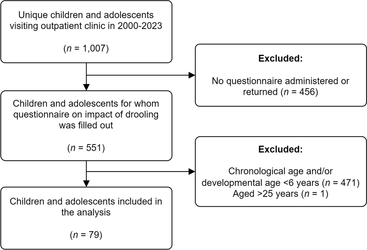
Patients
This prospective observational study was carried out at the third level Neonatal Intensive Care Unit (NICU) of the Careggi University Hospital of Florence after approval by the Pediatric Ethics Committee of Tuscany. Infants with gestational age < 32 weeks or birth weight < 1500 g and with a postnatal age > 7 days of life were enrolled in the study, after informed parental consent, if deemed suitable for KMC. Suitability for KMC was decided on the basis of good clinical condition and stability of vital parameters (body temperature, heart and respiratory rate, systemic blood pressure, SpO2 > 90% with FiO2 < 40%, absence of episodes of apnea in the previous 6 h). Exclusion criteria were major congenital malformations, chromosomal anomalies, intraventricular hemorrhage > 1st degree, and sepsis.
Study design
Enrolled patients were studied with NIRS (Root®Masimo Corporation, Irvine, CA, USA) for the measurement of rSO2L and rSO2C starting 30 min before the start of KMC and ending 60 min after its interruption, with a sampling interval of 6 s. rSO2 measurements obtained using NIRS technique reflect a combination of venous, arterial, and capillary oxygenated/deoxygenated intravascular hemoglobin in a ratio of approximately 75:20:5 [15].
Kangaroo mother care
For the purpose of this study, the duration of KMC was 2 hours (± 20 min). We chose to standardize the duration of KMC to limit possible biases due to shortening or prolongation. Mothers were seated in a reclining chair at a 60° angle wearing an open front blouse. Infants were placed naked, except for a diaper and hat, directly on the skin between the breasts and covered with a light blanket. Infants were fed 1 h before KMC. All infants were continuously monitored by electrocardiogram and their heart rate, systemic blood pressure, SpO2, and body temperature were measured hourly. KMC was discontinued in the event of thermal instability, food intolerance (i.e., regurgitation/vomiting), onset of apnea/tachypnea/dyspnea/bradycardia, or increase in FiO2 > 10% for > 10 min to maintain an SpO2 > 90%.
NIRS measurements
Near-infrared spectroscopy (NIRS) is a non-invasive tool allowing the measurement of regional tissue oxygen saturation (rSO2) which is the ratio between oxygenated hemoglobin and total hemoglobin. NIRS has been used in several studies to evaluate cerebral oxygenation and, to less extent, renal, hepatic, and splanchnic oxygenation [15,16,17,18,19]. However, although the penetration depth of 1.0–1.5 cm of the NIRS light [20] is appropriate for the study of the oxygenation of the lung parenchyma in preterm infants, this possible use has been poorly investigated [21].
Two self-adhesive optodes containing a light-emitting diode and two receiving sensors adequately spaced were applied to each patient. One will be positioned along the right mid-axillary line in correspondence with the 4th–6th intercostal space for the measurement of rSO2L [14], and the other on the forehead for the measurement of rSO2C [22]. All measurements were taken during calm phases or during newborn sleep to reduce NIRS artifacts.
Based on the rSO2S, rSO2C, and SpO2 measurements, we calculated the pulmonary (FOEL) and cerebral (FOEC) fractional oxygen extraction ratio, using the formula FOE = [(SpO2-rSO2)/SpO2]. This parameter reflects the balance between oxygen supply and consumption. Therefore, an increase in FOE suggests an increase in oxygen extraction by the tissues, due to the greater consumption of oxygen in relation to its supply, while its decrease suggests a lower use of oxygen compared to the supply [23, 24].
We then calculated the cerebro-pulmonary oxygenation ratio (CPOR: rSO2L/rSO2C), the ratio between the oxygen saturation of lung tissue compared to the brain tissue. As cerebral perfusion is self-regulating while lung perfusion is not, CPOR is reduced when there is a decrease in pulmonary blood flow, whereas it remains unchanged under normal conditions.
All NIRS data were recorded 30 ± 10 (Tbefore) min before KMC, 30 ± 10 (T30min), 60 ± 20 (T60min), 120 ± 20 (T120min) after it began, and 30 ± 10 (Tafter30min), 60 ± 20 (Tafter60min) min after its interruption together with SpO2. All patients were studied once only.
Data collection
For each patient studied, we reported gestational age, birth weight, sex, type of delivery, Apgar score at 5 min, antenatal steroids, age at the start of NIRS measurements, need for non-invasive and invasive respiratory support (mechanical ventilation ), early discontinuation of KMC and reasons for discontinuation, postnatal steroids, patent ductus arteriosus requiring treatment [25], bronchopulmonary dysplasia (BPD), necrotizing enterocolitis (NEC) requiring surgical treatment, sepsis, 1st degree intraventricular hemorrhage (IVH), ≥ 3rd degree retinopathy of prematurity (ROP), and mortality or length of hospital stay. BPD was diagnosed and defined as mild, moderate, or severe in agreement with Jobe and Bancalari [26]. Intraventricular hemorrhage and NEC were diagnosed according to Papile [27] and Bell [28] criteria, respectively. ROP was graded according to the international classification of retinopathy of prematurity [29].
Statistical analysis
The primary objective of the study was the measurement of changes in rSO2L during KMC in a cohort of preterm newborns using NIRS. Secondary objectives of the study were the measurement of changes in rSO2C, the calculation of pulmonary (FOEL) and cerebral (FOEC) tissue oxygen extraction fraction, and the calculation of the cerebro-pulmonary oxygenation ratio (CPOR) during KMC. Moreover, we compared NIRS variables in the subgroups of infants with or without BPD [26].
A sample size of at least 16 infants was calculated to detect a statistically significant 10% change in rSO2L (from 70 ± 10 to 60 ± 10%) measured before and after starting of KMC with 80% power at 0.05 level. Considering possible data loss, we planned to increase the sample size by 25% to 20 patients.
Clinical characteristics of patients will be described as mean ± SD, rate and percentage, or median and range. For each NIRS variable (rSO2L, FOEL, rSO2C, FOEC, CPOR), we calculated the mean (± SD) of selected 5-min periods which was chosen at the end of Tbefore, T30min, T60min, T120min, Tafter30min, and Tafter60min. We made this choice to obtain maximum stability of the NIRS signal. However, sometimes this was not possible due to the occurrence of unwanted artifacts (generally caused by patient movements): in this case, the 5-min artifact-free period closest to the end of the study period was selected.
Serial measurements of studied variables were compared with repeated-measures analysis of variance (ANOVA). A P < 0.05 will be considered statistically significant.


留言 (0)