
記住我


Disclaimer: Early release articles are not considered as final versions. Any changes will be reflected in the online version in the month the article is officially released.
Author affiliations: MediCheck Research Institute, Korea Association of Health Promotion, Seoul, South Korea (S. Hong, H. Shin, Y.-H. Lee, J.-Y. Chai, B.-K. Jung); Convergence Research Center for Insect Vectors, Incheon National University, Incheon, South Korea (S.-J. Hong); Dr. Song Jeong-Gil’s Internal Medical Clinic, Pyeongtaek, South Korea (S.-R. Kim, Y.-K. Kim, Y.-J. Son, J.-G. Song); Seoul National University College of Medicine, Seoul (J.-Y. Chai)
Echinostomes are zoonotic intestinal flukes infecting birds and mammals worldwide (1,2). Adult echinostomes generally inhabit the small intestines of the definitive host and attach to the mucosal surface, causing pathological changes that include inflammation of the mucosa, bleeding, and ulceration (1). Echinostoma cinetorchis infects humans, dogs, cats, rodents, chickens, and ducks in South Korea, Japan, China, Taiwan, and Vietnam (1,2). The second intermediate hosts—that is, the source of infection for definitive hosts—include freshwater snails, fish, and amphibians (1). Human E. cinetorchis infection has been relatively rare and reported in only a few patients who had abdominal pain, diarrhea, weakness, and weight loss (1,3). We report the case of a woman in South Korea infected with E. cinetorchis whereby adult flukes were recovered through colonoscopy and identified by morphologic and molecular analyses.
A 69-year-old woman with occasional gastrointestinal discomfort, indigestion, constipation, and diarrhea visited Dr. Song’s Internal Medicine Clinic, Pyeongtaek, Gyeonggi, South Korea, in October 2023. Laboratory examinations revealed overall blood counts, liver function markers, renal function indicators, and lipid profiles were within reference ranges. Feces examination revealed negative results for protozoa and helminths.
Figure 1
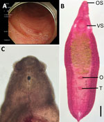
Figure 1. Analysis of a worm identified as Echinostoma cinetorchisremoved during colonoscopy from a 69-year-old woman in South Korea. A) Colonoscopy image showing a moving trematode in the mucosa of...
Colonoscopy showed 4 actively motile adult trematodes in the mucosa of the ileum, cecum, and ascending colon (Figure 1, panel A). A physician removed the worms with grasping forceps and transferred them to the MediCheck Research Institute, Korea Association of Health Promotion (Seoul, South Korea), for morphologic and molecular identification. Two of the 4 worms were intact, and the remaining 2 were broken during the clipping of the worms. Researchers observed the intact worms by using a light microscope after fixation with 10% formalin under coverslip pressure and stained with acetocarmine (Figure 1, panels B, C).
The worms were elongated and spindle-shaped, measuring ≈6.75 mm in length and 2.25 mm in width (both average measurements at the ovarian level. The worms had 37 collar spines (Figure 1, panel B), of which 24 were arranged in a single row, consisting of 6 corners and 6 laterals on each side, and the additional 13 dorsal spines were arranged in 2 alternating rows. The vitellaria were follicular and distributed mainly in lateral fields from the posterior margin of the ventral sucker to the posterior end of the body. One or both testes were absent in 3 of the 4 specimens (1 specimen had 2 testes), unlike other echinostome species, which usually have 2 testes. Intrauterine eggs (n = 10) were yellowish and operculated, measuring an average of 110 μm in length and 63 μm in width. The patient was prescribed a single dose of praziquantel (10 mg/kg).
Figure 2
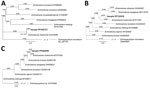
Figure 2. Phylogenetic trees of a worm identified as Echinostoma cinetorchisremoved during colonoscopy from a 69-year-old woman in South Korea (red text). Trees were based on nucleotide sequences of the...
We preserved the 2 broken worms in 70% ethanol for molecular studies. We isolated genomic DNA from the worm segments by using the DNeasy Blood and Tissue kit (QIAGEN, https://www.qiagen.com). We partially amplified (398 bp) the NADH dehydrogenase 1 (ND1) regions by using the standard PCR protocol with GenomicsOne 5X PCR Premix (GenomicsOne, https://www.donginbio.com) and 10 pmol of forward and reverse primers to detect Echinostoma spp. (4). We directly sequenced the PCR product at Macrogen Inc. (Seoul, Korea). Sequencing revealed 99.7% identity of our specimens GenBank (accession no. PP338757) with the sequences of E. cinetorchis deposited in GenBank (accession no. KU519289) (Figure 2, panel A). We obtained phylogenetic trees based on sequences of partial cytochrome c oxidase subunit 1 mitochondrial gene (CO1) (185-bp) and internal transcribed spacer (ITS) region (ITS1–5.8S-ITS2) (657-bp). Our sample for CO1 (accession no. PP710359) was 95.7% identical to E. cinetorchis (accession no. MT577587) (Figure 2, panel B), and our sample for the ITS region (accession no. PP683096) was 98.3% identical to E. cinetorchis (accession no. MT577832) (Figure 2, panel C).
In South Korea, few human infection cases with E. cinetorchis have been identified through adult worm recovery by purging with magnesium sulfate or through gastrointestinal endoscopy (1,3). Our diagnosis of E. cinetorchis infection was determined by adult worm recovery through colonoscopy, followed by morphologic and molecular analyses. Most adult echinostomes, such as Isthmiophora hortensis, reside in the upper portion of the small intestine or occasionally in the pyloric area of the stomach (5–10). In comparison, 2 endoscopy cases of E. cinetorchis infection (3), including our case, have identified the presence of worms in the colon. This unique location of echinostome flukes in humans might be a distinguishing feature for E. cinetorchis infection.
Freshwater snails are first as well as second intermediate hosts for E. cinetorchis (1). Large-sized snail species in particular (e.g., Cipangopaludina) and freshwater fish are potential sources of human infections. Our patient reported that she had sold snails and freshwater fish on the street and often consumed them raw or undercooked. Thus, the infection source for our patient might have been 1 or both kinds of intermediate hosts.
In countries where human echinostome infections are found, physicians should include echinostomiasis among the differential diagnoses of diseases causing nonspecific gastrointestinal problems. Public education regarding the hazards associated with consuming raw or undercooked snails or fish in these regions also would be useful in reducing E. cinetorchis infections.
Ms. Sooji Hong is a clinical pathologist and researcher at MediCheck Research Institute, Korea Association of Health Promotion, Seoul, Korea. Her primary research interest is parasite fauna study based on molecular methods, and she recently performed studies on the intestinal microbiome fauna of cats and dogs infected with parasites.
留言 (0)