
記住我


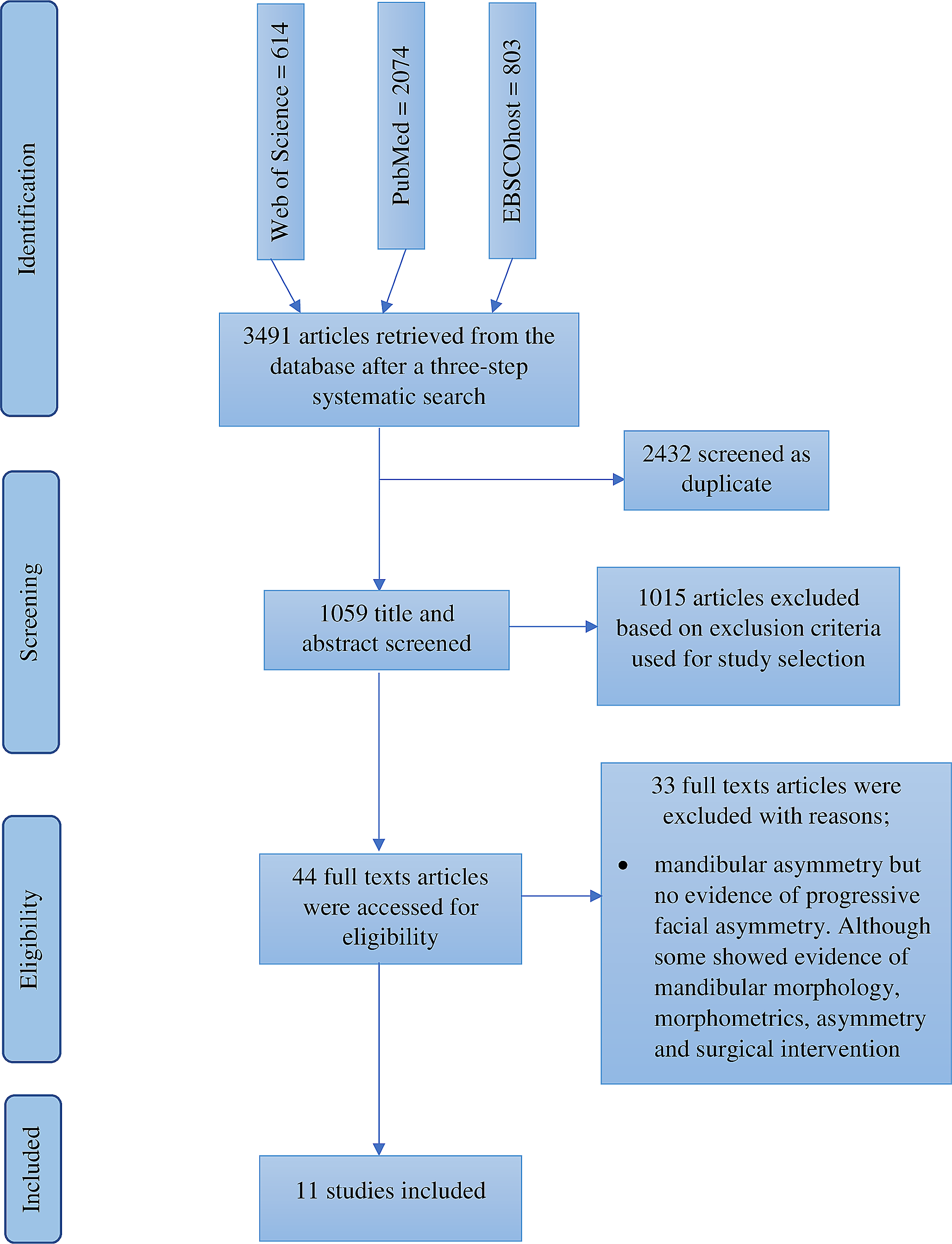
A total of 3491 articles were identified during the literature search, including research papers, reports, and books. Two thousand four hundred and thirty-two duplicates were removed. After screening titles and abstracts, 1015 articles were excluded based on the exclusion criteria. Forty-four full-text articles that met the eligibility criteria were reviewed. Thirty-three articles were excluded because they lacked evidence on the progression of FA, although they showed evidence of mandibular morphology, morphometrics, asymmetry, and surgical intervention. However, eleven articles were eligible and included in this review (Fig. 1).
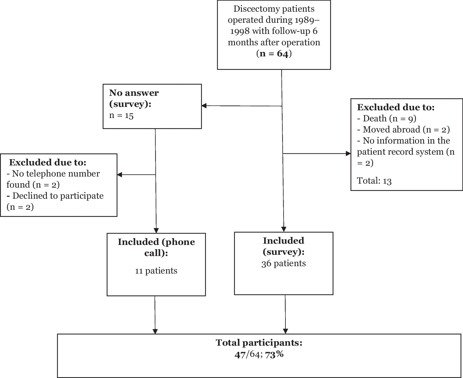
Fig. 1
Flow diagram of study selection
Table 2 summarises the included studies and their significant findings. Table 3 shows the frequency distribution of FA parameters in the included studies. Figure 2 shows the level of knowledge of included studies on the progression of FA in relation to Pruzansky-Kaban type classification, dentition age range and mandibular morphometrics. Figure 3 illustrates an assessment of all the included studies based on the facial asymmetry parameters in hemifacial microsomia patients. Most (55%) of the included studies were conducted in Europe [25, 35,36,37,38,39], 36% were conducted in the United States of America [24, 40,41,42], and 9% were conducted in the People’s Republic of China [43]. The largest sample size is 210 [43], while the lowest is 7 [40].
Table 2 Characteristics of included studiesTable 3 The frequency table shows the parameters of facial asymmetry in the eleven included studiesFig. 2
Number of studies suggesting the progression of facial asymmetry in relation to Pruzansky-Kaban type classification, dentition age range and mandibular morphometrics in the literature from 1969 to date. The X-axis shows the classification, dentition age and measurement of the deformed mandible, and the Y-axis illustrates the number of studies
Fig. 3
Line graph showing assessment of all the included studies based on the facial asymmetry parameters in hemifacial microsomia patients. The X-axis represents facial asymmetry parameters, and the Y-axis represents the number of studies showing the progression of facial asymmetry
Table 4 Mandibular parameters used to measure facial asymmetry and treatment in selected studiesAll included studies were observational; 82% were retrospective studies [24, 25, 36, 37], while 18% were prospective studies [35, 38]. Only three of the included studies (27%) were cross-sectional [24, 39, 43], while the remaining (82%) were longitudinal studies [25, 35,36,37,38,39,40,41,42] (Table 2). A total of 67% of the included studies described sex in their report [25, 36,37,38,39,40,41, 43]. Age grouping varied among the included studies; two (18%) utilised dentition age grouping [24, 39], two (18%) utilised infancy to childhood [41, 43], while others (64%) used varied age grouping [25, 35,36,37,38,39,40, 42]. Three (27%) of the included studies suggested the progression of facial asymmetry [24, 42, 43], while the remaining (73%) suggested that FA remains constant in HFM patients [25, 35,36,37,38,39,40,41].
The methodological quality of the included studiesThe eleven included studies were deemed good quality as they answered the first two screening questions and fulfilled at least three quantitative criteria of MMAT. Regarding the quantitative criteria, one study met three criteria- 60% and ten met four criteria- 80% (Table 2).
Table 3 shows the frequency distribution of FA parameters in the eleven included studies. The more common parameters used in the included studies were: 64% used ramus height [35, 36]. 27% used chin point [25, 40, 41], 27% used occlusal plane angle [24, 37, 38], 36% used gonial/intergonial angle [24, 39,40,41], 27% used total mandibular length [39,40,41], 27% used mandibular body length [41,42,43]. In addition, a few of the included studies discussed the treatment approach and outcome (Table 4).
Studies suggesting evidence of progressive facial asymmetry in relation to Pruzansky-Kaban type classification, dentition age range and mandibular morphometricsTen (91%) of the included studies utilised either Pruzansky [35, 39,40,41,42] or Pruzansky-Kaban [24, 25, 36, 37, 43] types to classify the mandibular asymmetry in HFM. Two (18%) of the included studies utilised dentition age [24, 39], two (18%) of the included studies utilised infancy to childhood stage [41, 43] for age grouping. The majority (91%) of the included studies showed evidence of knowledge of mandibular morphometry in HFM patients [24, 25, 35,36,37].
Assessment of all the included studies based on their conclusion on the progressiveness of FA in hemifacial microsomia patientsAn assessment of mandibular morphometric parameters was used to determine the progressiveness of FA in the HFM population in all (100%) of the included studies [24, 25, 35,36,37,38,
留言 (0)