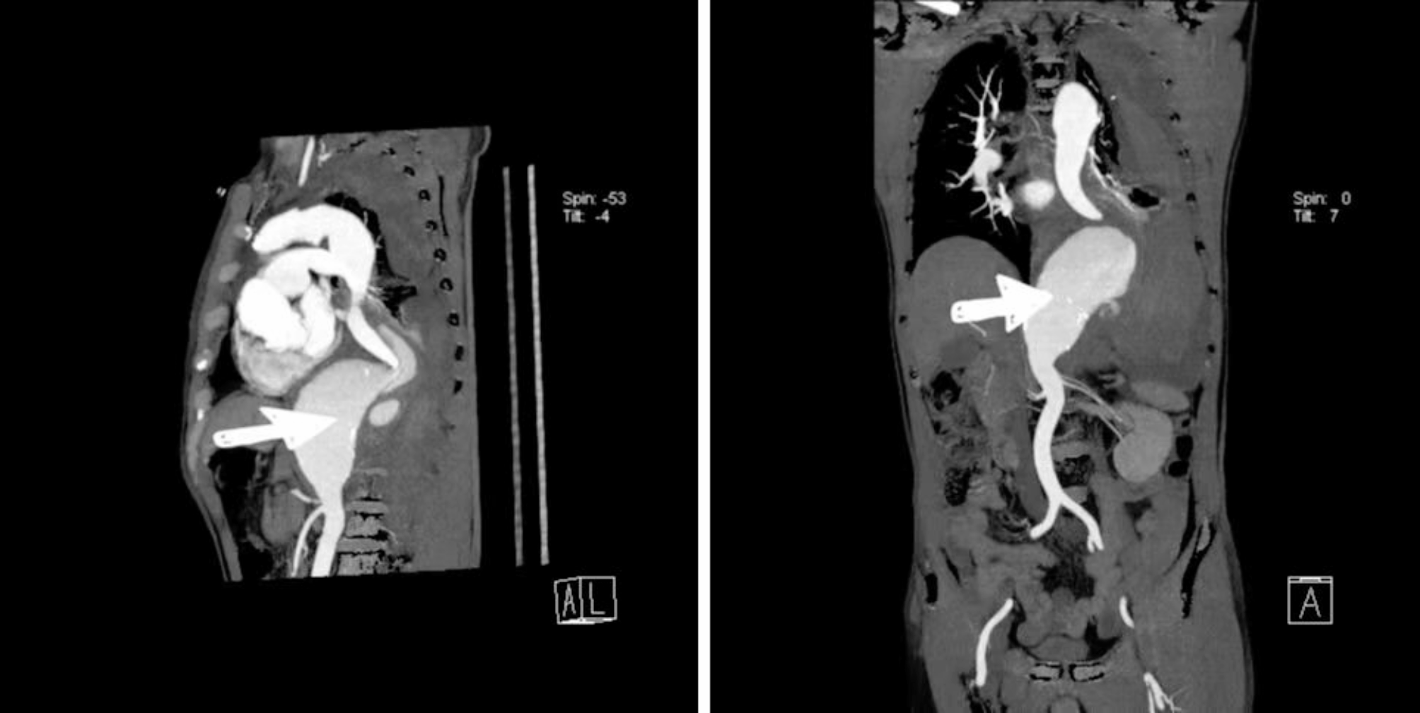
Osteochondroma is a benign tumor of the skeletal system that rarely occurs in the ribs. Patients with rib osteochondroma were usually asymptomatic, but occasionally felt pain, and those with progression to thoracic outlet syndrome (TOS) were even more so [5, 6]. Three-dimensional reconstruction of computed tomography can determine the size, location, and internal structure of the tumor, as well as the adjacent relationship with the neighboring organs, playing a guiding role in the scope of surgical resection of the tumor. Histologically, osteochondroma is divided into three layers: fibrous membrane, cartilaginous cap, and bone. The outermost layer is a thin fibrous membrane that continues with the periosteum of the basal bone. The middle layer is the cartilaginous cap, which is usually less than 2 cm thick. If the thickness is > 2 cm and the shape is irregular, the possibility of malignancy should be considered [7]. Surgery is the main treatment for osteochondroma, highlighting the importance of early detection and intervention to prevent tumor enlargement, functional impairment, and malignant degeneration.
The upper part of the first rib is the subclavian artery, vein, and brachial plexus nerve. The lower part is the thoracic roof. The clavicle shields the front, and the back is covered by the scapula. The inside and the sternostalk form the sternocostal joint, and the deep part is the lung tip. Therefore, the deep anatomical position of the first rib leads to its difficult exposure, leaving the surgical resection of the first rib tumor a difficult problem for thoracic surgeons. Importantly, surgeons must carefully select the proper surgical approach and determine how to expose the lesions while protecting the peripheral vital vessels and nerves. The methods for excising the first rib have been widely reported in the literature, including the subaxillary arc incision approach, supraclavicular incision approach, subclavian incision approach, combined cervicothoracic approach, endoscopic subaxillary incision approach and video-assisted thoracoscopic surgery [8, 9].
The patient underwent a first osteochondroma resection and thoracic reconstruction through an inverse L-shaped incision in the anterior chest and a longitudinal split of the thoracic bone. This approach provided good surgical exposure and sufficient operating space for complete resection of the tumor and peripheral vascular neuroprotection. The mass of the first rib was removed under direct vision to avoid massive bleeding induced by damage to the surrounding great blood vessels due to unclear surgical field and anatomy during free tumor resection. The patient had symptoms of brachial plexus injury after surgery, with numbness on the ulnar side of the forearm. The patient recovered gradually after 2 months with no residual symptoms. It is important to note that nerve injury symptoms can be attributed to excessive intraoperative stretching during tumor exposure. Therefore, gentle manipulation during exposure of the subclavian tissue, avoidance of excessive stretching, and limited use of electrocoagulation are recommended.
In summary, the authors suggested that an inverse L-shaped incision in the anterior chest approach is necessary for the complete excision of first rib tumors. This surgical technique provides effective exposure of the diseased tissue under direct vision while minimizing the risk of subclavian arteriovenous and nerve injury. However, due to the rarity of bone tumors at this site and the limited number of patients in this study, larger sample sizes and multi-center studies are warranted.


留言 (0)