
記住我


713 patients were included in the ALFA-0702 study and 576 reached CR/CRi in one course. Only 189 patients who reached CR (n = 179) or CRi (n = 10) after the first induction course (henceforth CR1) had available material for NGS analysis at both time points (Fig. 1). Comparison between the 189 patients and the 387 remaining patients with no material in CR was performed (supplementary table 2). Patients of the present study were more likely to harbor adverse or intermediate ELN risk (p = 0.043). There were no other major differences with other patients, and 2-years outcome was the same between the groups (PFS, OS, and CIR). (supplementary table 3). Eight patients (4%) had no identified molecular marker detected at diagnosis and could not be analyzed. Out of the 181 remaining patients, 15 had only one identified mutation, and 166 had at least two mutations. The main patient characteristics are described in Table 1, Supplementary Tables 4 and 5. Median age was 46 years old (18–60), and median leukocyte count was 7.9 × 10⁹/L (0.5–256). Karyotype was normal in 104 patients. ELN 2017 risk distribution was favorable in 52 (29%) patients, intermediate in 71 (40%), and adverse in 56 (31%). All patients received cytarabine consolidation courses, with (n = 73, 40%) or without (n = 108, 60%) clofarabine. Ninety-two (92) of the 127 intermediate/adverse risk patients received allo-SCT in CR1.
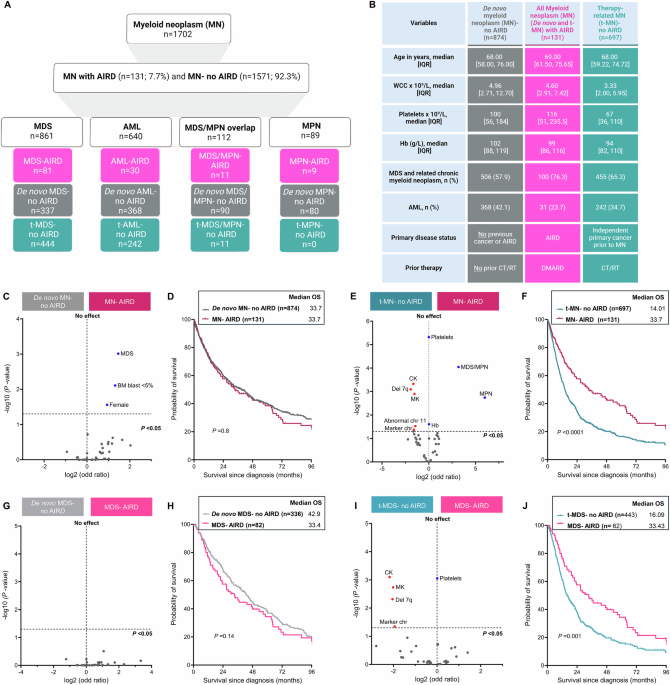
Fig. 1: Flow chart of the study.
CR1: Complete remission or complete remission with incomplete recovery; DTA: mutations in DNMT3A, TET2, or ASXL1; NGS-MRD: measurable residual disease using Next Generation Sequencing assay with a 0.1% threshold for positivity; neg: negative; pos: positive; NPM1-MRD: measurable residual disease for NPM1 mutation measured by Next Generation sequencing assays with a threshold of detection over 10−4 (see methods).
Table 1 Major patients’ characteristics.Mutational profile at diagnosis and CR1A total of 735 somatic mutations were identified at diagnosis in 181 patients. The median number of gene mutations was 4 (range 1–10). Four potential germline mutations (in CEBPA, NF1, DDX41, and RUNX1) were excluded from all analyses. Twenty-three VUS with persistence at a high level in CR were identified. As patients with only VUS persistence in CR1 had the same prognosis as negative MRD patients (not shown), VUS were excluded from further analyses. Detailed mutational data at diagnosis and CR1 are described in Fig. 2, supplementary Fig. 1, and Supplementary Table 4. The most frequently mutated genes detected were FLT3 (n = 56 ITDs and n = 52 other mutations), NPM1 (n = 67), NRAS (n = 57), DNMT3A (n = 50), and TET2 (n = 45).
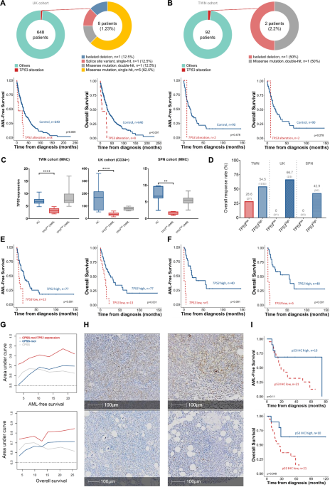
Fig. 2: Mutational profile at diagnosis and CR1 according to functional categories and gene mutations.
Diagnosis variant allele frequency (VAF) is plotted in x-axis (range 0–60% or 0–100% for genes with loss of heterozygosity) and VAF in CR1 in the y-axis (log-scale). The dotted red-line represents the retained threshold of detection of 0.1%. Mutations with VAF in CR1 under 0.01% were plotted at 0.01%. The “Epigenetic” plot summarizes data on DNMT3A, TET2, ASXL1 IDH1, IDH2, BCOR and ASXL2, ATRX, BCORL1, CTCF, CUX1, EP300, EZH2, KDM6A, KMT2C. The “Splice” plot summarizes data on SF3B1, SRSF2, U2AF1, and ZRSR2. The “Transcription” plot summarizes data on GATA2, RUNX1, CEBPA, WT1, PHF6, ETV6, and IKZF1. The “signaling” plot summarizes data on FLT3, KIT, MYC, PTPN11, NRAS, KRAS, NF1 and JAK2, JAK3, CHEK2, CSF3R, CBL and BRAF. The “All mutations” plot contains combined data of all mutated genes. A few mutations with uncertain VAFs at diagnosis or CR1 were not plotted (see methods).
The persistence of mutations in CR1 was variable according to gene’s identity and function. Regarding epigenetic regulators, some mutations were often detected in CR1, in particular mutations in DNMT3A (n = 42/50), IDH1 (n = 6/12), or BCOR (n = 9/15). This was less frequent for mutations in TET2 (n = 15/45) and was uncommon for mutations in some other genes such as IDH2 (n = 2/13) or EZH2 (n = 1/8). Other frequently persisting events included mutations in TP53 (n = 9/12) or in splice machinery components such as SRSF2 (n = 8/8) and U2AF1 (n = 3/5). Mutations in hematopoietic transcription factors, NPM1, or signal transduction-associated genes were infrequently detected in CR1 with the 0.1% threshold (Fig. 2 and supplementary Fig. 1).
Ninety-one patients had no mutation detected in CR1 (NGSNEG), and 90 had at least one mutation detectable, including 37 with only DTA mutations (NGSDTA) and 53 with at least one other gene mutation (NGSother). When comparing the main characteristics of the 3 groups (Table 1), NGSNEG patients were significantly younger (p = 0.0063). There was also a trend for the ELN2017 distribution to be different, with more favorable risk patients in the NGSDTA group, more adverse risk patients in the NGSother group (p = 0.08), and a trend for the NGSDTA group to harbor a higher initial leukocyte count (p = 0.054).
When separating patients according to Lindsley classification [37], NGS-MRD group was highly associated with AML ontogeny (p < 0.0001). De novo disease ontogeny was over-represented in the NGSDTA group (mainly due to NPM1 association), and under-represented in the NGSother group. In contrast, TP53 and secondary ontogeny genes were over-represented in the NGSother group (Supplementary Table 6).
NGS-MRD including DTA mutations is associated with poor prognosisWe first analyzed the prognosis of the 3 groups of patients (NGSNEG, NGSDTA, and NGSother). CIR was found significantly different between the three groups, with probabilities of 23% [13–33], 35% [19–51], and 51% [38–66] at 24 months for NGSNEG, NGSDTA, and NGSother groups, respectively (p = 0.0003). At 4 years, RFS estimates were 68% [58–80], 51%[38–70] and 39% [28–55] (p = 0.001), and OS estimates were 80% [73–89], 59%[46–78] and 54% [43–70] (p = 0.003), respectively, with prolonged survival in the NGSNEG group (Fig. 3A–C; Supplementary table 7). No significant differences were found when comparing CIR, RFS, and OS between NGSother and NGSDTA groups. The proportion of patients receiving CLARA as post-CR1 therapy was the same in the 3 groups.
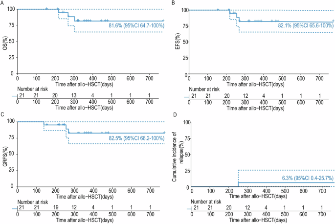
Fig. 3: Prognosis according to NGS-MRD.
Prognosis according to NGS status (A–C) and the number of persistent mutations (D–F). p-values are for log-rank tests for Relapse Free Survival and Overall Survival and for Gray test for Cumulative incidence of Relapse. Data were not censored at allogeneic hematopoietic stem cell transplantation. NEG no mutation detected in CR1, DTA detection of only DNMT3A, TET2 or ASXL1 mutation in CR1; other: detection of other mutation than DNMT3A, TET2 or ASXL1 in CR1.
In multivariate models adjusted with ELN2017 status and age, only NGSother was associated with higher CIR (HR = 2.73[1.48–5.03], p = 0.0013) when the trend for higher CIR in NGSDTA did not reach statistical significance (HR = 1.77[0.86–3.64], p = 0.12). NGSother (HR = 2.23[1.31–3.81], p = 0.0033) was predictive of worse RFS, but NGSDTA (HR = 1.79[0.97–3.30], p = 0.062) and ELN2017 did not reach statistical significance (p = 0.074). Both adverse ELN2017 risk (HR = 2.64[1.25–5.56], p = 0.011), NGSDTA (HR = 2.16[1.07–4.37], p = 0.032), and NGSother (HR = 2.26[1.21–4.24], p = 0.011) were associated with OS (Table 2).
Table 2 Multivariable analysis for cumulative incidence of relapse, relapse free survival and overall survival according to NGS MRD status.The number of NGS-MRD persisting events is associated with prognosisAs the detection of only one molecular event at MRD could be linked with CHIP-related pre-leukemic clones with blunted relapse-initiating capacity, we next investigated whether the persistence of multiple mutations (including DTA mutations) was associated with prognosis. All 166 patients with at least two gene mutations at diagnosis (Fig. 1) were included. Eighty-two patients had no mutation detected in CR (NGSnull), 48 had only one mutation (NGSone), and 36 had two mutations or more (NGSmore). Patients in the NGSDTA group harbored more often 1 mutation in CR than patients in NGSother (76% vs. 46%, respectively, p = 0.0099) (Table 1 and Supplementary Table 8).
CIR was significantly different when comparing patients from NGSnull, NGSone, or NGSmore groups, with 48-month estimates at 20%[10–31], 31%[18–45], and 58%[42–75], respectively (p = 0.0012). Likewise, at 48 months, probabilities of RFS were 70% [60–83], 62% [50–78], and 24%[14–44], respectively (p < 0.0001), and probabilities of OS were 82%[74–91], 72%[61–87], and 38%[26–59], respectively (p < 0.0001) (Fig. 3D–F). In univariate analysis of both RFS and OS, NGSone group was not significantly different from NGSnull group (HR = 1.66[0.91–3.04] and HR = 1.72[0.84–3.51] respectively).
In multivariate models adjusted with ELN2017 status and age, NGSone was marginally associated with higher CIR (HR = 1.92[0.94–3.92], p = 0.072), but was not associated with RFS nor OS. Conversely, NGSmore was associated with higher CIR (HR = 3.71[1.82–7.56], p < 0.0001) and shorter RFS (HR = 3.36[1.83–6.17], p < 0.0001) and OS (HR = 3.81[1.87–7.74], p = 0.00023) (Table 3).
Table 3 Multivariable analysis for cumulative incidence of relapse, relapse free survival and overall 608 survival including to the number of persisting mutations.Comparison of NGS-MRD and WT1-MRDWe then investigated whether NGS-MRD could add prognostic information when compared to other validated MRD strategies. In 100 patients with WT1 overexpression at baseline, both WT1 expression and NGS-MRD data were available in CR1 (Fig. 1). Eighty-four patients had low WT1 expression in CR1 (WT1low) including 37 with at least one marker detected by NGS-MRD (NGSPOS-19 patients with only DTA and 8 with multiple mutations) and 47 with no marker detected by NGS-MRD (NGSNEG). Sixteen patients harbored high WT1 expression at CR1 (WT1high) including 13 with NGSPOS, and 3 with NGSNEG (Fig. 1, supplementary Fig. 2). WT1high was associated with higher CIR at 4 years (p = 0.005) and shorter RFS (p = 0.0008) with a trend toward shorter OS (p = 0.06) (supplementary Fig. 3). We consequently focused on the WT1low patients. In these patients, NGSPOS was associated with higher CIR at 4 years (37% [22–54] vs. 22% [9–37] p = 0.04) but was not associated with RFS nor OS. (Fig. 4A–C). In multivariable analysis including NGS-MRD, WT1-MRD, and ELN2017, only WT1-MRD status was associated with RFS (HR = 3.21 [1.55–6.67], p = 0.0017) and OS (HR = 2.71 [1.13–6.49], p = 0.0025) (Supplementary Table 9).
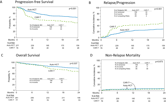
Fig. 4: Prognosis of NGS-MRD evaluation when compared to WT1 expression in CR and NPM1 MRD.
Panels A–C represent the cumulative incidence of relapse, relapse-free survival, and overall survival according to NGS-MRD in CR1 status in the 84 patients with low WT1 expression in CR1. Panels D–F represent the cumulative incidence of relapse, relapse-free survival, and overall survival according to NGS-MRD in CR1 and NPM1 MRD assessed by NGS in the 67 NPM1 mutated patients. p-values are for the log-rank test for RFS and OS and for the Gray test for CIR. Data were not censored at allogeneic hematopoietic stem cell transplantation.
Comparison of multi-target NGS-MRD and isolated NPM1-MRD evaluationsWe also compared NGS-MRD and specific NPM1-MRD performed with the NGS error-corrected assay in the 67 patients with NPM1 mutations. NPM1-MRD positivity was defined by the detection of at least one consensus read with error-corrected NGS (see methods). Forty-one patients had undetectable NPM1-MRD (NPM1NEG) including 21 NGSNEG and 20 NGSPOS. Twenty-six patients harbored detectable NPM1-MRD (NPM1POS) including 10 NGSNEG and 16 NGSPOS. All 5 patients with NPM1 mutation over 0.1% in CR1 have at least one other mutation detected in CR1. Detectable targets both in NPM1POS and NPM1NEG patients were mainly DNMT3A and TET2 mutations (Fig. 1, supplementary Fig. 5). NPM1POS was associated with higher CIR, and lower RFS and OS probabilities (supplementary Fig. 4). We divided the patient cohort into four groups according to NPM1- and NGS-MRDs. CIR was significantly different between groups with particularly high risk in double positive patients (5%[0–15] vs. 19%[2–36] vs. 30%[0–60] vs. 62%[37–88] at 4 years for NPM1NEGNGSNEG, NPM1NEGNGSPOS, NPM1POSNGSNEG and NPM1POSNGSPOS, respectively (p = 0.002). This was the same for RFS probabilities at 4 years with 90% [78–100], 71%[54–94], 50%[27–93] and 25%[11–58] respectively (p = 0.0004), and for OS probabilities at 4 years with 95%[86–100], 76%[60–97], 60%[36–100] and 37.5%[20–71], respectively (p = 0.004) (Fig. 4D–F).
We next performed a multivariable analysis (Supplementary Table 10) including NPM1-MRD, NGS-MRD, and ELN2017. NPM1- and NGS-MRD positivity were both significantly and independently associated with increased CIR (HR = 4.16[1.51–11.47] p = 0.0059, and HR = 3.37[1.09–10.39], p = 0.035, respectively). NPM1-MRD positivity was significantly associated with shorter EFS (HR = 3.55[1.53–8.25], p = 0.0032), with a similar trend for NGS-MRD positivity (HR = 2.33[0.96-5.77], p = 0.06). NPM1-MRD was the only variable associated with OS (HR = 2.97[1.15–7.67], p = 0.025), whereas NGS-MRD was not (HR = 2.3[0.81–6.54], p = 0.12).
Evaluation of NGS-MRD and allo-SCTFinally, we investigated whether NGS-MRD could be used to guide allo-SCT in CR1. We focused on the 127 patients with intermediate or unfavorable ELN2017, i.e. with standard allo-SCT indication. Ten patients relapsed before allo-SCT and ninety-two (72%) received allo-SCT in the first CR. The median time between allo-SCT and CR was 3.8 months [range 2.8–5.7]. Among these patients, 45 were NGSNEG and 47 were NGSPOS (including 16 NGS DTA). Considering allo-SCT as a time-dependent variable, both allo-SCT and NGS-MRD were predictive for relapse incidence and RFS. The interaction test between variables was not significant suggesting that NGSPOS at the time of CR does not identify a subset of patients with a specific benefit of allo-SCT. (supplementary Fig. 6 and Supplementary table 11).
留言 (0)