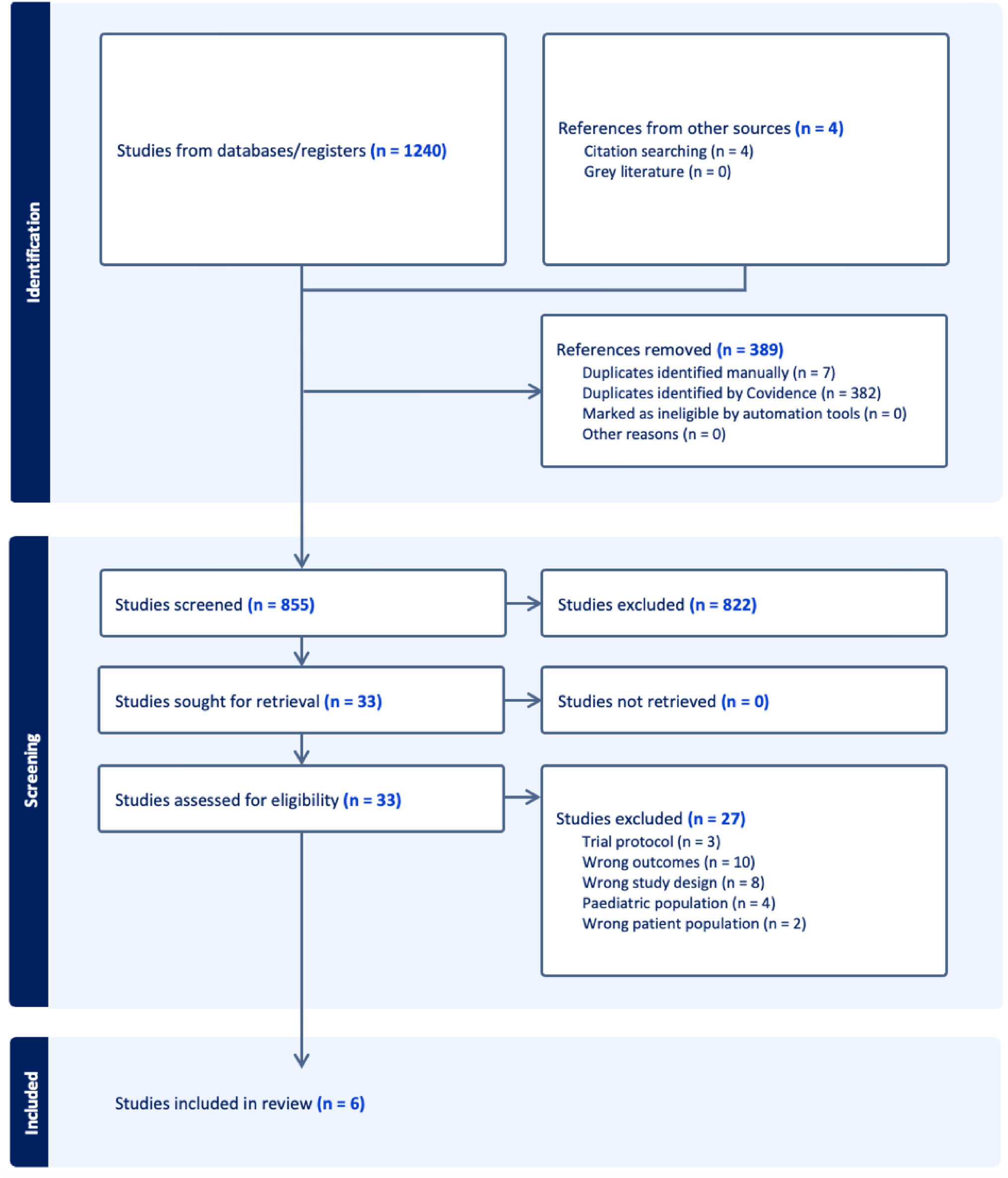
We performed a retrospective review of peripheral bronchoscopy using thin and ultrathin bronchoscopy with rEBUS and 2D fluoroscopy without any navigational system for peripheral lung lesions in our academic medical center from November 2015 through January 2021. The practice pattern was noticeably affected by the acquisition of an ultrathin bronchoscope (Olympus BF-MP190 with OD at tip 3.0 mm, WC 1.7 mm) on 3/4/2019 while the thin bronchoscope Olympus BF-P190 (OD 4.2 mm, WC 2.0 mm) continued to be used. We assessed diagnostic yield and the impact of different variables on outcomes. We compared the results between two groups of patients before and after employment of the ultrathin bronchoscope. Logistic regression models were employed to assess the independent associations of the most impactful variables.
Primary outcome: diagnostic yield
We used a strict definition for diagnostic yield. We also calculated the sensitivity for diagnosis of malignancy in our cohort. Diagnostic yield (DY) was calculated as the rate of true positive (TP) plus true negative (TN) results for malignancy divided by total number of bronchoscopies. TN results were concluded based on the following criteria:
1.
A specific benign (SPB) diagnosis was established (such as a granulomatous disease or a definitive infectious diagnosis).
2.
If a nonspecific benign (NSB) result was reported at index bronchoscopy, such as atypical or inflammatory cells, then the case was assessed longitudinally and categorized as TN (NSB-TN) only if subsequent biopsy (either bronchoscopic, CT-guided or surgical biopsy) or imaging confirmed a nonmalignant diagnosis or follow up imaging after at least 12 months showed stability or resolution of the lesion.
3.
The cases with normal lung/bronchi tissue reports from index bronchoscopy, and the cases in which a definitive diagnosis was not established because of lack of follow-up were counted as non-diagnostic bronchoscopy (ND). TN only included SBP and NSB-TN and not ND.
Sensitivity calculation
Sensitivity was calculated as the percentage of proven malignant lesions by index bronchoscopy out of total cases of malignancy. Total cases of malignancy included those proven via bronchoscopy or other methods, plus any case that received empiric treatment for malignancy (without tissue diagnosis), plus any case that was lost to follow up.
Secondary outcomes
1.
Achievement of concentric rEBUS view.
2.
Impact of addition of ultrathin bronchoscope on procedure outcomes.
3.
Correlation of radiographic characteristics of lesions with diagnostic yield; size, location, distance from closest visceral pleural surface, PET characteristics, border characteristics in lesions ≤ 15 mm (smooth or irregular), bronchus sign, appearance of lesion.
VariablesConcentric vs Eccentric rEBUS view
Concentric view is an ultrasonic image achieved when r-EBUS probe is positioned in the airways surrounded by the lesion, while eccentric view correlates with the images from airways adjacent to the lesion. Traditionally, concentric r-EBUS view correlates with higher diagnostic yield compared to eccentric view.
Lesion size
We measured the long axis (LA) diameter of the lesion in an axial plane of chest CT scan. We also measured a short axis which was defined as a perpendicular line crossing LA in the middle. Then the average of the long and the short axis was calculated.
Location
Based on the CT scan, the lesions in right upper lobe, right middle lobe, and left upper lobe were classified together compared to the lesions in right and left lower lobes.
Distance from closest visceral pleural surface
We used the distance from the center of the lesion to the closest visceral pleural surface (including fissure and mediastinal pleura) in the axial plane of the CT scan to objectively classify the location of the lesions relative to central airways. Due to lack of a standard definition for peripheral vs central lesion, we used this measurement to classify peripheral vs central lesions with a numerical value [32, 33]. The airways start branching from the lung hilum and extend to smaller airways in a semi-spherical pattern in each lobe. Visceral pleura marks the peripheral boundaries of each lobe, and the distance from the lesion to closest visceral pleural surface may best explain how peripheral the lesion is. This calculation takes into account the direction of airway extension in a spherical pattern. For example, a lesion located close to the midline of the body but close to the mediastinal pleura is called a peripheral lung lesion and may be challenging to reach via peripheral airways.
PET characteristics
The FDG avidity of the lesion reported by a PET/CT obtained 3 months before or after the index bronchoscopy was reported in numerical value.
Border characteristics
Based on the CT scan, the borders of the lesions were classified as smooth or irregular. Irregular border included the ones with spiculated or lobulated borders. Specifically in lesions ≤ 15 mm this characteristic was used to check for any possible correlation with diagnostic yield. We previously observed the smaller lesions with irregular borders to have a better chance of being detected by r-EBUS examination.
Bronchus sign
We reviewed the CT scan images in different planes and reported any visible air-filled airway which led to the targeted lesion. This is called a bronchus sign and previously has been reported to be associated with higher diagnostic yield [34, 35]. We examined whether the presence or lack of a bronchus sign had any impact on case selection by the bronchoscopist and if there was any correlation with diagnostic yield.
Nodule appearance
We reviewed the characteristics of the lesion in the CT scan images and based on a visual assessment, classified the lesions as solid, ground-glass, combined solid and ground-glass and cavitary.
Case Selection
Patients who were referred to the Interventional Pulmonary service for evaluation of a lung lesion identified by chest CT scan were included in this study. Bronchoscopy was performed for lesions with diameter of 1 cm and above with estimated intermediate pretest probability for malignancy (5–65%). Sub-centimeter nodules were typically followed by surveillance imaging, however a few nodules smaller than 1 cm were selected for bronchoscopy because of high suspicion for malignancy and lack of alternative diagnostic approach. Patients with a high (> 65%) probability of malignancy were referred for surgical resection, but bronchoscopy was performed if they were too unstable for surgery or biopsy prior to surgery was preferred. Patients who were at non-reversible and high risk of respiratory or cardiovascular failure were excluded from bronchoscopy.
Bronchoscopy procedure
CT scan images were reviewed before the procedure and were available for further review during the bronchoscopy if needed. In subjects with multiple pulmonary lesions, the decision about which and how many lesions to be sampled was made before bronchoscopy. The lung segment or segments containing the target lesion were identified based on airway anatomy and CT scan configuration. General anesthesia was administered by an anesthesiologist, with the majority of cases undergoing endotracheal intubation for the procedure. We advised our anesthesiologist against the use of paralytic agents. While deep sedation and general anesthesia were used, most of the cases were performed without apnea, even during the biopsy passes. The first part of the bronchoscopy included routine airway inspection to the segmental levels. Any patient with an endobronchial lesion was excluded from the study. We routinely used an Olympus BF-P190 bronchoscope (OD 4.2 mm, WC 2.0 mm) for airway inspection and clearance of airway secretions. After regular inspection, radial EBUS was employed to identify and localize the target lesion. Mediastinal staging using a linear array EBUS bronchoscope was performed when appropriate either before or after using rEBUS for a peripheral lung lesion.
A radial endobronchial ultrasound (rEBUS) probe (Olympus UM-S20–17S) with a wave frequency of 20MHz and OD 1.4 mm was inserted through the working channel of the flexible bronchoscope. Under 2D fluoroscopy the pre-identified lung segments which were assumed to contain the target lesion were examined. We rarely used a guide sheath for the rEBUS probe and no navigational platform was utilized. Every small branch of the airways in the relevant area was examined to find the lesion. This peripheral airway survey was continued until the best rEBUS view (preferably a concentric view) of the target was obtained. A rEBUS guide sheath could be used and worked as an extended channel when the tip of the scope was not parked close to the target, but it was rarely needed in our practice. After the ultrathin bronchoscope became available (3/4/2019), our bronchoscopy technique was modified. If a concentric rEBUS view was not obtained, if the target was not found at all, or if the distance from the tip of scope to the target was too long which could potentially cause redirection of the biopsy tools into wrong airways in subsequent passes, then the bronchoscope was switched to an ultrathin Olympus BF-MP190 bronchoscope (OD at tip 3.0 mm, WC 1.7 mm). The ultrathin bronchoscope could visualize smaller branches of the airways and get closer to the smaller lesions, leading to more concentric rEBUS view captures.
Specimen collection
Once the optimal view of the lesion was identified, the rEBUS probe was removed while the tip of the bronchoscope remained in position. Trans-bronchial needle aspiration (TBNA) was performed using an Olympus PeriView FLEX or Olympus NA-1C-1 21-gauge needle. The needle specimens were reviewed by a cytotechnician during the procedure and depending on their feedback, further sampling vs. adjustment of the bronchoscope before further sampling was done. Then trans-bronchial biopsy was done using either a disposable Radial Jaw™ 4 Boston Scientific Pulmonary Standard Capacity 2.0-mm or Olympus FB-231D oval cup 1150mm X 2.0mm disposable biopsy forceps with thin scope. Olympus FB-433D disposable oval cup 1.5 mm biopsy forceps was used with ultrathin scope. Routinely 5 passes with needle and 5 passes with forceps were executed. Further sampling including trans-bronchial needle, forceps, brushing, and broncho alveolar lavage were performed at the discretion of the bronchoscopist.
Post-bronchoscopy follow up
A biopsy that resulted in a specific diagnosis, either malignant or benign, was counted as a successful bronchoscopy (diagnostic). If no specific diagnosis was made based on the index bronchoscopy results, then the case was discussed among the multidisciplinary lung cancer team members which included an interventional pulmonologist, thoracic surgeon, medical and radiation oncologist, chest radiologist, pathologist, and nuclear medicine specialist. The following pathways were pursued based on the consensus recommendation:
Excisional biopsy or CT-guided biopsy
If it showed a similar result, then the bronchoscopy was counted as successful.
Surveillance imaging for 12 months or longer
If it showed stability or resolution of the lesion, then the bronchoscopy was counted as successful (excluded the cases in which cytology and pathology reports from the index bronchoscopy showed a normal lung or bronchial tissue).
Repeat bronchoscopy
If the lesion remained suspicious and risk of the above two pathways considered to outweigh their benefits, then a second diagnostic bronchoscopy was considered.
Empiric treatment
After exhaustion of the above procedures and if no diagnosis was made but the concern of the treating physicians was a malignant lesion, empiric treatment including stereotactic body radiation therapy (SBRT) was considered.
Complications
Adverse event such as pneumothorax, significant bleeding or any other significant event during bronchoscopy which required escalation of care such as hospital admission of an out-patient procedure or ICU transfer of a patient admitted on medical floor were documented. Every bronchoscopy was followed by a portable chest x-ray to rule out pneumothorax. Chest ultrasonography was performed in some cases to exclude pneumothorax. The number of cases requiring intervention, such as chest tube placement, was reported.
Statistical methods
Continuous variables were compared between groups using the Wilcoxon rank sum test. The chi-square or Fisher’s exact test was used to compare categorical variables. Logistic regression models were also fit to the data to assess the independent associations of concentric rEBUS view, solid appearance, upper/middle lobe location, and larger nodule size with successful bronchoscopy in the group after the ultrathin scope became available. A two-sided P < 0.05 was considered statistically significant. All analyses were conducted using SAS 9.4.


留言 (0)