
記住我


Disclaimer: Early release articles are not considered as final versions. Any changes will be reflected in the online version in the month the article is officially released.
Author affiliation: Centers for Disease Control and Prevention, Atlanta, Georgia, USA
In recent years, clade 2.3.4.4b highly pathogenic avian influenza A(H5N1) viruses have exhibited substantial host expansion, geographic spread, and reassortment with other circulating influenza A viruses (IAVs) in birds, resulting in epornitics on all continents and virus detection in an expanding group of mammals (1). Human cases of H5N1 clade 2.3.4.4b virus infection have been reported, typically following direct exposure to infected animals, contaminated environments, or both (2). A 2.3.4.4b highly pathogenic avian influenza A(H5N1) virus was isolated from a human patient in Chile during 2023 (A/Chile/25945/2023 [Chile/25945]) (3) and caused severe and fatal disease in ferrets intranasally inoculated with 106 PFU of virus. Transmission of virus to animals housed in close contact was also reported (3), highlighting the pandemic potential of clade 2.3.4.4b viruses.
Although the eyes represent a secondary mucosal surface that is susceptible to respiratory virus exposure (4), as evidenced by recent reports of conjunctivitis in 2 dairy workers exposed to clade 2.3.4.4b H5N1 virus (5), risk assessment approaches for clade 2.3.4.4b H5N1 viruses to date have been limited to standard intranasal inoculation (3,6) and have not evaluated the capacity of those viruses to cause disease after alternative portals of entry. To investigate relative similarities between ocular and respiratory exposure to H5N1 virus, we assessed the severity and kinetics of disease after ocular exposure of ferrets to Chile/25945 virus and compared our findings with a previously published assessment of animals intranasally inoculated with this virus at the Centers for Disease Control and Prevention in 2023 (3).
Figure 1
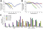
Figure 1. Disease severity and systemic spread of Chile/25945 after ocular inoculation of ferrets. Ferrets were inoculated by the ocular route as previously described (7) with a high (106...
To assess disease severity and transmissibility under different exposure concentrations, we inoculated ferrets by the ocular route with either a high (106 PFU) or low (103 PFU) dose of Chile/25945 virus (7). At either challenge dose, all ferrets inoculated by the ocular route became productively infected, reaching mean maximum weight loss of 12.7% (high dose) and 13.2% (low dose) and mean maximum rises in temperature of 2.4°C (high dose) and 2.0°C (low dose). Humane endpoints were reached on postinoculation days 5–7 in 3/3 (high dose) and 2/3 (low dose) animals (Figure 1, panel A). During necropsy, we detected infectious virus throughout the respiratory tract and in several extrapulmonary tissues (including from the gastrointestinal tract, central nervous system, and ocular system) (Figure 1, panel C), consistent with the highly virulent phenotype observed after high-dose intranasal inoculation of ferrets (3). One ferret survived the challenge with a serologic titer of 160.
Figure 2
Figure 2. Transmission of Chile/25945 virus after ocular inoculation of ferrets. Ferrets were inoculated by the ocular route as previously described (7) with a high (106PFU) or low...
To assess if ferrets inoculated by the ocular route were as capable as intranasally inoculated ferrets to transmit Chile/25945 virus in a direct contact setting (3), we cohoused a serologically naive ferret with each inoculated ferret 24 hours after inoculation. To assess virus replication within and beyond the respiratory tract, we collected nasal wash, conjunctival wash/swab, and rectal swab samples from inoculated and contact animals. All ferrets inoculated by the ocular route with a high dose of virus had detectable infectious virus in nasal wash (mean peak titer 5.3 ± 0.2 log10 PFU/mL), conjunctival wash/swab (4.4 ± 0.9 log10 PFU/mL), and rectal swab (3.6 ± 0.3 log10 PFU/mL) samples (Figure 2, panel A). The magnitude and frequency of viral titers in these specimens was reduced, but still present, in animals inoculated with a low dose of virus (Figure 2, panel B). Infectious virus was detected in either nasal or rectal wash samples in all contact animals on at least 1 day; infectious virus was not detected in conjunctival wash samples from any contact animal, possibly resulting from less overall virus shedding than in inoculated animals. Humane euthanasia because of severe disease was warranted for 4/6 contact animals (Figure 1, panel B); the other 2 survived, 1 of which exhibited a low level of seroconversion (hemagglutination inhibition titer 20).
Our finding that a clade 2.3.4.4b H5N1 virus isolated from a human can exhibit a virulent and transmissible phenotype after nontraditional inoculation, even with a low dose of inoculum, underscores the public health threat posed by those IAVs. The ocular surfaces may be exposed to infectious virus from the environment by several means (e.g., airborne particles, physical transfer from contact with fomites, and splashing liquids). Furthermore, circulation of tear fluid between ocular and nasopharyngeal tissues via the lacrimal duct offers an opportunity for infectious virus to spread from the respiratory tract to the ocular system (8), in agreement with successful H5N1 viral isolation from both conjunctival and nasopharyngeal swab specimens from a human with conjunctivitis (5). Conjunctivae may be exposed to virus by direct contact (e.g., virus-contaminated hands), indirect contact (e.g., virus-contaminated fomites), or after deposition of virus-laden droplets or aerosols onto the ocular surface (4), permitting opportunities for H5N1 virus to establish a productive infection in humans even in the absence of an ocular tropism. Considering the myriad ways humans may be exposed to IAVs, our study supports the need to consider nontraditional inoculation modalities in risk assessment activities and supports consideration of using eye protection in potentially contaminated environments (9).
Dr. Belser is a microbiologist in the Influenza Division, National Center for Immunization and Respiratory Diseases, Centers for Disease Control and Prevention, Atlanta, GA. Her research interests include the pathogenicity, transmissibility, and tropism of influenza viruses, with an emphasis on ocular tropism and ocular exposure.
留言 (0)