
記住我


All experimental protocols were approved by the institutional review board at Akita University Hospital (approval number: 2679). All data were collected under this IRB Protocol, which allows collection of medical record with consent or waiver of consent when no personalized health information is required, as was the case in this study. An opt-out approach was used for this retrospective study.
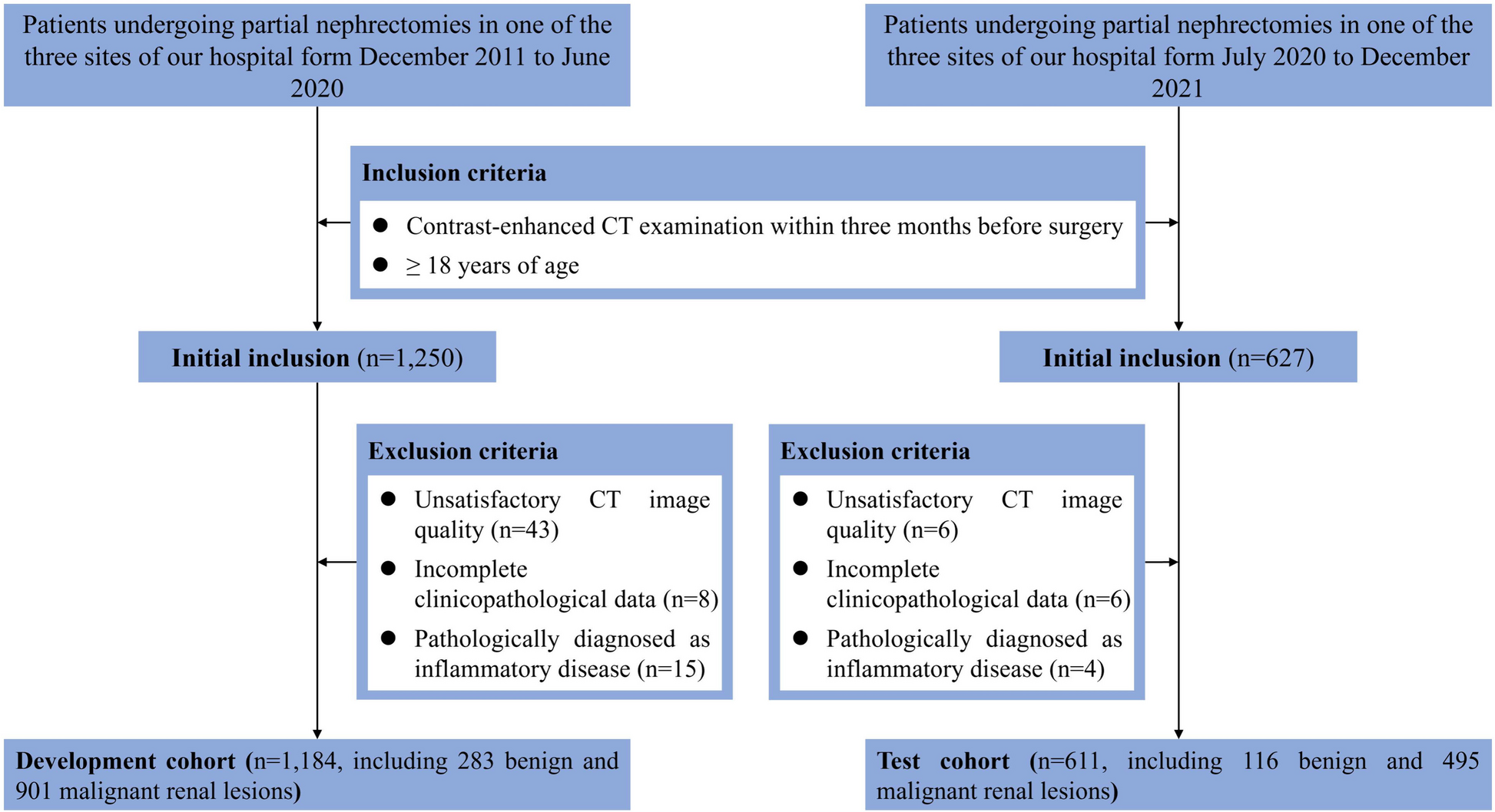
PatientsThis was a single-center study of initial staging using imaging tests in patients who had undergone complete surgical resection (R0 resection) for clinical T1N0 Stage IAs NSCLC. The medical records of 344 clinical T1N0 NSCLC patients who underwent lobectomy or segmentectomy at our institute between January 2017 and December 2022 were retrospectively reviewed. Of those, 71 cT1N0 patients who did not receive pretreatment screening brain MRI and/or BS were excluded. The remaining 273 participants, who had cT1N0 GGNs, including part-solid nodules (GGN group, n = 183), or cT1N0 solid tumors (ST group, n = 90) were deemed eligible for investigation and comparison. The patients’ characteristics are listed in Table 1. A diagram of the process by which cases were selected for study is shown in Fig. 1.
Table 1 Characteristics of patients with clinical T1N0 non-small cell lung cancerFig. 1
Flow chart illustrating the subject enrollment protocol
Preoperative imaging for determining clinical stagingIn addition to chest CT, all patients underwent brain MRI and planar BS and/or whole-body 18F-FDG PET/CT as routine procedures within 3 months before surgery.
Revolution CT (GE Healthcare) or other model was used for preoperative testing. Typical scan parameters were 60 keV and auto-mA. The scan area always included the chest, whereas inclusion of the abdomen to the pelvic region varied by case. The scans included both contrast-unenhanced and contrast-enhanced CT (pulmonary arterial and venous phase and equilibrium phase). Five-mm- and 0.625-mm-thick axial and 2mmthick coronal and sagittal sections were reconstructed. High-resolution computed tomography reconstructed 1.25-mm axial sections with a field of view of 180 mm.
Contrast-enhanced brain MRI to search for brain metastases was performed using a 3T system (Vantage Cencurian, Canon, or Discovery MR750, GE Healthcare) or other systems using a standard head coil. Examinations included axial T2-weighted images with turbo spin-echo, axial fluid-attenuated inversion-recovery images, axial contrast-unenhanced T1-weighted images with spin-echo (SE), axial and coronal contrast-enhanced T1-weighted images with SE and a contrast-enhanced three-dimensional gradient-echo pulse sequence. Contrast-enhanced sequences were obtained at least 5 min after intravenous injection of the contrast agent. MR angiography of the head and neck to detect arterial stenosis was added at the discretion of the radiologist.
BS was performed using a dual head gamma camera (Symbia Evo or Symbia E Dual Head System, Siemens Healthcare GmbH) with low-energy and high-resolution collimator. 900–1000 MBq of 99 m-Technetium (99mTc)-Methylene diphosphonate (MDP) (PDRadiopharma Inc.) or 99mTc-Hydroxy methylene diphosphonate (HMDP) (Nihon Medi-physics Co., Ltd.) was injected intravenously. Data acquisition was started after 3–4 h. The imaging parameters were a matrix size of 1024 × 256 and a bed speed of 9–12 cm/min. A subsequent tomography (single-photon emission computed tomography) was performed as needed.
PET/CT image of 18F-FDG was obtained using Biograph Vision 600 (Siemens Healthcare GmbH) or Discovery ST Elite 16 (GE Healthcare). 3.7 MBq/kg of 18F-FDG (Nihon Medi-physics Co., Ltd.) was injected venously and data acquisition began 60 min later. The PET imaging range was from the top of the head to the proximal 1/3 of the femur. The collection method was either whole-body dynamic imaging at 3 mm/sec for 4 times and additive reconstruction, or 3 min/bed (8–9 beds). A diagnostic CT scan for fusion was obtained using a standard protocol without intravenous contrast (120 kV; auto mA range, 20–666 mA; thickness, 3–3.75 mm; pitch, 1.2–1.75).
Co-registered images were displayed and analyzed using a high-speed 3D-image analysis system that enabled visualization of medical images in 3D for diagnosis and surgical simulation (SYNAPSE VINCENT, Fujifilm Corporation, Tokyo, Japan).
All patients had CT, brain MRI, and BS. 126 of 183 patients (68.9%) in the GGN group and 68 of 90 patients (75.6%) in the ST group had PET/CT. For evaluations using CT, MRI, BS and/or PET/CT, tumor size, lymph nodes, distant metastasis and staging were classified based on their location (i.e., mediastinal or hilar) and the 8th edition of the Union Internationale Contre le Cancer (UICC)-TNM staging system [15]. Board-certified thoracic surgeons (KI, NK, ST, SK, RD, HS, YH, TF, SS, AW, YN, YS, and YM) and radiologists (MK, NM and colleagues) evaluated the results of these preoperative tests for clinical staging (and detection of other diseases). Incidental findings are defined as incidentally discovered masses or lesions detected for an unrelated reason [16, 17].
Surgical procedure and follow-upAll patients received standard pre- and intraoperative care, and radical segmentectomy/lobectomy plus systemic node dissection. Pathological staging was also based on the 8th edition of the UICC-TNM classification [15]. Although the follow-up schedule after surgery varied, it usually entailed a chest CT every 3–6 months and others every 6–12 months for the first 2 years. If recurrence was suspected, the follow up schedule was tightened.
Statistical analysisClinical characteristics were statistically compared between the GGN and ST groups. Continuous variables were investigated using unpaired t tests or the Wilcoxon/Kruskal-Wallis test, while categorical variables were investigated using the Chi-squared test with continuity correction or Fisher’s exact test, as appropriate. 5-year overall survival (OS), relapse-free survival (RFS) and disease-free survival (DFS), and compared between the GGN and ST groups using the log-rank test. The RFS was calculated as the time from surgery of disease to any event, irrespective of cause, except for any second primary cancers. The DFS was the time from random assignment to cancer recurrence or death from any cause. Patients known to be alive or lost to follow-up on the date of last contact were treated as censored. Univariate and multivariate logistic regression analyses assessed the relationship between key variables and incidental findings. We calculated odds ratios along with 95% Confidence Interval (95%CI). All statistical analyses were performed using JMP IN 17.0.0 software (SAS Institute, Cary, NC, USA). P-values were 2-sided and considered significant if less than 0.05.
留言 (0)