
記住我


 Figure
 Box 1
Box 1Elbow injuries are a common orthopedic complaint among children seen in EDs, urgent care centers, and primary care offices.1 Diagnoses vary widely based on the mechanism of injury; however, one common cause of elbow injuries in children is radial head subluxation, also called a nursemaid's elbow or a pulled elbow injury.2 This injury typically is seen in children ages 6 months to 6 years, with a recent study showing the highest prevalence among children ages 1 to 2 years.3 Anecdotally, caregivers often are alarmed by the child's sudden lack of use of the affected arm and frequently seek care for the child soon after the injury occurs. Apparent cases often are easily corrected right in the examination room, and with timely reduction, complications are rare. This article reviews the epidemiology, pathophysiology, evaluation, and management of radial head subluxation injuries.
EPIDEMIOLOGYRadial head subluxation is a common elbow injury in younger children, with an overall annual rate of about 8 cases per 10,000 children age 6 years or younger.3 It is more commonly seen in younger children (ages 6 months to 6 years) than older children.3 There is a slight predominance in females, and patients tend to be classified in higher weight percentiles for their age and sex.1 The left arm is more likely to be affected, which is thought to be due to the high prevalence of right-hand dominance, both in caregivers, who are more likely to grasp a child's left hand, as well as faster development of the child's right arm due to the aforementioned dominance.4
 Box 2PATHOPHYSIOLOGY
Radial head subluxation typically results from a sudden pull or traction force on an extended, pronated arm.5 This results in the radial head slipping under the annular ligament.5 However, a history of nonaxial traction-related injury, such as falls or direct blows to the elbow, does not exclude nursemaid's elbow from the differential diagnosis, especially in younger children.6
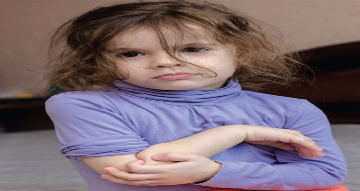
CLINICAL PRESENTATIONChildren typically present to the clinic with a history of a pulling injury to the arm. A common example is the caregiver holding the child's hand when the child attempts to abscond. On presentation, the child usually holds the arm in adduction, slightly flexed at the elbow, and pronated. Depending on the child's age, they may point anywhere along the upper extremity as to the location of pain, but with no swelling or tenderness to palpation present. If the presentation fits the classic picture and the child has no history of direct trauma, radiologic evaluation generally is unnecessary.
A full musculoskeletal evaluation of both upper extremities is important to avoid missed fractures or injuries elsewhere, as well as to evaluate the unaffected upper extremity to assess baseline range of motion. In particular, evaluation of the clavicle, especially in toddlers, is paramount to evaluate for occult fracture. To aid in a smooth examination, seat the child on the caregiver's lap, and sit at the patient's level if able. Distraction with toys or other devices also may be helpful to get a true sense of any tenderness to palpation.
IMAGINGRadial head subluxation is a clinical diagnosis, and imaging is not necessary unless the mechanism of injury is unclear. Obtain plain radiographs if a fracture is suspected or if closed reduction attempts are unsuccessful.7
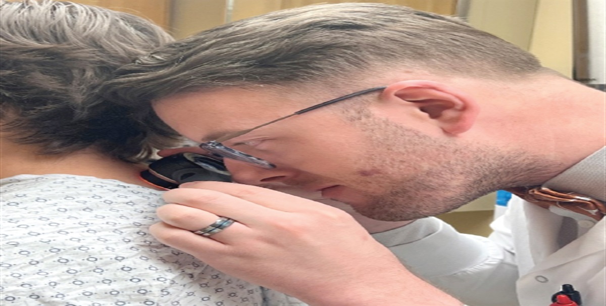
Bedside ultrasound has been increasingly used recently to evaluate elbow injuries in children, including radial head subluxation, in cases where the clinical picture may not be clear (Figures 1 and 2).8 The presence of synovial fringe enlargement on ultrasound has a high correlation with nursemaid's elbow.9,10 In a study by Tsai and Chiang, a partial eclipse sign was identified on ultrasound, showing the posterior synovial fringe caught between the annular ligament and the radial head.10 After reduction, ultrasound showed resolution of this partial eclipse sign in all 13 patients.10 Furthermore, Varga and colleagues showed 83% sensitivity and 100% specificity with two-plane ultrasound in a study of 205 cases to diagnose radial head subluxation with the presence of synovial fringe enlargement (Figure 3).9
 FIGURE 1.:
 Ultrasound showing the supinator muscle with a sharp edge (A, arrow) and the annular ligament (A, arrowhead) on the unaffected side in a different patient. Before reduction, an entrapped supinator muscle is seen in the radiocapitellar joint (B, bold arrow) and the annular ligament is absent (B, arrowhead); an enlarged synovial fringe is visible deep to the radiocapitellar joint (C, arrow) and olecranon fossa effusion. The degree of swelling of the supinator was evaluated by comparing the width of the supinator muscle (dashed line) at the radial neck area between the unaffected side (A) and affected side (B). C, capitellum; R, radial head.Reprinted with permission from Lee SH, Kim SG, Kwak D, et al. The usefulness of ultrasound and the posterior fat pad sign in pulled elbow. Injury. 2019;50(6):1227-1231.
 FIGURE 2.:
 Ultrasound before reduction (A). After reduction (B), ultrasound shows a disentangled swollen supinator muscle with a sharp edge (bold arrow), subsupinator effusion (arrow), and a restored annular ligament (arrowhead).Reprinted with permission from Lee SH, Kim SG, Kwak D, et al. The usefulness of ultrasound and the posterior fat pad sign in pulled elbow. Injury. 2019;50(6):1227-1231. doi:10.1016/j.injury.2019.04.026.
 FIGURE 3.:
 Ultrasound of normal and enlarged synovial fringe in the radiocapitellar joint. Comparative ventral longitudinal views. Arrow shows the interposed soft tissue.Reprinted with permission from Varga M, Papp S, Kassai T, et al. Two-plane point of care ultrasonography helps in the differential diagnosis of pulled elbow. Injury. 2021;52(suppl 1):S21-S24.
In a study by Rabiner and colleagues, 83% of patients with a clinical diagnosis of radial head subluxation were found to have no fat pad sign on ultrasound, with all patients having a successful reduction subsequently.11 This lack of fat pad sign, combined with the detection of synovial fringe enlargement, indicates a high likelihood of a radial head subluxation injury.9
Although the role of bedside ultrasound in elbow injuries warrants additional study, it can help children avoid the ionizing radiation of radiographs and may reduce time to injury reduction.
REDUCTION TECHNIQUESReduction of a radial head subluxation generally is achieved through either a supination-flexion maneuver, in which the forearm is rotated laterally at the elbow, bringing the palm face up, and then brought directly up toward the humerus (Figure 4), or a hyperpronation maneuver, in which the forearm is rotated medially at the elbow with the palm facing downward (Figure 5).12 Many conflicting studies exist about the superiority of hyperpronation compared with supination flexion; however, both techniques are effective.13-16
 FIGURE 4.:
 Supination-flexion techniqueReproduced with permission from Ulici A, Herdea A, Carp M, et al. Nursemaid's elbow—supination-flexion technique versus hyperpronation/forced pronation: randomized clinical study. Indian J Orthop. 2019;53(1):117-121.
 FIGURE 5.:
 Hyperpronation techniqueReproduced with permission from Ulici A, Herdea A, Carp M, et al. Nursemaid's elbow—supination-flexion technique versus hyperpronation/forced pronation: randomized clinical study. Indian J Orthop. 2019;53(1):117-121.
A click or pop may be heard or felt during reduction, and if successful, children generally can use the arm within 5 to 10 minutes.17 Anecdotally, a toy or treat can be used to help confirm reduction during reevaluation of the patient: Hold the reward far enough away for the child to need to fully extend the affected arm out to grab, which, if performed, is a sign of a successful reduction. If the child is unable to fully extend the arm, further imaging is warranted. If no abnormalities are found on imaging, splint the child's arm and send the child to orthopedics for further evaluation.2
RECURRENCE AND COMPLICATIONSThe prognosis for children who have a successful reduction is excellent. Children who demonstrate that they can fully extend their arm are ready for discharge home. Recurrence occurs in about a quarter of patients, due to ligamental laxity created by the initial injury and reduction.4 Complications are rare with prompt reduction; however, children with radiocapitellar instability caused by repeated radial head subluxations have a higher incidence of osteochondritis dissecans.18 Consider osteochondritis dissecans in the differential diagnosis for this population if they subsequently present with new or worsening elbow pain.18
CAREGIVER EDUCATIONPostreduction education is imperative to preventing recurrence. Caregivers should be instructed not to swing children by the hands or pick them up by the hands; instead, children should be lifted by placing the caregiver's hands under the axillae.19
CONCLUSIONRadial head subluxation is a common orthopedic condition, and children can present to a variety of medical settings. Careful history-taking and examination may allow for clinical diagnosis; however, further imaging should be obtained if the mechanism of injury is unclear. Hyperpronation and supination-flexion reduction techniques are effective for treating the injury. Ultrasound is a newer modality being used to identify and confirm treatment of this condition, and it reduces the amount of ionizing radiation a child receives while providing a better diagnostic picture when the diagnosis of radial head subluxation is not clear.
REFERENCES 1. Vitello S, Dvorkin R, Sattler S, et al. Epidemiology of nursemaid's elbow. West J Emerg Med. 2014;15(4):554–557. 2. Baiu I, Melendez E. Nursemaid's elbow (elbow subluxation). JAMA. 2018;319(5):515. 3. Zhou JJ, Shah NV, Scheer RC, et al. Trends and epidemiology of radial head subluxation in the United States from 2004 to 2018. Eur J Orthop Surg Traumatol. 2022;32(6):1137–1144. 4. Wong K, Troncoso AB, Calello DP, et al. Radial head subluxation: factors associated with its recurrence and radiographic evaluation in a tertiary pediatric emergency department. J Emerg Med. 2016;51(6):621–627. 5. Griffin ME. Subluxation of the head of the radius in young children. Pediatrics. 1955;15:103–106. 6. Li N, Khoo B, Brown L, Young T. Nonaxial traction mechanisms of nursemaid's elbow. Pediatr Emerg Care. 2021;37(6):e292–e294. 7. Cohen-Rosenblum A, Bielski RJ. Elbow pain after a fall: nursemaid's elbow or fracture. Pediatr Ann. 2016;45(6):e214–e217. 8. Lee SH, Kim SG, Kwak D, et al. The usefulness of ultrasound and the posterior fat pad sign in pulled elbow. Injury. 2019;50(6):1227–1231. 9. Varga M, Papp S, Kassai T, et al. Two-plane point of care ultrasonography helps in the differential diagnosis of pulled elbow. Injury. 2021;52(suppl 1):S21–S24. 10. Tsai C-C, Chiang Y-P. The usefulness of dynamic ultrasonography in nursemaid's elbow: a prospective case series of 13 patients reconsideration of the pathophysiology of nursemaid's elbow. J Pediatr Orthop. 2023;43(6):e440–e445. 11. Rabiner JE, Khine H, Avner JR, Tsung JW. Ultrasound findings of the elbow posterior fat pad in children with radial head subluxation. Pediatr Emerg Care. 2015;31(5):327–330. 12. Ulici A, Herdea A, Carp M, et al. Nursemaid's elbow—supination-flexion technique versus hyperpronation/forced pronation: randomized clinical study. Indian J Orthop. 2019;53(1):117–121. 13. Bexkens R, Washburn FJ, Eygendaal D, et al. Effectiveness of reduction maneuvers in the treatment of nursemaid's elbow: a systematic review and meta-analysis. Am J Emerg Med. 2017;35(1):159–163. 14. Bertucci N, Cowling K. Is hyperpronation more effective than supination for reduction of a radial head subluxation. Ann Emerg Med. 2018;72(5):586–587. 15. Krul M, van der Wouden JC, van Suijlekom-Smit LW, Koes BW. Manipulative interventions for reducing pulled elbow in young children. Cochrane Database Syst Rev. 2012;1(1):CD007759. 16. Porozan S, Forouzan A, Hassanzadeh R. Hyperpronation versus supination-flexion in radial head subluxation reduction: a randomized controlled trial. J Pediatr Intensive Care. 2020;9(4):256–260. 17. Heydari F, Masoumi B, Samsamshariat S. Radial head subluxation: possible effective factors on time to re-use the affected limb. Adv J Emerg Med. 2018;2(2):e19. 18. Jarrett DY, Walters MM, Kleinman PK. Prevalence of capitellar osteochondritis dissecans in children with chronic radial head subluxation and dislocation. AJR Am J Roentgenol. 2016;206(6):1329–1334. 19. Nemours Children's Hospital. Nursemaid's elbow. https://kidshealth.org/en/parents/nursemaid.html. Accessed December 20, 2023.
留言 (0)