
記住我


An autopsy was performed almost a year (357 days) after the man’s death.
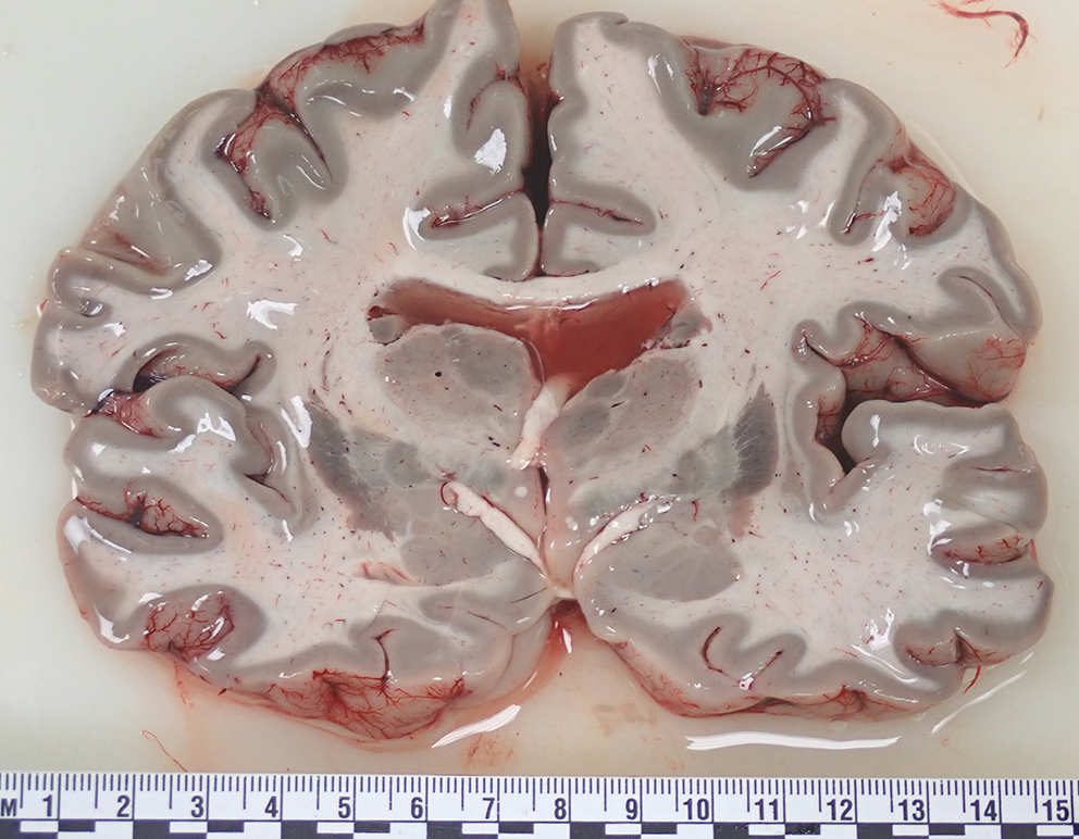
An external examination showed conspicuous putrefactive phenomena. The corpse was oily and soggy, widely covered in brownish and yellowish liquid due to the high post-mortem interval. The body height was 175 cm. An internal thoracic examination showed the presence of putrefactive liquid in pleural spaces and multiple, strong, extensive and diffuse fibrotic adhesions involving both lungs, especially on the diaphragmatic surface. In the right cavity, we found pleural-costal adhesions on the lateral posterior side of ribs I, II, III, IV and V and pleural diaphragmatic adhesions along the lung base, all covered in vegetative neoformations. In the left cavity, we found pleural-costal adhesions between the paravertebral line and the scapular line (from rib I to V), up to the axillary line (from rib III to V), all covered in vegetative neoformations (Fig. 3).
Fig. 3
Macroscopic examination of the ribcage: A, B, C, D: evidence of multiple fibrotic adhesions along the right and left ribs, all covered in vegetative neoformations (blue arrows)
The thoracic organ block (Ghon’s technique) was removed together with the diaphragm due to significant basal and bilateral adhesions. No other significant findings were observed during autopsy.
Thick and irregular neoformations were observed on the diaphragmatic sides of both lungs’ after the removal of thoracic organ block (A for the left lung, B for the right lung) (Fig. 4).
Fig. 4
Macroscopic examination of thoracic organs bloc district: A, B evidence of pleural plaques (forceps) at both lungs’ bases
The evidence of these fibrotic adhesions was confirmed during macroscopic examination in both ribcages after fixation (10% buffered formalin solution, 43 days after autopsy), which were especially prominent along the paravertebral line after ribcage isolation (Fig. 5).
Fig. 5
Macroscopic examination of left (A) and right (B) hemicostates after formalin fixation: evidence of multiple, gray and irregular vegetative neoformations strictly adhered to the hemicostates (pleural plaques)
HistopathologyPathological features were estimated using histological sections stained with hematoxylin–eosin, (H&E), and trichrome stains (Masson, Van Gieson). Histological examination of parietal pleural samples showed acellular hyalin collagen fibers in a reticular pattern, including areas with calcium salts deposit (Fig. 6). Visceral pleural samples showed multiple areas with fibrotic thickening (Fig. 7).
Fig. 6
Parietal pleura (A, H&E, 10X; B, H&E, 20X): fibrotic plaques with acellular hyalin collagen fibers in a reticular pattern, including areas with calcium salt deposits
Fig. 7
Visceral pleura: A thickening caused by fibrotic phenomena (Masson’s trichrome, 2.5X); B pleural thickness = 1.5 mm (yellow line) (H&E, 20X)
Histological examination of pulmonary samples showed fibrosis of respiratory bronchiole walls, extending to alveolar ducts and adjacent alveoli (Fig. 8). There was fibrotic thickening of the inter-alveolar septa between two or more contiguous respiratory bronchioles, with higher representation in subpleural districts (Figs. 9 and 10). In some areas, fibrosis was thicker and more intense, obliterating the surrounding alveoli. The examination also showed areas with a rare honeycomb pattern with no significant interstitial inflammatory infiltration and centriacinar emphysema.
Fig. 8
A Fibrosis of respiratory bronchiole walls (H&E, 20X), extending (B) to alveolar ducts and adjacent alveoli (Masson trichrome, 20X)
Fig. 9
A Fibrosis of respiratory bronchiole wall (Van Gieson trichrome, 20X); B fibrosis of alveolar ducts and alveoli walls (Masson trichrome, 20X)
Fig. 10
A Fibrosis of alveolar ducts and alveoli walls (Masson trichrome, 20X); B fibrotic thickening of the inter-alveolar septa between two or more contiguous respiratory bronchioles (Van Gieson’ trichrome, 20X)
Observing samples processed with either hematoxylin-eosin or Perls Prussian blue showed evidence of multiple fusiform structures consistent with the presence of asbestos fibers inside and at a distance from the peribronchial interstitium (Fig. 11).
Fig. 11
Amorphous fusiform structure compatible with asbestos fiber (yellow arrow), located in thealveolar space with fibrotic thickening of the wall. (Masson trichrome, 100X)
Pulmonary samples were submitted to an immunochemistry (IHC) investigation, performed using the following antibodies:
-BAP-1 (“BRCA1 Associated Protein-1, located on chromosome 3p21.1” for the differentiation of Mesothelioma from benign mesothelial proliferation); calretinin “Calcium-binding protein” for the differentiation (as part of a panel) of pleural Mesothelioma (positive) from lung adenocarcinoma (negative); cytokeratins 5/6 (“Basic (type II) cytokeratins of molecular weight 58 kDa (CK5) and 56 kDa (CK6)”) to distinguish epithelioid Mesothelioma (CK5/6 + in 83%) from lung adenocarcinoma (CK5/6- in 85%); WT-1 (“WT1 gene encodes for Wilms tumor protein located on chromosome 11p13” to differentiate malignant mesothelioma (WT1+) from non small cell lung carcinomas (WT1-); D2-40 “D2-40 Podoplanin is a 40 kDa, transmembrane, oncofetal, O linked sialoglycoprotein (mucin-type) found on lymphatic endothelium, mesothelium, and fetal testis” to differentiate Mesothelioma (+), even in effusions, versus adenocarcinoma (-); HBME-1 (”Marker of mesothelial cells, named after the laboratory of Dr. Hector Battifora and MEsothelioma”) to label mesothelial cells, both benign and malignant (malignant Mesothelioma), and for the identification of Mesothelioma.
The results were negative.
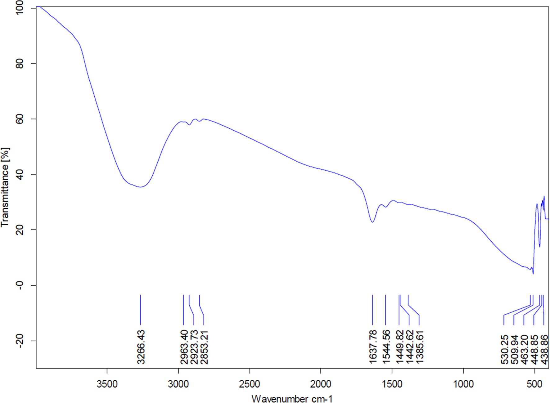
Scanning electron microscopy (SEM)Pulmonary samples were examined using SEM for research and identification of asbestos fibers (Fig. 12) as previously described [15]. Analyses were carried out at the Electron Microscopy Center of the Environmental Protection Agency of Lombardy Region (CRME). The results are described in Table 1. The margin of error is expressed as the bounds of the 95% confidence interval. The limit of detection is defined as the bound of the 95% confidence interval of the Poisson distribution and serves as an indicator of the analysis’s sensitivity. The percentage of commercial amphibole asbestos (amosite + crocidolite) is 100%. The average length and diameter of the asbestos fibers are 3.22 microns and 0.22 microns, respectively.
Table 1 Results of quantitative analysis of asbestos fibers through scanning electron microscopy (SEM)Fig. 12
Image of an asbestos fiber (yellow arrow) detected during the analysis (magnification 12000X)
Death was attributed to the underlying diseases, given the patient’s age, his medical history (cardiac diseases, old age, loss of weight) an the absence of clinical evidence suggestive of a severe asbestos-related cardiopulmonary disease. Therefore, a causal relationship between the death and the asbestosis was excluded. Asbestosis diagnosis was confirmed based on findings from the the investigations. The diagnosis of mesothelioma was excluded.
留言 (0)