
記住我

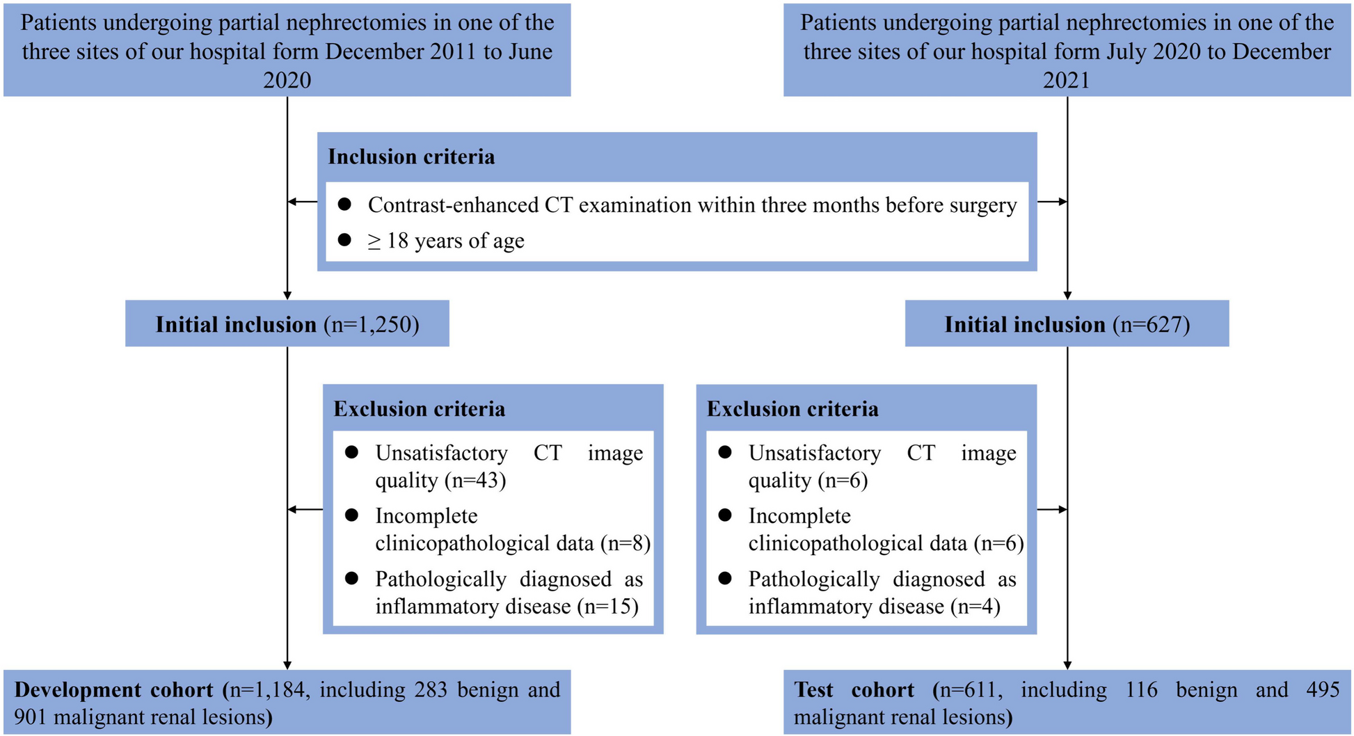


The Ethics Committees of Beijing Chao-Yang Hospital, Capital Medical University (no. 2021-ke-443) approved this retrospective, single-center study and waived the requirement for informed consent. A total of 797 consecutive patients with a histologically proven diagnosis of primary NSCLC underwent chest high-resolution CT (HRCT) between January 2013 and October 2018 at our hospital. The exclusion criteria were as follows: (i) previous thoracic surgery, radiation therapy, or chemotherapy; (ii) severe tuberculosis or pneumoconiosis; (iii) history of collagen vascular disease; (iv) preexisting ILD; and (v) missing clinical data. The HRCT images of these patients were reviewed retrospectively by two pulmonary radiologists (X-L X and Y-L G, with 10 and 25 years of experience, respectively) to identify the radiological evidence of concomitant ILA. According to the criteria of the Fleischner Society, ILAs are diagnosed as non-dependent ground-glass opacities (GGO), reticular abnormalities, architectural distortion, non-emphysematous cysts, honeycombing, and traction bronchiectasis that affect > 5% of any lung zone [4, 15]. Finally, 109 patients with concomitant lung cancer and ILA were included in this study (Fig. 1). Histological diagnosis was based on the World Health Organization classification published in 2021 [16] and TNM staging of lung cancer was according to the eighth edition [17], respectively. Molecular analysis of the EGFR mutation status was performed using polymerase chain reaction-based amplification-refractory mutation system analysis with the Human EGFR Gene Mutations Detection Kit (Beijing ACCB Biotech, Beijing, China). Smoking status was determined by reviewing medical records and was quantified by pack-years.
Fig. 1
Flowchart of the selection of the study population and the exclusion criteria. CT, computed tomography; HRCT, high-resolution CT; ILA, interstitial lung abnormality; ILD, interstitial lung disease; NSCLC, non-small cell lung cancer
CT evaluation by radiologistsCT images were obtained for all patients in the supine position at the end of inspiration using a variety of CT units. Images were reconstructed with contiguous 1–2 mm sections using a high-resolution reconstruction algorithm for analysis. Two chest radiologists independently evaluated the CT images in random order without any clinical or pathologic information. Image analysis was performed using the indicated window settings (lung: width, 1500 HU; level, -400 HU; mediastinum: width, 400 HU; level, 40 HU) for axial images on a picture archiving and communication system; radiologists were allowed to moderately change the default window settings for ease of assessment.
ILAs were subcategorized as non-subpleural, subpleural nonfibrotic, and subpleural fibrotic ILA [4], with subpleural fibrotic ILA corresponding to a probable usual interstitial pneumonia pattern [18]; non-subpleural ILA and subpleural nonfibrotic ILA were defined as non-fibrotic ILA in this study. The distribution, overall extent of the lesions, and extent of each ILA lesion were further evaluated, and disagreements concerning the scores were resolved by consensus. The predominant distribution included peripheral, peribronchovascular, and mixed patterns. The extent of CT findings of ILA in each case was scored using a four-point scale (score of 1, 5–25% involvement; score of 2, 26–50% involvement; score of 3, 51–75% involvement; and score of 4, 76–100% involvement) [1]. The extent of the ILA findings was further assessed in six lung zones (upper, middle, and lower zones of both lungs) on CT images. The division of the upper, middle, and lower lung zones was determined based on the levels of the inferior aortic arch and right inferior pulmonary vein [4]. Readers scored the lung fields that showed abnormalities in each of the six zones based on GGO, reticulation, and honeycombing. The average of the six lung zones was used to calculate the percentage of the whole lungs. Traction bronchiectasis was assessed by summing the number of bronchiectasis-affected pulmonary segments [19].
Morphological CT features of lung tumors were also analyzed, including location, size (maximum long-axis diameter), shape (round, somewhat irregular, irregular), density (solid, part-solid, GGO), margin (lobulation, spiculation), internal (vacuole sign, lumen, cavity, air bronchogram, bronchial cut-off sign), surrounding structures (vascular convergence, pleural traction, halo sign), and associated findings (pleural fluid). Previously published evaluation methods were used in this analysis [10, 20]. Disagreements regarding the CT features of lung cancer were resolved by consensus.
Statistical analysisAll statistical analyses were performed using SPSS version 20.0. Data are presented as means, medians, counts, and percentages, where appropriate. Clinical and CT feature variables between the EGFR mutation and wild-type groups were compared using X2 or Fisher’s test, Student’s t-test, or the Mann–Whitney U test, where appropriate. Multiple logistic regression analysis was used to assess the value of clinical features, tumor CT findings, and ILA CT features in predicting EGFR mutations. Significant factors in the univariate analysis were identified as potential covariates in a multivariate logistic regression model using the forward likelihood ratio test. A receiver operating characteristic (ROC) curve was used to estimate the significant predictors of EGFR identification. The area under the curve (AUC) was calculated to determine the predictive capability. A p value of less than 0.05 was considered to indicate a significant difference.
The probabilities of OS at three and five years after diagnosis were estimated using the Kaplan–Meier method. Multivariate analysis was performed using the Cox proportional hazards model to evaluate the hazard ratios (HR) for OS probabilities with 95% confidence intervals (CI).
留言 (0)