
記住我


To construct the rolling machine, a fraction of CEA aptamer was bound with walker DNA, forming a "DNA gate" that effectively secured the DNAzyme cleavage site. The DNA named Track DNA was modified to Fe3O4@Au NPs by the Au–S bond as the substrate of DNAzyme. Figure 1 illustrates the three stages of the cascade amplification assay, including (1) the activation of CEA-activated DNA walking on AuNPs; (2) the rolling of Au-walker on Fe3O4@Au-track surface and releasing DNA fragments (T0); (3) T0 activated the trans-cleavage of the ssDNA reporter by the Cas12a-crRNA complex. In the presence of the target, the CEA hybridizes with aptamer and releases Walker DNA, which then recognizes and digests the Track DNA by DNAzyme from the Walker. As the digestion of the track DNA progresses, the Walker DNA is released and forms a bond with an adjacent track. The presence of numerous walking DNA strands acting as legs on the surface of AuNP results in the Au-walker rolling instead of walking on the Fe3O4@Au NPs surface. This rapid rolling process releases a significant amount of target DNA of CRISPR-Cas12a to initiate the Rolling Machine-CRISPR/Cas12a Cascade Amplification Assay (Roller-Cas12a reaction), which can be visually observed or quantified using fluorescence detection methods.
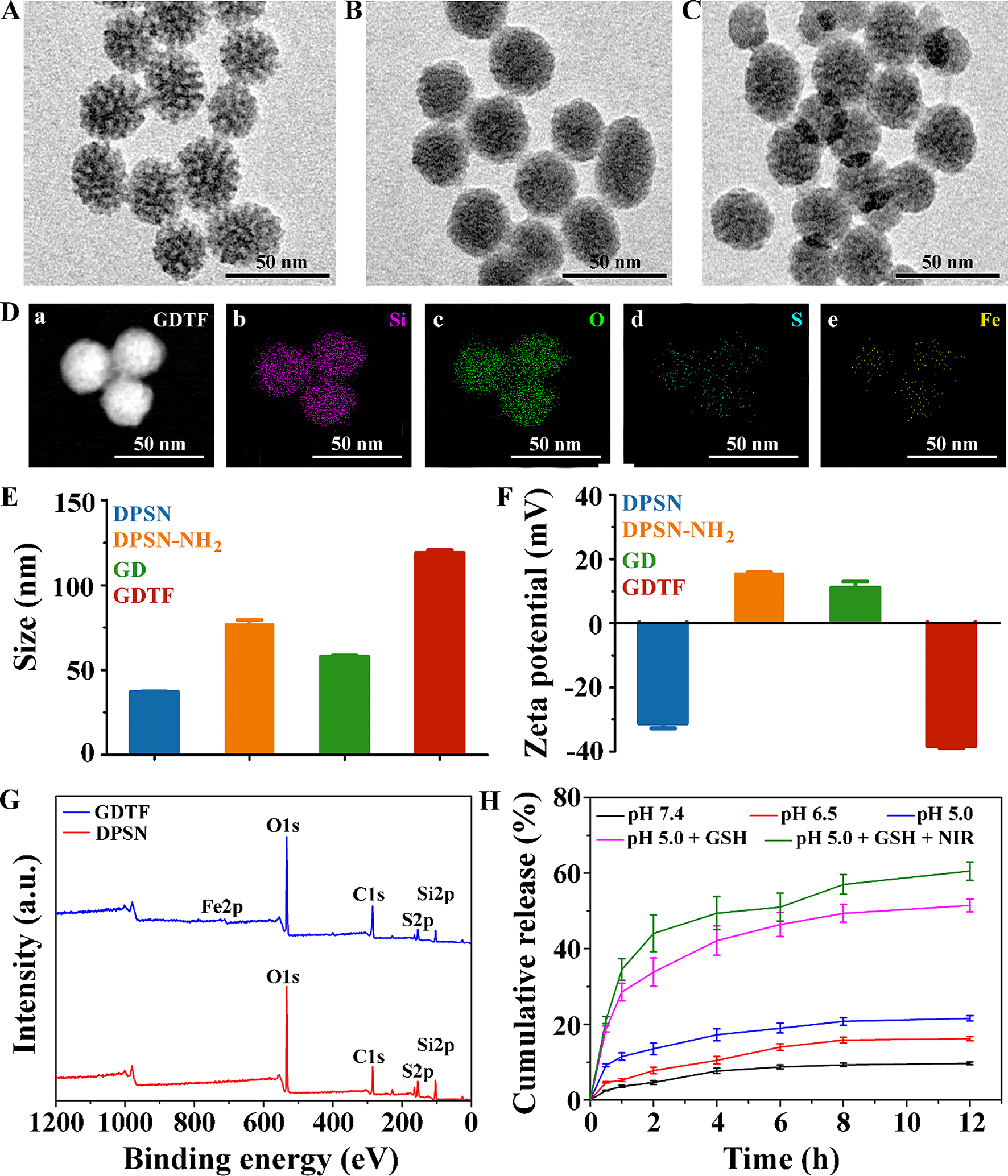
Characterization of Au-walker and Fe3O4@Au-trackMorphological features of Au-Walker and Fe3O4@Au-Track were characterized by Transmission electron microscope (TEM), Dynamic Light Scattering (DLS), and Zeta potential investigations. To confirm the attachment of Walker DNA on the surface of AuNPs, we characterized Au-walker by TEM. Fig. S1A reveals that AuNPs were well dispersed with a uniform diameter of 15.3 ± 0.2 nm (by measuring the diameters of ~ 50 particles), respectively. After being decorated with the Walker and aptamer DNA, the size of AuNPs under TEM remained unaltered (Fig. S1B), whereas the hydrated particle size of Au-Walker increased from 21.0 ± 2.3 nm (Fig. S1C) to 68.1 ± 1.2 nm (Fig. S1D). According to Fig S1E, the UV–vis absorption peak of AuNPs at 520 nm was shifted to 525 nm, which illustrated that AuNPs are successfully decorated with DNA [24,25,26].
As shown in Fig. 2A, Fe3O4 nanocores were spherical, with an average size of 483.4 ± 33.0 nm. The prepared Fe3O4@Au nanoparticles are composed of Fe3O4 nanocores with gold seeds (Fig. 2B). The gold seeds (~ 15 nm) were uniformly and randomly attached to the surfaces of the Fe3O4 nanocores (Fig. 2C). Gold seeds appeared black, and Fe3O4 nanocores presented a light color in the TEM images, this can be attributed to the higher electron density of Au in comparison to that of Fe3O4. The TEM-mapping results further confirmed the coexistence of Fe3O4 and Au. After the modification of Track DNA, the morphology and size did not change significantly (Fig. 2D), the hydrated particle size increased from about 531.0 nm to 615.0 nm (Fig. S2B, C), which illustrated that Fe3O4@Au NPs were modified by DNA. Zeta potential results of Au-walker and Fe3O4@Au-Track were -38.4 ± 1.8 mV (Fig. S1F) and − 34.0 ± 0.08 mV (Fig. S2D), respectively. These results suggest that Au-walker and Fe3O4@Au-Track were synthesized and modified successfully and can be used for further experiments.
Fig. 2
Characterization results of DNA rolling machine. TEM image of A Fe3O4 nanocores, B Fe3O4@Au NPs, C single Fe3O4@Au NPs, D Fe3O4@Au NPs-Track. TEM elemental mapping of Fe3O4@Au NPs-Track for E Fe, F Au, and G O
Viability analysisAs previously stated, the Roller-Cas12a reaction involves two components: the CEA-mediated operation of the 3D rolling machine and the subsequent automated CRISPR-Cas12a reaction. The confirmation of walker release and DNAzyme cleavage of Track in the presence of Mn2+ was initially verified through agarose gel electrophoresis. According to Fig. S3, the presence of a band with a length of ~ 100 bp (Lane 3) corresponding to the hybridization of aptamer with walker. Due to the affinity of aptamer and CEA being greater than that of DNA complementary [27], the band at 100 bp became lighter (Lane 4) when the CEA was added, which indicates the CEA binds to the aptamer and dissociates the aptamer-walker complex. We further confirmed that the aptamer-walker complex had no DNAzyme activity that could not trigger the rolling process with Mn2+. As shown in Fig. S4, aptamer or aptamer-Walker complex and Track did not interact with each other in the absence of CEA (lane 6, 7, and 10), as no new band appeared. However, fast migration bands were observed after Track DNA was incubated with single Walker DNA and Mn2+ (lane 9), or aptamer-Walker complex, CEA, and Mn2+ (lane 11), indicating that it was effectively cut by the DNAzyme of Walker. These results demonstrate that the 3D rolling machine can only be activated in the presence of CEA and Mn2+.
The working principle was further interrogated through fluorescence analysis. Without adding CEA or Mn2+, the signal was negligible (Fig. 3, red and purple curve). Conversely, a significant rise in fluorescence signal was detected when the target was present (Fig. 3, blue curve). In addition, due to the modification of Track DNA on Fe3O4@Au through Au–S bonds, the DTT treatment was performed to reduce disulfide bonds. According to the green curve in Fig. 3, the strong fluorescence proves the successful preparation of the track components of the rolling machine. The above results demonstrated the expected viability of this strategy by showing that our Roller-Cas12a reaction was successfully activated exclusively in the presence of the CEA, which is consistent with Agarose gel electrophoresis.
Fig. 3
Viability analysis of DNA rolling machine. Fluorescence spectrum of rolling machine, rolling machine + PBS with Mn2+, rolling machine + 10 ng/mL CEA with Mn2+, rolling machine + 10 ng/mL CEA without Mn2+and rolling machine + DTT
Furthermore, to investigate the impact of nanoparticle characteristics on the rolling machine, we respectively modified DNA sequences on AuNPs and Fe3O4@Au NPs to form Au-Walker, Au-Track, Fe3O4@Au-Walker, and Fe3O4@Au-Track, and combine them in pairs to build the rolling machines. According to Fig. S5, in the presence of CEA, the Rolling machine I (Au-Walker and Fe3O4@Au-Track) and Rolling machine II (Au-Walker and Au-Track) exhibits obvious fluorescence intensity difference compared to PBS. However, rolling machine II requires centrifugation to obtain T0, which may hinder development of fast and convenient detection methods.
Comparison between a DNA walking machine and a DNA rolling machineAccording to previous work [28], increasing the local concentration of Track DNA effectively improves the walking efficiency of nanomachines, which can also improve signal output and equilibrium time. Therefore, we initially assessed the modification efficiency of the Track DNA in both the walking and rolling machines. An equal amount of Track was used for synthesizing the walking machine and rolling machine, and the prepared nanomachines were treated with DTT to reduce the Au–S bond, thereby releasing modified Track DNA. The ligation efficiency (LE) was calculated as follows:
$$\mathrm=\frac_}\times 100\%$$
F0 represents the fluorescence intensity of the original Track DNA, while F represents the fluorescence intensity of the nanomachines treated with DTT. As shown in Fig. S6, the rolling mode exhibits higher ligation efficiency and releases more fluorescence intensity with CEA. The ligation efficiency of the rolling machine was 21%, compared to only 14% for the walking machine, indicating that our rolling machine has enhanced the local concentration of Track DNA.
The kinetics of the second-stage rolling process were compared with those of the traditional DNA walking machine, confirming the improved walking efficiency and signal amplification capabilities of the 3D DNA rolling machine. The Walker strand was initially attached to AuNPs utilizing the same experimental setup as that for the construction of Au-Walker to evaluate the rolling kinetics. The Au-Walker was then added in Fe3O4@Au-Track solutions along with Mn2+. The fluorescence signal of the supernatant was measured at different time intervals through magnetic separation. All experimental conditions for the walking mode were identical, except that an equal amount of walker strand and Track strand was modified on the same AuNPs to construct the traditional DNA walking machine. According to Fig. 4, our DNA rolling machine completed the reaction in only 50 min, compared to at least 100 min for a traditional DNA walking machine, effectively doubling the walking speed of the rolling machine relative to the walking machine. The highest fluorescence intensity of the rolling machine is twice that of the traditional walking machine, indicating that rolling nanomachines more readily release Track DNA. This could be because modifying Walker and Track DNA on different nanoparticles improves DNA binding and release, thereby demonstrating great potential for CEA detection.
Fig. 4
Kinetics comparison between rolling machine (blue curve) and walking (red curve). Consistent experimental parameters were applied to both the rolling and walking processes: Au-Walker (10 μL), Fe3O4@Au-Track (30 μL), Mn2+ (10 mM, 10 μL), and CEA (10 ng/mL, 10 μL). The concentration of Walker, aptamer, and Track conjugated with AuNPs was adjusted to an equivalent level (detailed procedure was described in SI)
Optimization of the conditionsWe optimized key experimental conditions to achieve optimal analytical performance, including the categories of Walker, the molar ratio of Au-Walker and Fe3O4@Au-Track, the concentration of Mn2+, the temperature, pH value, and reaction time. The impact of the aforementioned conditions on the change in fluorescence signals (F–F0) is depicted in Fig. S7, with detailed descriptions provided in the Supplementary Information (SI). Ultimately, medium binding length (9 nt) of Walker, 1:3 of Au-Walker to Fe3O4@Au-Track, pH 7.4, 10 μL 15 mM of Mn2+, incubation of 50 min at 25 ℃ were determined to be the optimal conditions and were utilized in subsequent experiments.
Analytical performanceUnder the optimal experimental conditions, the fluorescence intensity was recorded by a series concentration of CEA. In response to the absence of literature on the upper limit of serum CEA concentration, we extended the test range of CEA (0, 0.1, 0.2, 0.4, 0.5, 1, 5, 10, 20, 30, 40, 50, 60, 70 and 80 ng/mL) to accommodate clinical samples with abnormal concentrations. As demonstrated in Fig. 5A, the fluorescence intensity (FI) exhibited a gradual increase corresponding to the rising concentrations of CEA, ranging from 0.2 ng/mL to 20 ng/mL. When CEA concentration exceeds 20 ng/mL, FI reaches the plateau as CEA concentrations increase gradually. Green fluorescence was visible with a handheld UV lamp at concentrations as low as 0.2 ng/mL, establishing the detection limit of our assay at 0.2 ng/mL. Consequently, the limit of detection (LOD) for our visual assay was determined to be 0.2 ng/mL (the minimum concentration can be distinguished by naked eyes as criterion). Therefore, our assay indicated a “Yes” results when the concentration of CEA was higher than 0.2 ng/mL. In addition, the analytical performance of the traditional DNA walking machine integrated with CRISPR-Cas12a was evaluated. Based on Fig. S8, the DNA walking machine method exhibits a distinct fluorescent signal when CEA ≥ 10 ng/mL compared to blank controls. Therefore, the LOD of the walking machine coupled with CRISPR/Cas12a is 10 ng/mL (the minimum concentration can be distinguished by naked eyes as criterion). Our assay, based on the rolling machine, demonstrates high sensitivity and significantly reduces the LOD of the traditional DNA walking machine by 50-fold to 0.2 ng/mL, making it more suitable for developing methods for detecting CEA in clinical samples.
Fig. 5
Sensitivity, selectivity, and stability test of Roller-Cas12a. A Fluorescence intensity responses of our assay for CEA at varying concentrations (bottom to top: 0, 0.1, 0.2, 0.4, 0.5, 1, 5, 10, 20, 30, 40, 50, 60, 70 and 80 ng/mL). B The fluorescence intensity versus the various concentrations of CEA. Insert: Images of the reaction system rolling machine combined with CRISPR-Cas12a. C Fluorescence intensity in the presence of different reagents (1 → 7: PBS, BSA, PSA, AFP, CEA, BSA + PSA + AFP, and BSA + PSA + AFP + CEA). The concentration of BSA, PSA, and AFP was each at 50 ng/mL. The concentration of CEA was 10 ng/mL and 10 mM PBS buffer was the blank control. D Stability of this assay within 9 days. F–F0 value of CEA at 10 ng/mL on 1, 3, 5, 7, and 9 days. Error bars represent the standard deviation of 3 replicates. *P < 0.05, “ns” means not significant (P > 0.05). Statistical analysis compares with 0 ng/mL (B), PBS (C), and 1st day (D), respectively
The specificity of the assay for CEA was further assessed by measuring bovine serum albumin (BSA), prostate-specific antigen (PSA), and alpha-fetoprotein (AFP). As depicted in Fig. 5C, BSA, PSA, and AFP exhibited fluorescence output at nearly background levels, underscoring the excellent selectivity of our assay.
Finally, to investigate the stability of the rolling machine combined with the CRISPR-Cas12a assay, three consecutive tests with 10 ng/mL CEA were conducted in parallel (Fig. 5D). Over a period of 9 days, this assay consistently exhibited a relative standard deviation (RSD) of 3.6%, demonstrating its good stability. These findings reveal that, in comparison to some earlier publications for biomarkers detection (Table S2), the rolling machine coupled with the CRISPR-Cas12a system has a similar detection limit and a shorter detection time.
Detection of CEA in serum samplesTo evaluate the practicability of the rolling machine combined with CRISPR-Cas12a assay in complex samples, 15 clinical human serum samples are analyzed, and the results were compared with those obtained using the Human CEA ELISA Kit (D711374, Sangon Biotech). According to previous reports, the concentration of CEA in the healthy human serum is less than 5 ng/mL [29, 30]. Therefore, a method that provides a simple Yes/No answer with a detection limit of 5 ng/mL is more advantageous for early tumor screening. In clinical sample testing, we optimized the concentration of gRNA (Fig. S8) to develop a straightforward, equipment-free, visual, and semi-quantitative assay suitable for on-site and point-of-care testing (POCT). The optimized is capable of producing "danger" or "safe" results based on the observable fluorescence intensity to the naked eye, enabling the differentiation between healthy individuals and those suspected of having tumors. As shown in Table 1, when the concentration of CEA is exceeds 5 ng/mL, our detection system shows strong fluorescence, indicating a potential tumor risk, and the diagnostic consistency between our assay and the ELISA kit was found to be 100%. When the concentration of CEA in human serum is approximately 5 ng/mL, the system shows weak fluorescence emission, suggesting a potential tumor presence. Expanding the scope of clinical screening in this way is advantageous for the early detection, diagnosis, and treatment of tumors. For lower serum CEA concentrations, our assay exhibits no fluorescence, indicating a "safe" status for tumor screening. For clinical serum samples with CEA concentrations below 5 ng/mL, the diagnostic consistency between our assay and the ELISA kit was 73%. Compared to the commercial ELISA kit, our assay features a shorter detection time (over 200 min for ELISA vs. 75 min for our assay) and is easy to operate. Additionally, compared to existing methods (Table S2), our assay evaluated clinical serum samples instead of spiked serum samples, offering better anti-interference capabilities and practicality.
Table 1 Comparison of commercial ELISA kit and our assay (n = 3, \(\overline\pm s\))
留言 (0)