
記住我


Wearing a facemask in professional settings is a usual procedure mainly for surgical purposes. The COVID-19 pandemic rendered the long-term use of facemasks a daily reality for the vast majority of citizens. This was the case in professional and even private interactions but also in MRI scanning facilities. This practice was widely accepted and well tolerated to limit the propagation of the virus mainly in vulnerable groups. Under normal conditions and mild exercise, wearing a facemask does not affect sensorial processing, motor or high-level cognitive performances (Morris et al., 2020; Slimani et al., 2021; Wells et al., 2023). However, some lines of evidence indicated that facemasks may alter concentration and visual attention in maximal running aerobic tests (Slimani et al., 2021). By impacting social interaction, facemasks modify the neural responses to recognition of facial cues but also pivotal human abilities serving our daily behavior such as emotion recognition that stimulate empathic responses, perceived closeness, trust attribution and even re-identification of unmasked faces (Ferrari et al., 2021; Grundmann et al., 2021; Marini et al., 2021, 2022; Proverbio and Cerri, 2022; Tsantani et al., 2022; Proverbio et al., 2023). In contrast to behavioral changes, data on the effect of wearing a facemask on brain activation remain rare. Two previous studies indicated that this practice has a subtle but still significant effect on cerebral blood flow and oxygen saturation but also BOLD (blood oxygenation level dependent) baseline signals. This BOLD effect is the basis of functional MRI. In short, local neuronal activity is associated with increased demands on oxygen and associated with a local vascular response which in turn modifies the local concentration ratio of oxygenated versus deoxygenated hemoglobin. This modifies the local magnetic properties that can be assessed using a dedicated MRI pulse sequence. The BOLD response is therefore an indirect vascular response that can be measured in fMRI. In a previous study, we also reported a small yet significant alteration in at rest functional connectivity limited to higher-level salience network in an independent sample of community-dwelling healthy controls (Haller et al., 2022). Functional connectivity fMRI is based on the principle assumption that if spontaneous fluctuations in the BOLD signal in two regions are not random but correlated, then these two regions are probably functionally connected. The more similar the BOLD signal fluctuations over time in two regions, i.e., the higher the correlation between two regions, the more likely is the functional connectivity between them. This study included only at rest fMRI analysis and cannot thus define whether brain activation during highly demanding cognitive tasks may be affected by wearing a facemask. To address this issue, we performed a cross-sectional study comparing the behavioral and fMRI patterns when wearing or not a tight KN95/FFP2 facemask during the performance of a 2-back working memory task in healthy controls. The 2-back working memory task is well established for fMRI studies and previously used by our group (Haller et al., 2013, 2014; Sinanaj et al., 2015; Haller et al., 2017; Zanchi et al., 2017). Since we anticipated only subtle effects related to the wearing of a facemask, the selection of the highly demanding 2-back condition, that implies the mobilization of working memory and not only attentional resources, allowed for obtaining the strong and reproducible fMRI activations needed for the detection of small group differences.
2 Materials and methods 2.1 ParticipantsThis prospective study was approved by the Ethics Committee of the University Hospitals and University of Geneva, Switzerland, the study was in accordance with the Declaration of Helsinki, and all participants gave written informed consent. The study included 24 community-dwelling male healthy control participants (mean age ± SD = 37.6 ± 12.7 years) recruited via advertisements in local media. The following exclusion criteria were applied: a. presence or history of a chronic psychiatric disorder (psychosis, bipolar disorder) b. history of loss of consciousness lasting longer than 30 min, c. history of head injury or post-concussion symptoms, d. history of auditory or visual deficits, seizure and neurological disorders, and e. regular use of psychotropic medications and alcohol. The exclusion of acute psychiatric disorders was confirmed by the Mini Neuropsychiatric Interview (Stanga et al., 2019).
2.2 Working memory taskWe used a classical fMRI experiment in order to explore the presence of subtle differences in the spatial distribution of brain activation between MASK and NOMASK conditions. The fMRI technic was selected since it makes possible to examine the functional reactivity of the human brain facing increased cognitive demands and in particular working memory activation. Previous contributions established in detail the patterns of brain activation during the successful performance of the 2-back task both in controls and clinical populations (Haller et al., 2013, 2014; Sinanaj et al., 2015; Haller et al., 2017; Zanchi et al., 2017). Briefly, a sequence of letters was presented visually on an MR compatible canvas in the MRI scanner. In the active 2-back condition (high WM demand), targets are letters that are identical to the letter presented two items ago (e.g., “a f h f”). In the control condition 0-back (visual processing with minimal WM requirements), the target is a pre-defined letter (e.g., “x”). Both conditions are contrasted to evaluate the effect of WM demand in 2-back versus the control condition 0-back.
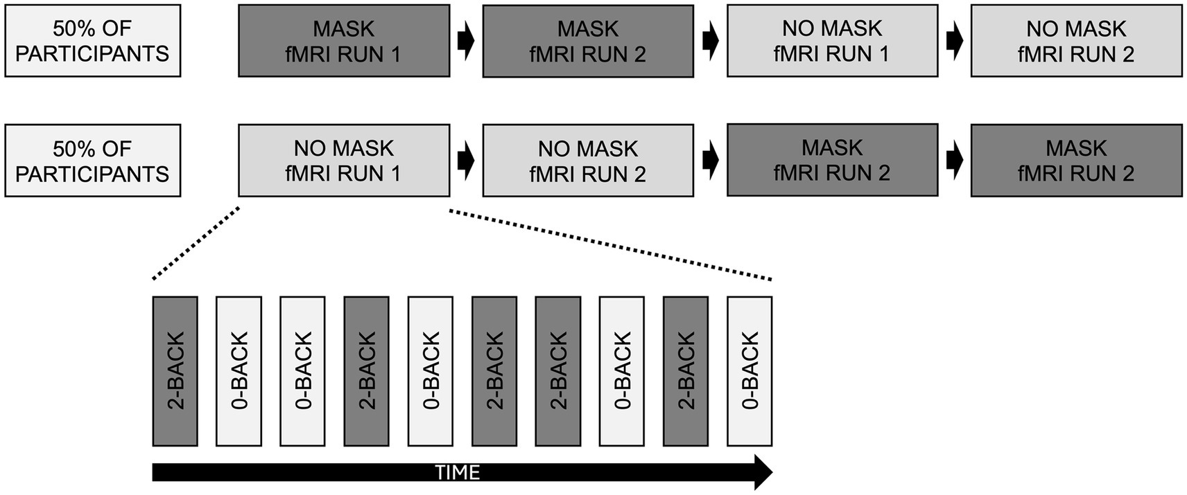
For the fMRI experiment, we used a cross-over design: half of the participants had first MASK then NOMASK condition, the other half had the inverse order. All participants had the same KN95/FFP2 facemask (a commercial model without metal to be compatible with the MR scanning). First, participants were familiarized with the task demands outside the MRI using a training session. The actual fMRI protocol consisted of four runs (2 × MASK and 2 × NO MASK). Each run included alternation blocks of 35 s each for conditions 2-back and 0-back with interleaving rest conditions of 15 s to allow the hemodynamic response to recover from the previous block. Each n-back condition (0-back or 2-back) was repeated five times in a pseudo-randomized order. Participants provided response (target versus no target) via an MR compatible response box for targets (33% of trials) and another button for non-targets (Figure 1).
Figure 1. Schematic illustration of the experimental setup. In a cross-over design, half of participants first performed 2 fMRI runs MASK and then 2 fMRI runs NO MASK, while the other half of participants performed the opposite order. Each run consisted of 10 blocks, 5 blocks of 0-back and 5 blocks of 2-backs in pseudo-randomised order.
The entire MRI scanning lasted approximatively 1 h. We made sure that participants had the facemask on for 10 min before the start of MASK condition, and no facemask for 10 min before NOMASK condition. To avoid potential bias of the resting fMRI results due to basic physiologic parameters, we monitored breathing and heart rate during the fMRI runs.
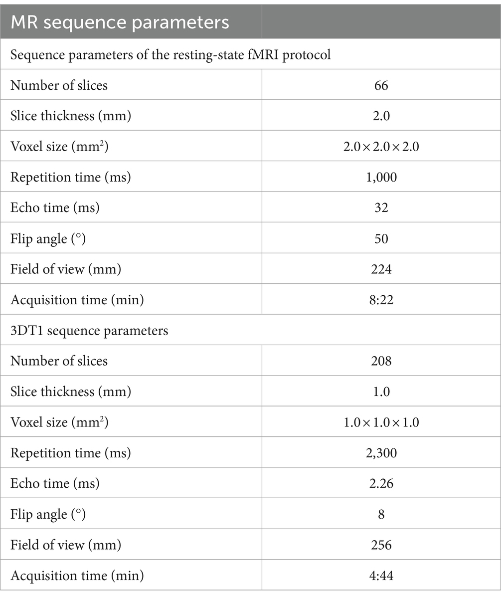
2.3 MR imagingMR images were acquired using a 3 T MRI scanner (MAGNETOM PRISMA, Siemens) at Campus Biotech Geneva. MR sequence parameters are listed in Table 1. Functional echo-planar imaging had the following essential parameters: 66 slices, slice thickness = 2.0 mm, voxel size = 2.0 mm × 2.0 mm × 2.0 mm, repetition time = 1,000 ms, echo time = 32 ms, flip angle = 50°, and field of view = 224 mm, resulting in 8.22 min per fMRI run. Each participant performed 2 runs in a crossover design, once with and once without an FFP2/KN 95 facemask. An additionally acquired 3DT1 sequence (208 slices, slice thickness = 1.0 mm; voxel size = 1 × 1 × 1 mm; repetition time = 2,300 ms; echo time = 2.26 ms; flip angle = 8°; field of view = 256 mm) was used for spatial normalization and registration.
Table 1. MR sequence parameters.
3 Statistical analysis 3.1 Behavioral measures statistical analysisThe behavioral measures, notably reaction time and number of errors, were analyzed using Graphpad Prism Version 9 using repeated measures parametric t-test for MASK versus NOMASK without correction for multiple comparisons (to make sure that eventual small changes are not masked by multiple comparison corrections).
3.2 Image analysis 3.2.1 Task-related fMRI analysisTask related fMRI analysis was performed in FSL version 5.0.10 using the standard processing pipeline FEAT as described in detail (Jenkinson et al., 2012), equivalent to previous analyses (Haller et al., 2014; Sinanaj et al., 2015; Haller et al., 2017; Zanchi et al., 2017).
The main contrast of interest was MASK versus NOMASK, which was analyses for 2-back only, 0-back only and 2-back versus 0-back. AGE and gender were used as non-explanatory co-regressors. A statistical threshold was defined as corrected p < 0.05 using the false discovery rate (FDR) (Genovese et al., 2002).
3.2.2 Resting fMRI analysisResting fMRI analysis was performed in FSL version 6.0.6.1 using the standard processing pipeline MELODIC as described in detail (Jenkinson et al., 2012). First, a tensorial independent component analysis (TICA) was performed using 20 independent components. Then, the s-modes, a unitless measure of the activations strength of each component, was compared for MASK versus NOMASK using parametric tests. Finally, a dual regression analysis was performed using the same setup as above, i.e., first for 2-back only, then 0-back only and 2-back versus 0-back. Again, age and gender were used as non-explanatory co-regressors with a statistical threshold of corrected p < 0.05 FDR (Genovese et al., 2002).
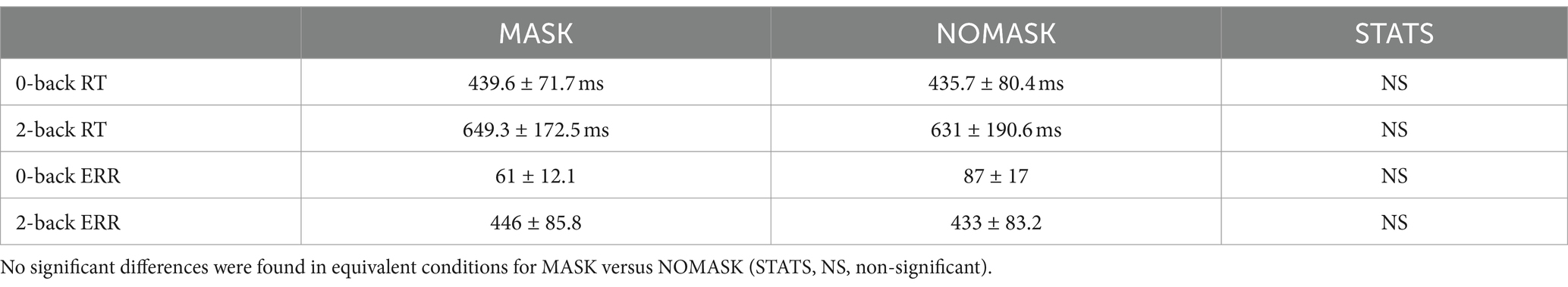
4 Results 4.1 Behavioral dataThere was no different in the behavioral data, notably reaction time and number of errors for MASK versus NOMASK during both conditions 0-back and 2-back (Table 2).
Table 2. Analysis of behavioral measures notably reaction time (RT) and number of errors (ERR) for the conditions 0-back and 2-back.
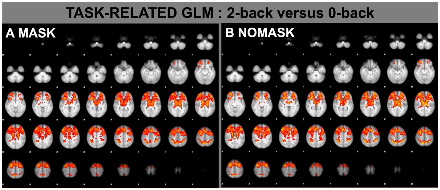
4.2 Task-related activationThe main effect of 2-back versus 0-back resulted in the typical and well-established working-memory pattern of activations for both only MASK and only NOMASK (Figure 2). The direct comparison of MASK versus NOMASK as well as NOMASK versus MASK yielded no supra-threshold activations.
Figure 2. Task-related fMRI analysis (FEAT) for the contrast of 2-back versus 0-back while wearing a mask (A) and without wearing a mask (B). p < 0.05 corrected FDR.
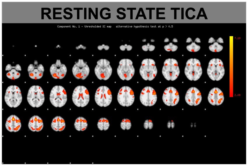
4.3 Resting fMRI activation (TICA and dual regression)The TICA resting fMRI analysis of both conditions MASK and NOMASK resulted in typical resting state networks (RSNs) (Figure 3). The direct comparison of the s-modes (a parameter of activation strength of those RSNs) for MASK versus NOMASK yielded no supra-threshold activations.
Figure 3. Example of a functional connectivity component based on a TICA (tensorial independent component) analysis.
Moreover, the dual regression yielded no supra-threshold differences for MASK versus NOMASK (or inverse).
5 DiscussionThe present study shows that wearing a facemask does not impact on both performances and brain activation patterns during a highly demanding 2-back working memory task. From a cognitive viewpoint, this finding is in line with several observations under normal conditions, high temperatures or mild exercise that demonstrated that the wearing of facemask does not or affect minimally the cognitive performances (Morris et al., 2020; Slimani et al., 2021; Wells et al., 2023). Only one study reported deficits in concentration and visual attention in the context of maximal running aerobic tests (Slimani et al., 2021). It is thus likely that, when wearing a facemask, a deleterious effect on cognition can be observed only under condition of motor or emotional stress.
Imaging data relative to the wearing of a facemask remain rare. In a series of 13 young individuals wearing a FFP2 facemask, Fischer et al., reported a 6.5% increase of cerebral blood flow and a 0.9% increase of oxygen saturation measured by transcranial hybrid near-infrared spectroscopy. The first fMRI study available in this field assessed the effect of wearing a facemask on functional MRI focusing on a basic sensory-motor task designed to activate visual, auditory, and sensorimotor cortices in eight middle-aged participants (Law et al., 2021). The authors reported no significant impact of facemask on task-related activation of sensorimotor areas. More recently, we analyzed the functional connectivity in a resting-state fMRI analysis of an independent sample of 23 healthy controls (Haller et al., 2022). This study reported no significant effect of wearing facemask on the functional connectivity of lower-level sensorimotor or visual networks but found a subtle impact on the interaction between the salience network as the seed region and the left middle frontal and precentral gyrus. More recently, Wu et al. examined the amplitude of low frequency fluctuation (ALFF) and functional connectivity at rest in 15 middle-aged healthy subjects wearing a KN95 mask and natural breathing. In contrast to the previous observations, they reported significant increases and decreases of ALFF as well as significant alterations of functional connectivity of posterior cingulate and medial prefrontal areas when wearing masks (Wu et al., 2023). These first fMRI studies in this field were performed with no or no significant cognitive challenge so that it was not possible to comment on a possible deleterious effect of the facemask in highly demanding situations. Because of its implication in numerous cognitive and cognitive-motor tasks, working memory is called upon in a wide range of activities. We decided to focus our analysis on this cognitive function since it is of key importance in daily life interactions and its brain correlates were very well established across the lifespan (Maillet and Rajah, 2014; Andre et al., 2016). Our findings indicate that wearing of the facemask does not change the functional connectivity patterns even when the demand of cognitive resources is high. These observations parallel the preliminary data of Klugah-Brown and collaborators (Klugah-Brown et al., 2022) who examined the effect of simple surgical mask on fMRI activation patterns during finger tapping, emotional face matching, working memory tasks with negative conclusions.
Some limitations should be considered when interpreting the present findings. To avoid the well-documented gender-related differences in functional connectivity (Murray et al., 2021), female participants were not included in this study. Moreover, we deliberately used a tight FFP2/KN95 facemask that has been the standard of reference the COVID-19 pandemic. It is, however, highly unlikely that the wearing of the less tight surgical facemasks led to significant changes in brain activation patterns. Last but not least, one should keep in mind that the 2-back task is a classical paradigm of working memory that involves attention and executive components but not emotional processing. This latter seems to be the most vulnerable domain of human cognition when wearing a facemask (Ferrari et al., 2021; Grundmann et al., 2021; Marini et al., 2021, 2022; Proverbio and Cerri, 2022; Tsantani et al., 2022; Proverbio et al., 2023). Future fMRI studies including tasks of social cognition are warranted to explore whether the wearing of facemask modifies brain activation patterns when dealing with emotional processing in complex environments.
Data availability statementThe raw data supporting the conclusions of this article will be made available by the authors, without undue reservation.
Ethics statementThe studies involving humans were approved by University of Geneva, Switzerland. The studies were conducted in accordance with the local legislation and institutional requirements. The participants provided their written informed consent to participate in this study. Written informed consent was obtained from the individual(s) for the publication of any potentially identifiable images or data included in this article.
Author contributionsM-LM: Writing – review & editing, Writing – original draft, Visualization, Validation, Supervision, Software, Resources, Project administration, Methodology, Investigation, Funding acquisition, Formal Analysis, Data curation, Conceptualization. SH: Writing – review & editing, Writing – original draft, Visualization, Validation, Supervision, Software, Resources, Methodology, Investigation, Formal Analysis, Data curation, Conceptualization. CR: Writing – review & editing, Writing – original draft, Project administration. FH: Writing – review & editing, Writing – original draft, Formal Analysis. PG: Writing – review & editing, Writing – original draft, Visualization, Validation, Supervision, Software, Resources, Project administration, Methodology, Investigation, Funding acquisition, Formal Analysis, Data curation, Conceptualization.
FundingThe author(s) declare financial support was received for the research, authorship, and/or publication of this article. This research was funded by the Swiss National Foundation (Grant number 320030_189247).
AcknowledgmentsWith contributions of the Clinical Research Center, Geneva University Hospitals and Faculty of Medicine of University of Geneva, Geneva, Switzerland. The study was supported by the Human Neuroscience Platform, Foundation Campus Biotech Geneva, Geneva, Switzerland.
Conflict of interestThe authors declare that the research was conducted in the absence of any commercial or financial relationships that could be construed as a potential conflict of interest.
The author(s) declared that they were an editorial board member of Frontiers, at the time of submission. This had no impact on the peer review process and the final decision.
Publisher’s noteAll claims expressed in this article are solely those of the authors and do not necessarily represent those of their affiliated organizations, or those of the publisher, the editors and the reviewers. Any product that may be evaluated in this article, or claim that may be made by its manufacturer, is not guaranteed or endorsed by the publisher.
Footnotes ReferencesAndre, J., Picchioni, M., Zhang, R., and Toulopoulou, T. (2016). Working memory circuit as a function of increasing age in healthy adolescence: a systematic review and meta-analyses. Neuroimage Clin 12, 940–948. doi: 10.1016/j.nicl.2015.12.002
PubMed Abstract | Crossref Full Text | Google Scholar
Ferrari, C., Vecchi, T., Sciamanna, G., Blandini, F., Pisani, A., and Natoli, S. (2021). Facemasks and face recognition: potential impact on synaptic plasticity. Neurobiol. Dis. 153:105319. doi: 10.1016/j.nbd.2021.105319
PubMed Abstract | Crossref Full Text | Google Scholar
Genovese, C. R., Lazar, N. A., and Nichols, T. (2002). Thresholding of statistical maps in functional neuroimaging using the false discovery rate. NeuroImage 15, 870–878. doi: 10.1006/nimg.2001.1037
PubMed Abstract | Crossref Full Text | Google Scholar
Haller, S., Montandon, M. L., Rodriguez, C., and Giannakopoulos, P. (2022). Wearing a KN95/FFP2 facemask induces subtle yet significant brain functional connectivity modifications restricted to the salience network. Eur Radiol Exp 6:50. doi: 10.1186/s41747-022-00301-0
PubMed Abstract | Crossref Full Text | Google Scholar
Haller, S., Montandon, M. L., Rodriguez, C., Moser, D., Toma, S., Hofmeister, J., et al. (2014). Acute caffeine administration effect on brain activation patterns in mild cognitive impairment. J. Alzheimers Dis. 41, 101–112. doi: 10.3233/JAD-132360
PubMed Abstract | Crossref Full Text | Google Scholar
Haller, S., Montandon, M. L., Rodriguez, C., Moser, D., Toma, S., Hofmeister, J., et al. (2017). Caffeine impact on working memory-related network activation patterns in early stages of cognitive decline. Neuroradiology 59, 387–395. doi: 10.1007/s00234-017-1803-5
PubMed Abstract | Crossref Full Text | Google Scholar
Haller, S., Rodriguez, C., Moser, D., Toma, S., Hofmeister, J., Sinanaj, I., et al. (2013). Acute caffeine administration impact on working memory-related brain activation and functional connectivity in the elderly: a BOLD and perfusion MRI study. Neuroscience 250, 364–371. doi: 10.1016/j.neuroscience.2013.07.021
PubMed Abstract | Crossref Full Text | Google Scholar
Jenkinson, M., Beckmann, C. F., Behrens, T. E., Woolrich, M. W., and Smith, S. M. (2012). Fsl. NeuroImage 62, 782–790. doi: 10.1016/j.neuroimage.2011.09.015
Crossref Full Text | Google Scholar
Klugah-Brown, B., Yu, Y., Hu, P., Agoalikum, E., Liu, C., Liu, X., et al. (2022). Effect of surgical mask on fMRI signals during task and rest. Commun Biol 5:1004. doi: 10.1038/s42003-022-03908-6
PubMed Abstract | Crossref Full Text | Google Scholar
Maillet, D., and Rajah, M. N. (2014). Age-related differences in brain activity in the subsequent memory paradigm: a meta-analysis. Neurosci. Biobehav. Rev. 45, 246–257. doi: 10.1016/j.neubiorev.2014.06.006
PubMed Abstract | Crossref Full Text | Google Scholar
Marini, M., Ansani, A., Paglieri, F., Caruana, F., and Viola, M. (2021). The impact of facemasks on emotion recognition, trust attribution and re-identification. Sci. Rep. 11:5577. doi: 10.1038/s41598-021-84806-5
PubMed Abstract | Crossref Full Text | Google Scholar
Marini, M., Paglieri, F., Ansani, A., Caruana, F., and Viola, M. (2024). Facial impression of trustworthiness biases statement credibility unless suppressed by facemask. Curr Psychol. 43, 13072–13082. doi: 10.1007/s12144-022-03277-7
Crossref Full Text | Google Scholar
Morris, N. B., Piil, J. F., Christiansen, L., Flouris, A. D., and Nybo, L. (2020). Prolonged facemask use in the heat worsens dyspnea without compromising motor-cognitive performance. Temperature 8, 160–165. doi: 10.1080/23328940.2020.1826840
Crossref Full Text | Google Scholar
Murray, L., Maurer, J. M., Peechatka, A. L., Frederick, B. B., Kaiser, R. H., and Janes, A. C. (2021). Sex differences in functional network dynamics observed using coactivation pattern analysis. Cogn. Neurosci. 12, 120–130. doi: 10.1080/17588928.2021.1880383
PubMed Abstract | Crossref Full Text | Google Scholar
Proverbio, A. M., Cerri, A., and Gallotta, C. (2023). Facemasks selectively impair the recognition of facial expressions that stimulate empathy: an ERP study. Psychophysiology 60:e14280. doi: 10.1111/psyp.14280
PubMed Abstract | Crossref Full Text | Google Scholar
Sinanaj, I., Montandon, M. L., Rodriguez, C., Herrmann, F., Santini, F., Haller, S., et al. (2015). Neural underpinnings of background acoustic noise in normal aging and mild cognitive impairment. Neuroscience 310, 410–421. doi: 10.1016/j.neuroscience.2015.09.031
PubMed Abstract | Crossref Full Text | Google Scholar
Slimani, M., Miarka, B., Znazen, H., Moalla, W., Hammami, A., Paravlic, A., et al. (2021). Effect of a warm-up protocol with and without facemask-use against COVID-19 on cognitive function: a pilot, randomized counterbalanced, cross-sectional study. Int. J. Environ. Res. Public Health 18:18. doi: 10.3390/ijerph18115885
Crossref Full Text | Google Scholar
Stanga, V., Turrina, C., Valsecchi, P., Sacchetti, E., and Vita, A. (2019). Well-being in patients with schizophrenia, mood and personality disorders attending psychiatric services in the community a controlled study. Compr. Psychiatry 91, 1–5. doi: 10.1016/j.comppsych.2019.02.001
PubMed Abstract | Crossref Full Text | Google Scholar
Tsantani, M., Podgajecka, V., Gray, K. L. H., and Cook, R. (2022). How does the presence of a surgical face mask impair the perceived intensity of facial emotions? PLoS One 17:e0262344. doi: 10.1371/journal.pone.0262344
PubMed Abstract | Crossref Full Text | Google Scholar
Wells, A. D., Mermier, C. M., Bellovary, B. N., Deyhle, M. R., Hsiao, Y. Y., and Amorim, F. T. (2023). The physiological, perceptual, and thermoregulatory responses to facemask use during exercise. J. Sports Med. Phys. Fitness 63, 264–272. doi: 10.23736/S0022-4707.22.14032-6
PubMed Abstract | Crossref Full Text | Google Scholar
Wu, X., Ma, L., Yin, Q., Liu, M., Wu, K., and Wang, D. (2023). The impact of wearing a KN95 face mask on human brain function: evidence from resting state functional magnetic resonance imaging. Front. Neurol. 14:1102335. doi: 10.3389/fneur.2023.1102335
PubMed Abstract | Crossref Full Text | Google Scholar
Zanchi, D., Montandon, M. L., Sinanaj, I., Rodriguez, C., Depoorter, A., Herrmann, F. R., et al. (2017). Decreased fronto-parietal and increased default mode network activation is associated with subtle cognitive deficits in elderly controls. Neurosignals 25, 127–138. doi: 10.1159/000486152
留言 (0)