
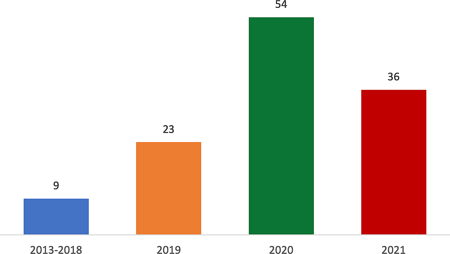
記住我


Cell lines, including MCF-10A, MCF-7, T-47D, and MDA-MB-231 were cultured and maintained in DMEM-F12 and DMEM media with FBS and antibiotics at 370C with 5% CO2. MTT assay was performed in these breast normal and cancer cell lines using Resveratrol. After seeding 10,000 cells in each well of 96 well plates and incubated overnight, cells were treated with different concentrations (10-50μM) of Resveratrol. MTT dye (5mg/mL) was added to each well and incubated for 4 hours followed by lysis solution was added and incubated for another 1 hour before being read at a wavelength of 570nm. From the absorbance, calculated the percentage of viable cells in these cell lines and found that 30μM concentration of Resveratrol gives 50% viable cells. MCF-10A cell line showed no effect of resveratrol up to 50μM concentration (Fig. 1A).
Fig. 1
A MTT assay of Resveratrol was done on MCF-10A, MCF-7 , MDA-MB-231 & T47D Breast normal and cancer cells. % of viable cells Vs. Resveratrol concentration calculated and plotted as histogram. B-D High CpG rich and transcription factor binding site Promoter sequences of BRCA1, BRCA2 & p16 gene was identify by Insilico using CpG plot and Tfscan online database tool and used for EMSA analysis as biotin labeled probe for protein binding
Consensus CpG rich promoter sequence retrieved by database by Insilco methodsThe promoter sequences of BRCA1, BRCA2 and p16 were retrieved from the eukaryotic promoter database. A high CpG island was identified in these promoter sequences using the EMBOCpG plot. Bases with more than 40% of CpG sequences were considered for CpG island promoter sequence for EMSA analysis. The Tf sites scan tool was used to identify the high transcription factor binding site of this methyl-CpG promoter sequence. A 35-38 base sequence from high transcription factor binding site of this methyl-CpG sequence taken for EMSA analysis as forward and reverse probe for BRCA1, BRCA2 & p16 genes (Fig. 1B-D).
MBD proteins bind to the promoter sequence of the BRCA1 gene in breast cancer cellsEMSA was performed to check the DNA binding activity of MBD proteins on BRCA1, BRCA2, and p16 gene promoter in breast cancer. We performed EMSA analysis using biotin labeled promoter sequence of BRCA1, BRCA2, and p16 gene retrieved from EMBOCpG plot for MBD1, MBD2, and MeCP2 proteins binding isolated from MCF-10A, MCF-7, T-47D & MDA-MB-231 human breasts normal and cancer cell line. EMSA results revealed the presence of active MBD proteins binding on BRCA1 promoter sequence in all cell lines using a specific primary antibody. Shift of bands, as well as a supershift of MBD1, MBD2 & MeCP2, proteins and BRCA1 promoter along with protein specific primary Antibody complexes in an PAGE gel, confirmed the MBD proteins binding on BRCA1 promoter sequence in MCF-10A, MCF-7, T-47D & MDA-MB-231 breasts normal and cancer cell line but no binding was observed on BRCA2 and p16 promoter sequences as there was no bands observed on gel in the cell lines mentioned above (Fig. 2A-I).
Fig. 2
A-I Promoter binding of MBD1, MBD2 & MeCP2 proteins on BRCA1, BRCA2 & p16 genes were analyzed by EMSA assay on 8% PAGE gel and transferred in nylon membrane then exposed in X-ray film. Shifting of bands was observed by using protein specific primary antibody arrow showing in between image indicate the bands. J Chromosome immune precipitation of BRCA1, BRCA2 & p16 gene was done by ChIP assay using MBD1, MBD2 & MeCP2 primary antibody and amplification of these genes were done by PCR and run on agarose gel and bands was observed. K Methylation immune precipitation of BRCA1, BRCA2 & p16 genes were done by MeIP assay using 5mC methylation specific primary antibody and amplification of these genes were done by PCR and run on 10% agarose gel to observed bands
BRCA1 gene was identified by ChIP assay in immuno-precipitation complex in breast cancer cellsChIP assay was performed to confirm MBD proteins binding on BRCA1, BRCA2, and p16 gene promoters in MCF-10A, MCF-7, T-47D & MDA-MB-231 cell lines using ChIP assay kit. First cross-linked the proteins with DNA using formaldehyde and sonicated to breakdown the DNA into the fragments. Precipitated the bound DNA using MBD1, MBD2 & MeCP2 proteins specific primary antibody which was amplified by PCR using forward and reverse primers and found that only BRCA1 was amplified by PCR and bands were observed in the agarose gel but no amplification was observed for BRCA2 and p16 genes which confirm MBD proteins binding on BRCA1 gene promoter sequence (Fig. 2J).
MeIP assay identified BRCA1 gene presence in the methylated immune precipitation complex in breast cancer cellsThe Abcam MeIP test kit was used to perform the methylation immune precipitation experiment using a 5mC primary antibody. DNA isolated from MCF-10A, MCF-7, T-47D & MDA-MB-231 cell lines and precipitated by 5mC primary antibody. Precipitated DNA was used to amplification of BRCA1, BRCA2, and p16 genes using forward and reverse primers and after running PCR product in agarose gel we found that only the BRCA1 gene was amplified by PCR but no amplification was observed for BRCA2 and p16 genes. This demonstrated that the BRCA1 promoter sequence was substantially methylated, as shown by the presence of a 5mC primary antibody (Fig. 2K).
MBD proteins regulate the BRCA1 gene expression in resveratrol treated brest cancer cellsReal-time gene expression analysis was done to check gene expression in correlation with resveratrol treatment. We have performed Real-time PCR of MBD1, MBD2, MeCP2, BRCA1, BRCA2, p16, and GAPDH genes, in MCF-7, MDA-MB-231 & T-47D breast cancer, and MCF10A breast normal cell lines treated with different concentrations of resveratrol. MBD1, MeCP2, BRCA1, BRCA2, and p16 genes expression were up-regulated in MCF-10A cells with increasing concentrations of resveratrol, with showing extremely significant results at 40µM (BRCA1 t-test P<0.0007, One way ANOVA P<0.00018; p16 t-test P<0.0045, One way ANOVA P<0.0006). MBD2 gene expression was down regulated along with increasing concentrations of resveratrol in the MCF-10A breast epithelial cell line. MBD2, MeCP2, and p16 genes expression were up-regulated with increasing resveratrol concentrations in the MCF-7 cell line, with the highest levels at 40µM (MBD2 t-test P<0.0124, One-way ANOVA= P<0.0001, MeCP2 t-test P<0.0007, One-way ANOVA= P<0.0001 and p16 t-test P<0.0001, One-way ANOVA= P<0.0001). However, at 50µM, the expression of BRCA1 (t-test P<0.0133, One-way ANOVA=P<0.0227) & BRCA2 (t-test P<0.0025, One-way ANOVA=P<0.0043) genes were down-regulated and significant. In the MDA-MB-231 cell line, BRCA1 gene expression was up regulated along with the increasing concentrations of resveratrol and has a significant high at 40µM (t-test P<0.0001, One-way ANOVA=P<0.0371) concentration. MBD1 (P<0.0047) MBD2 (P<0.0001), BRCA2 (P<0.0044) genes were up-regulated up to 30µM and MeCP2 (P<0.0004), & p16 (P<0.0008) up to 40µM then down-regulated. However in T-47D cell line MBD1 (P<0.0023), MBD2 (P<0.0001), MeCP2 (P<0.0001) genes expression up-regulated up to 30µM and BRCA2 (P<0.0001) gene up to 50µM concentration and has significant at this concentration, whereas BRCA1 & p16 genes expression is down-regulated along with the increasing concentrations of resveratrol (Fig. 3).
Fig. 3
Real time gene expression analysis of MBD1, MBD2, MeCP2, BRCA1, BRCA2 & p16 gene were done in MCF-10A, MCF-7, MDA-MB-231 & T-47D breast normal and cancer cell lines treated with different concentration of resveratrol. CT value of each gene was calculated by using relative quantitative methods and normalized with housekeeping GAPDH gene and plotted as bar graph
Correlation analysis also revealed that in MCF-7 BRCA1 & BRCA2 negatively correlated with resveratrol treatment and BRCA1 has significant (Pearson r = -0.9581, p<0.0026). In MDA-MB-231 all genes are positively correlated except p16, whereas BRCA1 significantly positively correlated (Pearson r = 0.9257, p<0.0081) to resveratrol. BRCA2 significantly positively correlated in T-47D (Pearson r = 0.9366, p<0.0059) breast cancer cells and BRCA1 & p16 negatively correlated but not significant. However in MCF-10A breast normal cell line all gene except MBD2 has positively correlation, out which MeCP2 (Pearson r = 0.9380, p<0.0056), BRCA1 (Pearson r = 0.9338, p<0.0064) and p16 (Pearson r = 0.9225, p<0.0088) have significantly correlated to resveratrol treatment.
MBD proteins expression and their regulation on the BRCA1 protein expression in resveratrol treated breast cancer cellsWestern blotting analysis was performed In MCF-7, MDA-MB-231, T-47D breast cancer, and MCF10A breast normal cell lines, to check protein expression of MBD1, MBD2, MeCP2, BRCA1, BRCA2, p16, and β-actin genes in relation to resveratrol treatment. We discovered that in MCF-7 cells MBD1, MBD2, MeCP2 & p16 protein expression were up-regulated and significant at 40µM (P<0.0001) and BRCA1 protein expression down regulated with increasing concentrations of resveratrol and significant at 50µM (P<0.0001) concentration. Also BRCA2 protein expression down regulated up to 50µM concentration but not significant. ANOVA analysis also showed that change in protein expression was significant P<0.0001 concerning resveratrol treatment in all genes. In MDA-MB-231 breast cancer cells MBD1 (P<0.0001), MBD2 (P<0.0003), MeCP2 (P<0.0003), protein expression were down-regulated and significant at 40µM and BRCA2 at 50µM (P<0.0033) concentration of resveratrol. Whereas BRCA1 (P<0.0172) and p16 (P<0.0003) protein expression were up-regulated and significant at 50µM concentration of resveratrol. Also ANOVA analysis revealed that all gene showed significant P<0.0001 change in protein expression. In T-47D cell line MBD1, MBD2, MeCP2 (40µM P<0.0001) protein expression up-regulated and significant at 50µM for MBD1 (P<0.0002) & MBD2 (P<0.0005), whereas MeCP2 at 40µM (P<0.0001). BRCA1, BRCA2 and p16 protein expression down-regulated along with increasing concentrations of resveratrol and significant at 50µM concentration. ANOVA analysis in T-47D cells though showed significant but only MBD1 & MBD2 genes have highly significant P<0.0001 change in protein expression concerning resveratrol. In the MCF-10A protein expression is upregulated up to 30µM of resveratrol treatment, however, there are no significant effects of resveratrol on protein expression of MBD1, MBD2, MeCP2, BRCA1, BRCA2, p16 genes observed in MCF-10A cell breast normal cell line (Fig. 4).
Fig. 4
Protein expression of MBD1, MBD2, MeCP2, BRCA1, BRCA2 & p16 genes were done by western blotting in resveratrol treated MDA-MB-231, MCF-7, T-47D & MCF-10A breast normal and cancer cell lines. Bands were transferred to PVDF membrane and exposed to X-ray film. Densitometry analysis was done by MyImage analysis software (thermo Scientific) to quantify the bands intensity and normalized with housekeeping β-actin gene protein and bar graph plotted as fold change in protein expression vs. concentration of resveratrol
The association between protein expression and resveratrol levels was also investigated using correlation analysis. MBD1 (Pearson r 0.9626, P<0.0021), MeCP2 (Pearson r 0.8903, P<0.0174), and p16 (Pearson r 0.9131, P<0.0110) were substantially favorably connected with resveratrol therapy in MCF-7 cells, whereas BRCA1 (Pearson r -0.9683, P<0.0015) was adversely correlated. We have also found a negative correlation between MBD proteins and BRCA1 protein expression in MCF-7 cells where MBD1 & MeCP2 has significant negative correlation with BRCA1 but MBD2 not significant negative correlation. In MDA-MB-231 cells MBD1, MBD2, MeCP2 & BRCA2, negative correlated to resveratrol and BRCA1 & p16 positively correlated with resveratrol treatment. We have also checked the correlation between MBD proteins and BRCA1 protein expression and found that it is negatively correlated but not significant. In the T-47D cell line only MeCP2 (Pearson r 0.8449, P<0.0342) positively correlated to resveratrol treatment and there is a negative correlation between MBD proteins and BRCA1 protein expression and MBD1 (Pearson r -0.9688, P<0.0014) & MBD2 (Pearson r -0.9153, P<0.0105) has significant correlation but MeCP2 not significantly correlated.
Resveratrol inhibited proliferation and colony formation in breast cancer cellsClonogenic assay was performed to elucidate colony formation capacity of MCF-10A, MCF-7 & MDA-MB-2321 cells after resveratrol treatment. Cells were counted and 1000 cells were seeded in each well of 6 well plates and allowed cells to adhere on the surface. Cells were treated with different concentrations of resveratrol (5µM, 10µM, 15µM, 20µM, 30µM) and allowed cells to grow until two colonies keep minimum distance in control sample. Media was removed and cells were fixed with fixation solution (Methanol & Glacial Acetic acid 7:1) for 30 minutes and stained with crystal violet for 30 minutes again and washed with tap water and let it dry (Fig. 5A). Images were captured and colonies were counted using Image-J software and survivals frequency was calculated using the formula. From the above results we discovered that survival frequency decreased as resveratrol concentration increasing in MCF-7 (One-way ANOVA P<0.0001) and MDA-MB-231 (One-way ANOVA P<0.0001) breast cancer cell lines, with 50% survival at 20µM (t-Test P<0.0001) and 30µM (t-Test P<0.0015) respectively, but no effect observed on MCF-10A breast normal cell line (Fig. 5B-D).
Fig. 5
A-D Clonogenic assay of MCF-10A, MCF-7 & MDA-MB-231 cells treated with different concentration of resveratrol was done on 6 well plates and stained with crystal violet after fixing the cells and images were captured using scale bar (10X resolution, 2.3mm) in fluorescent inverted microscope. Colonies were counted by Image J software and their plating efficiency and survival frequency was calculated using formula and plotted as bar graph. E-J Migration assay was done by wound healing assay on MCF-10A, MCF-7 & MDA-MB-231 cells treated with different concentration of resveratrol and images were captured using scale bar (10X resolution, 2.3mm) in fluorescent inverted microscope and gap of wound closure was calculated by Image J software and plotted as bar graph
Resveratrol inhibited the migration of breast cancer cells at a high concentrationTo confirm the anti-metastatic property of resveratrol in breast cancer cells. We have performed migration assay also called wound healing assay in MCF-10A, MCF-7 & MDA-MB-231 Cell lines. Cells were cultured and enough cells were seeded in each well of 6 well plates. After 100% confluence, media was removed and washed with 1x PBS, the scratches were done using pipette tips and washed out. Different concentrations (5µM, 10µM, 15µM, 20µM, 30µM) of resveratrol were treated to each well, and images of the scratches were taken every 6 hours from 0-24 hours time duration and migration distances were measured and calculated. We observed that after 24 hours treatment, migration of MCF-7 (P<0.0271) and MDA-MB-231(P<0.0111) cell lines were reduced with the increasing concentration of resveratrol and significant at 30µM concentration, and no migration was observed on MCF10A breast normal cell line (Fig. 5E-J).
Resveratrol inhibited the sphere formation of breast cancer cellsSphere formation assay was performed to demonstrate that the size of spheres in MCF-10A, MCF-7, and MDA-MB-231 & T-47 D Cell lines treated with increasing concentrations of resveratrol. We have performed sphere formation assay in an ultralow adherent plate with serum-free media containing antibiotics along with the different concentrations of resveratrol and we found that at 5th-day sphere size of MCF-7, MDA-MB-231 & T-47D (One-way ANOVA: P<0.0001) cells were reduced along with the increasing concentrations of resveratrol and have significant at 50µM concentration (t-test; MCF-7 P<0.0002, MDA-MB-231 P<0.0036, T-47D P<0.0001). No sphere formation was observed in MCF-10A breast epithelial cells (Fig. 6A-B).
Fig. 6
A-B Sphere formation assay of MCF-10A, MCF-7, MDA-MB-231& T-47D breast normal and cancer cell line were done on 96 well plates treated with different concentration of resveratrol and Images were taken at scale bar (10X resolution, 2.3mm) by inverted fluorescence microscope and spheres size were calculated by imaging software (Rad scientific) and presented graphically as bar graph as sphere size vs. resveratrol concentration. C-D Sphere of MCF-7, MDA-MB-231 & T-47D cells were treated with different concentration of resveratrol and apoptotic cells were stained with acridine orange/Etbr staining dye. Images were taken at scale bar (10X resolution, 2.3mm) by inverted fluorescence microscope and % of apoptotic cells were counted using Image-J software and graphically presented by bar graph. E-F Apoptosis assay was performed onMCF-10A, MCF-7, and MDA-MB-231 & T-47D breast normal and cancer cells treated with different concentration of resveratrol. Images were taken at scale bar (10X resolution, 2.3mm) by inverted fluorescence microscope and apoptotic cells were counted using Image-J software and graphically presented by bar graph as % of apoptotic cells vs. resveratrol concentration
Resveratrol increased apoptotic cells no. in the sphere at high concentration in breast cancer cellsTo perform apoptosis in sphere model, we have treated spheres of breast cancer cells with different concentrations of resveratrol and stained them with acridine orange/ EtBr staining dye after 24 hours. We observed apoptotic cells in sphere by fluorescence microscope and found that higher concentrations of resveratrol increasing the apoptotic cells in the spheres of breast cancer cells (One-way ANOVA: MCF-7 P<0.0001, T-47D P<0.0001; MDA-MB-231 P<0.0019) and significant at 30µM (P<0.0004) in MCF-7. In T-47D and MDA-MB231 30µM to 40 µM (P<0.0006), and 30µM (P<0.0076) to 50 µM (P<0.0011) significant respectively (Fig. 6C-D).
Resveratrol increases apoptosis at high concentration in cells monolayer of breast cancer cellsThe cytotoxicity of resveratrol was assessed by apoptosis analysis on MCF-10A, MCF-7, MDA-MB-231, and T-47D cells monolayers of breast normal and cancer cell line. We observed that after 24hr resveratrol treatment apoptotic cells number were increasing along with the increasing concentrations of resveratrol in MCF-7, MDA-MB-231 & T-47 D (One-way ANOVA: P<0.0010; P<0.0001; P<0.0002 respectively) breast cancer cell line and has significant at 40µM in MCF-7 (P<0.0021), and MDA-MB-231(P<0.0070), 50µM in T-47D (P<0.0032) breast cancer cell line, however, no effects was observed on MCF10A breast normal cells (Fig. 6E-F).
留言 (0)