
記住我


A total of 77 articles were initially identified from three different databases. After the removal of duplicates, 44 articles underwent a title and abstract screening. Subsequently, 23 articles were subjected to a full-text review, and 2 full-text articles were included for evaluation. Five articles were excluded for not meeting the eligibility criteria. Ultimately, 16 articles were deemed eligible. Figure 2 shows the flow chart of the screening process in the current study, generated using the PRISMA Flow Diagram tool [14]. A manual search was performed, but no additional articles meeting the inclusion criteria were found.
Fig. 2
Flow chart of literature screening process
Of the 16 included studies, 13 were in-vitro investigations, and 3 were clinical investigations (level 2b) [15,16,17]. Notably, no randomized clinical trials (RCTs) were identified. The included studies explored various factors influencing the accuracy of digital implant scans. The factors investigated can be broadly categorized as follows: ISB Position- Influence of Palatal Area Stitching- Bevel Orientation, Placement, and Implant Angulation- Effect of Operator on Scan Precision- Effect of Scan Pattern- Impact of Implant Angulation and Depth- Effect of ISB Material- Comparison of Different IOS Devices- Scan Aids- Comparison of Digital and Conventional Impressions (Table 1).
Table 1 Summary of reviewed studies of factors affecting the accuracy of SBsQuality assessment of studiesFor each study, two independent reviewers (M.R., P.G.) assessed the risk of bias across the following domains: randomization process, deviations from intended interventions, missing study outcome data, measurement of outcomes, selection of the reported result, and overall risk of bias. Disagreements were resolved through discussion and consensus. The risk of bias was rated as low, high, or unclear for each domain, and an overall risk of bias was assigned for each study. Studies with a high or unclear risk of bias were excluded from the final analysis. To assess the impact of the high-risk studies on the overall results, an additional sensitivity analysis was carried out. The risk of bias quality assessment was conducted to ensure the validity and reliability of the included studies and to provide a clear understanding of the strength of the evidence base (Table 2).
Table 2 Example of ‘Risk of bias’ table for a single studyParameters of scan bodies influencing the accuracy outcome of intraoral scansThe ISB position was found to be a relevant factor affecting the accuracy of digital scans. An in-vitro study showed that distance (P < 0.001) and angular (P < 0.001) deviation values are parameters that significantly influence the trueness of ISB positions [18]. In addition, it has been reported that accuracy is unaffected by whether the palatal area of a maxillary scan was stitched or unstitched [18].
Additionally, in-vitro studies revealed that the orientation of the bevel on ISBs (the angle at which the scan body’s bevel is positioned), their placement within the dental arch, and implant angulation significantly influenced the precision of digital scans. Notably, a considerably higher level of accuracy was achieved when the implant was positioned lingually, as opposed to random, distal, mesial, or facial locations. The results demonstrated that the lingually positioned bevel exhibited distinct differences in linear measurements compared to other orientations (F = 7.92, P < 0.001), with an explanation of 2.80% of the variation [19].
A study on a dentate maxillary model using a combined healing abutment-scan body (CHA-SB) system and implants at three different sites reported that implant location could affect scan accuracy (trueness: P < 0.001, precision: P < 0.020). This study also evaluated whether a different operator could affect scan precision. However, the effect of the operator on scanning accuracy was found to be insignificant (P > 0.051) [8]. Using a similar CHA-SB system, four types of scan patterns were investigated. The results of this in-vitro study showed that the scan accuracy could be affected by the scan pattern selected. It was evident that the scan pattern exerted a significant influence on precision, particularly evident when considering angular deviation data (F = 6.227, df = 3, P = 0.002) [20].
A study on a master cast indicated that the accuracy of digital impressions is not related to angulation and implant depth. Interestingly, inexperienced operators performed better in this study, and camera position was one of the key factors that could improve accuracy [9].
In another ex vivo investigation, the primary aim was to evaluate the accuracy of five intraoral scanners in replicating ISBs and soft tissues within an edentulous maxilla, considering the influence of operator experience. The outcomes exhibited notable disparities in implant platform deviation between inexperienced and experienced operators following complete surface alignment. It is noteworthy that after alignment of the ISBs, no significant inter-operator variation was observed for the selected scanners. The scanner rankings displayed variability based on operator experience. Furthermore, the study uncovered a tendency for mucosal alignment to overestimate the platform deviation. These findings emphasize the critical role of operator expertise and meticulous scanner selection in achieving precise and reliable intraoral scanning outcomes for edentulous cases [21].
Trials evaluating the 3D positional accuracy of ISBs and IOS devices reported that the selected system could significantly affect the 3D positional accuracy. Six types of ISBs—Straumann RC, Core 3D, Straumann CARES Mono, Amann Girrbach, Sirona InPost, Nobel Procera Pos Locator—and four kinds of IOS devices—Straumann RC, Core 3D, Straumann CARES Mono, Medentika L-Series—were utilized. Straumann RC demonstrated the lowest accuracy for both ISBs and IOS [22].
Five types of ISB systems—AF (IO-Flo; Dentsply Sirona), NT (NT-Trading GmbH & Co KG), DE (DESS-USA), C3D (Core3Dcentres), and ZI (Zimmer Biomet Dental)—and four types of scanning techniques—no modification, glass beads, pressure indicating paste, and floss—were evaluated in an in-vitro model. As a result, the authors demonstrated that both ISBs and scanning techniques could significantly affect the accuracy of digital implant scans [23].
The accuracy of using four different types of IOS devices in an in-vitro model was investigated. Primescan and iTero devices showed superior digital scans with slight errors than Medit i500 and Vatech EZ scans (p < 0.05) [24].
Another in-vitro study used dome-shaped and cuboidal ISBs on a master model of an edentulous maxilla. The authors stated that the virtual alignment of ISBs could significantly affect the precision of digital scans (up to ∼ 30 μm/0.09°). The cuboidal ISBs in this study demonstrated larger deviations rather than dome-shaped ones [25].
ISB material and implant angulation were investigated in an in-vitro model. The results showed that titanium ISBs outperformed polyetheretherketone ISBs in terms of accuracy. In terms of angulations, mesially tilted distal implants exhibited better accuracy regardless of the type of intra-oral scanners [26].
Another in-vitro study using an ISB on a single implant in the right first molar position showed that the chosen measurement technique could affect the accuracy of digital scans. Three experienced operators performed evaluations using two different approaches: circle-based and point-based. Results displayed that the circle-based method had a significantly higher deviation than the point-based technique (P = 0.001) [10].
Scan aids can help to improve the accuracy of implant scans. Various designs - irregular, square, circular - and materials - white, gray, beige - of scan aids have been studied in-vitro. Findings showed beige color and irregular design have the highest precision, but their poor strength hinders the clinical use of this type. The clinically applicable form was gray in color and irregular in design [27].
One in-vitro study compared the accuracy of digital and conventional implant impressions. No significant differences in accuracy were found when scanning short spans, but when scanning long spans, digital impressions were significantly less accurate compared to traditional analog impressions. These results suggest that the scan span and implant position should be considered when choosing between digital and conventional impressions [28].
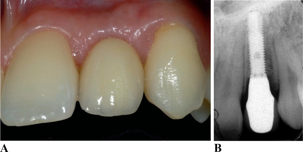
Clinical studies on the impact of ISBs on the scan outcome are scarce. The following three clinical trials were conducted with differing objectives related to ISBs. A recent clinical trial of 30 patients evaluated the accuracy of digital versus conventional impressions. The study demonstrated that digital scanning and the use of ISBs could potentially facilitate implant restorative treatment for practitioners and patients. Yet ISB misfit can occur based on the location of the respective implant. The lowest misfit was found for single molar implants (40.5 ± 18.9 μm) and the highest for distal three-unit implants (80.3 ± 12.4 μm) [16]. In a clinical study of 14 patients (8 women and 6 men), Gherlone et al. evaluated the survival rate of implants with digital impressions. After a follow-up period of 6–12 months, the survival rate was 100% for all implants examined. This study suggests that digital impressions provide accurate models that facilitate prosthetic work and satisfy the dental team [17].
In a retrospective clinical investigation of 35 patients, the effect of ISB design and number of implants were evaluated. First, the study showed that digital scans resulted in an acceptable fit of the implant superstructure with an accuracy of 86.70%. Second, the influence of the ISB design (P = 0.005) and the number of implants (P = 0.039) on the accuracy of fit was significant. Cylindrical ISBs on 4 implants exhibited better accuracy than polygonal-shaped ISBs [29].
留言 (0)