
記住我
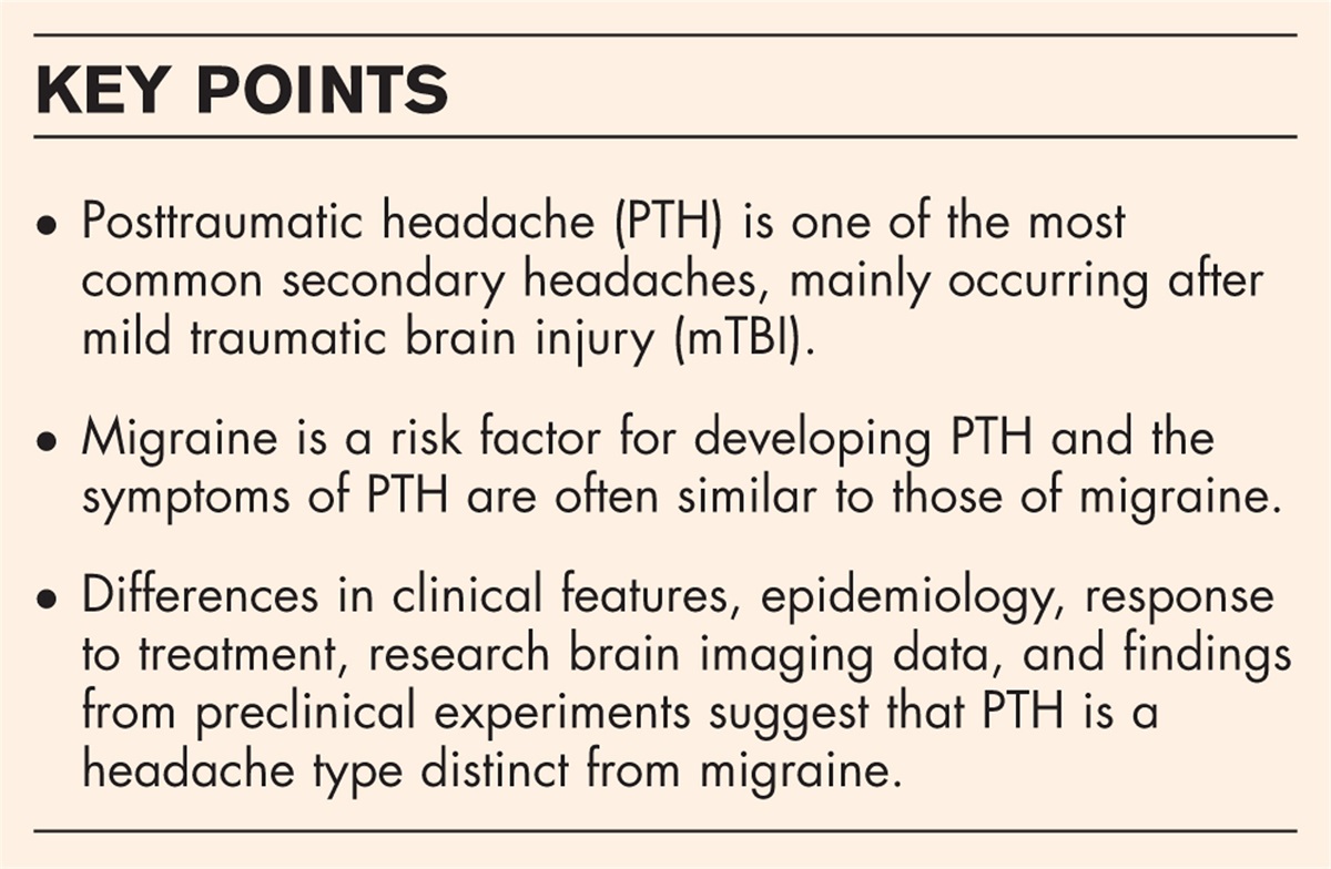
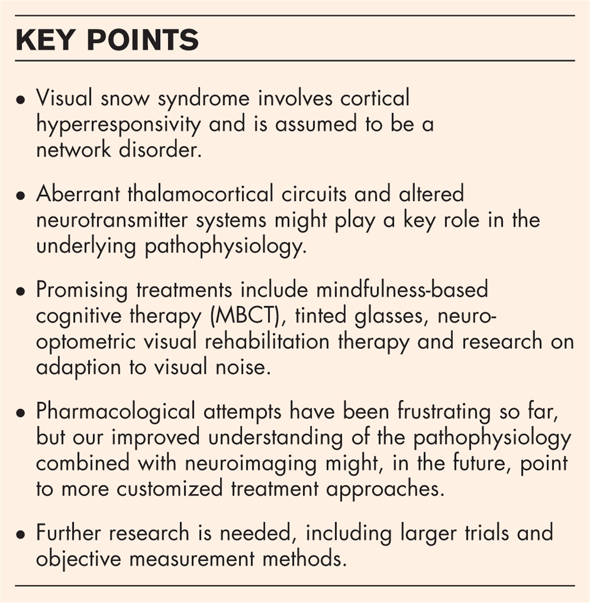


Visual snow syndrome is characterized by a distinctive combination of visual disturbances, including the visual snow phenomenon (manifesting as visual static), afterimages (palinopsia), heightened perception of entoptic phenomena, impaired night vision (nyctalopia), and persistent photophobia [1]. It is frequently associated with tinnitus, migraine, and affective disorders [2,3]. Initial electrophysiological studies have indicated cortical hyperresponsivity in visual brain areas [4,5], suggesting a disturbance in the processing within higher-order visual brain areas.
Structural and functional MRI studies have further implicated cortical visual brain areas, attentional networks, and limbic structures [6–9]. So far, a consistent focal disease could not be identified, and neurologically seen, it could not even account for the clinical presentation. Consequently, a disturbance within the interaction of various brain regions, indicative of a network disorder, is suspected [10]. One potential explanation for the diverse findings and symptoms, indicating involvement beyond the visual system, is the concept of thalamocortical dysrhythmia, leading to a deficiency in inhibitory modulation [11].
Although there are established criteria for the diagnosis of VSS [2], there is currently no objective measurement for the symptoms. Self-reports and questionnaires are used to measure characteristics and severity. One study showed that subjective severity is associated with visual allodynia including photophobia and correlates negatively with time [12]. Similarly, a follow-up study by Graber et al.[13▪▪] demonstrated that the perception of symptom severity might fluctuate or adapt, but typically persist over many years.
The treatment approaches until now have primarily focused on pharmacological attempts, employing substances known to influence cortical excitability, such as antiseizure medications, especially lamotrigine, or those used in migraine prophylaxis. While there is currently no definitive evidence supporting the efficacy of certain treatments for VSS, particularly not at the level of randomized and controlled trials [14,15], substances like recreational drugs and alcohol can worsen VSS symptoms [14]. Specifically, hallucinogens and stimulants, such as ecstasy, should be avoided, as they may alter the brain's visual pathways and potentially lead to symptomatic VSS, that is, hallucinogen persisting perception disorder (HPPD) [16].
Although the underlying pathophysiology is not entirely understood and there is no established treatment concept yet, the research landscape has evolved significantly in the past years. This review article focuses on the knowledge gained from electrophysiological and imaging studies over the past two years, as well as the first treatment trials that have been carried out to investigate neuromodulatory and neurobehavioral treatment approaches.
 Box 1:
 no caption available
NEUROSCIENTIFIC RESEARCH TECHNIQUESThis section focusses on recent neuroscientific insights into the pathophysiology of visual snow syndrome, specifically functional and structural aberrations, neurotransmitter involvement, and effects of drugs.
ImagingA recent 7-Tesla MRI study found cortical and thalamic microstructural differences (lower T1 signal). The thalamic changes could be correlated to the severity of VSS symptoms (number of symptoms and perceived impairment). These findings might support the hypothesis of the involvement of thalamocortical circuits in the pathophysiology of VSS [17]. In a second study, the group also examined changes in functional MRI connectivity. The local interaction between nodes and organization was decreased in several parietooccipital brain regions [18]. Van Laere et al.[19] conducted an FDG-PET/MRI study demonstrating hypermetabolism in secondary visual brain areas (lingual gyrus and cuneus), aligning with previous findings [8,9], as well as increased gray matter volume in the left secondary and associative visual cortex and in the left lingual gyrus. Additionally, hypometabolism in the mesiotemporal cortex and increased gray matter volume in frontotemporal areas, including components of the limbic system, were found [19].
A study by Puledda et al.[20▪▪] indicated that both glutamatergic and serotoninergic neurotransmission might be involved in the pathophysiology of VSS using Receptor-Enriched Analysis of Functional Connectivity by Targets (REACT) combined with resting functional MRI data. Changes in glutamatergic-enriched connectivity in the dorsal anterior cingulate cortex/middle cingulate cortex (ACC/MCC) might be linked to deregulation of normal attentional functions and sensory processing integration in VSS. Further, reduced connectivity in certain brain regions points to a possible role in modulating the altered connectivity in areas of the visual motion network in VSS. The results of these studies emphasize that various networks and neurotransmitters might be involved in the pathophysiology of VSS. In the future, a more specific knowledge in this respect might offer a targeted pharmacological approach in contrast to the currently applied unspecific one.
ElectrophysiologyOne study investigated cortical oscillatory patterns in VSS using magnetoencephalography (MEG). The main finding revealed that VSS patients exhibited significantly increased gamma power in the primary visual cortex and reduced phase-amplitude coupling compared to control participants, indicating hyperactive and disorganized cortical activity during early visual processing [21]. This is consistent with the concept of ‘thalamocortical dysrhythmia’ [11] which suggests that dysfunctional cortical oscillations may play a role in the pathophysiology of VSS. In the context of inhibition, the observed dysrhythmic cortical activity in VSS may be associated with alterations in inhibitory processes within the visual cortex, potentially contributing to the hyperactivity and disorganization observed in VSS. This is supported by previous research suggesting alterations in inhibitory neurotransmission in other disorders associated with cortical dysrhythmias, such as migraine [22]. Further research into the specific inhibitory mechanisms underlying these dysrhythmic activities could provide a deeper understanding of the pathophysiology of VSS and may lead to the development of more targeted treatments for this disorder.
Neurobehavioral and psychophysicsAssuming a hyperexcitability in the visual cortex in patients with VSS, Brooks et al.[22] tested the two competitive approaches of increased neural noise (elevated spontaneous neural activity) and increased neural gain (stronger neural response to visual input) by visual contrast detection. Individuals with VSS exhibited increased contrast gain for increments in the putative parvocellular on-pathway, assumed to be responsible for fine spatial detail. Further, an increased contrast sensitivity for decrements was found, which might be inferred from the magnocellular off-pathway. Contrast gain refers to the amplification of visual signals in the brain, essentially how strongly the brain responds to visual stimuli. On the other hand, contrast sensitivity involves the ability to discern differences in luminance, enabling one to detect objects against a background that may not be distinctly outlined [23]. The study showed that both these aspects are abnormally increased, indicating heightened visual processing in certain pathways. These findings suggest abnormalities in the processing of luminance contrast, potentially contributing to the visual disturbances associated with VSS [22]. Contrary to VSS, migraine was not associated with neuronal hyperreactivity to contrast, indicating differences in the pathophysiology of the two neurological disorders, even if the subjective hyper-sensitivity is similar. Further, the study did not find any evidence of abnormal neural noise levels in VSS, indicating that the condition is not associated with increased internal noise in the visual system [22]. On the other hand, a study on adaption demonstrated that visual snow disappeared after presenting visual noise which indicates that spontaneous neuronal activity in the visual pathways is indeed necessary to generate the visual perception of snow [24▪]. This underlines the hypothesis of inadequate filtering or suppression processes in VSS which may be due to thalamocortical dysrhythmia [10,25].
Clinical studiesA retrospective overview over 400 cases and treatment responses to different medications could not identify any drug to be helpful in a significant portion of patents, most had no effect. Single cases of amelioration under lamotrigine or benzodiazepines were found [14]. On the other hand, there is evidence of developing persistent visual problems in association with serotonin reuptake inhibiting antidepressants (SSRIs) like citalopram [26]. A study examined patient-reported visual problems associated with SSRIs and found 124 reports from 18 countries [27▪]. The most commonly reported side effects included blurred vision, night blindness, and floaters with significant impact on daily life and work for the affected patients. Remarkably, some patients reported persistent visual problems even after stopping the drug. The study highlights the need for healthcare professionals to be aware of these potential side effects. Interestingly, Naguy et al.[28] described a case of a child with ADHD who developed visual snow phenomenon, photopsia, and tinnitus under treatment with methylphenidate, a noradrenaline and dopamine reuptake inhibitor. The symptoms were reversible during tapering and appeared to correlate with the dosage [28].
In a case series, three patients presented with visual snow associated with acute ischemic stroke, two of whom had a history of migraine with visual aura. The ischemic strokes affected different areas involved in visual processing including the lingual gyrus suggesting that the lingual gyrus is important for visual snow. However, these patients did not show the full picture of VSS underscoring the complexity of VSS as a network disorder [29].
Oculomotor assessmentA retrospective analysis of 40 patients with VSS identified a high frequency (up to 50%) of occurrence of oculomotor deficits such as accommodative insufficiency, convergence insufficiency and saccadic deficits [30]. Another retrospective study also showed a very high rate (26/27 patients) of comorbid binocular abnormalities in the VSS population such as oculomotor dysfunction (OMD) (59.3%), convergence insufficiency (51.9%), and accommodative insufficiency (54.5%) among other eye movement disorders [31]. VSS manifests a wide range and high prevalence of oculomotor-based dysfunctions and shows related reading problems [32]. There are also promising findings that the deficits in eye movement patterns in VSS are independent of psychiatric comorbidities and therefor are useful in diagnostic contexts [33].
THERAPEUTIC ATTEMPTSThere are currently no objective markers for VSS symptoms. To facilitate communication for patients, therapists and physicians could utilize artificial intelligence. Freely available text-to-image programs are promising tools to provide patients with the ability to visualize their symptoms [34]. The following section discusses recent promising treatment approaches for VSS. Figure 1 depicts an attempt to link these approaches to our current pathophysiological understanding.
 FIGURE 1:
 In recent years, there has been tremendous progress in our understanding of the pathophysiology of visual snow syndrome. Several treatment approaches can be linked to these findings and, in return, will provide further insights in the disorder.
Chromatic filter and oculomotor therapyA retrospective analysis of 40 individuals with VSS found that the use of tinted glasses, particularly those that reduce transmission in the blue range of the visible spectrum, reduced the perceived intensity, duration, and frequency of visual snow in 80% of patients [30]. These tints may have a dual effect by reducing overall luminance and further reducing luminance due to the chromatic distortion of the filter. The tinted glasses were also effective in reducing other related visual symptoms such as palinopsia or entoptic phenomena and could be incorporated into glasses correction or used as clip-ons [30].
Another retrospective study underlines a reduction of VSS when using preferred (i.e. self-selected) filters [31]. Twenty-seven participants reported a lowering of at least 50% in frequency and intensity of visual snow. Using chromatic filter combined with specific oculomotor-based saccadic therapy reduced palinopsia in both frequency and intensity [31]. While these results are promising, further research with larger samples and standardized protocols is needed to validate the long-term efficacy and adaptive phenomena associated with chromatic tints.
Behavioral approachesThe first study to assess the effect and feasibility of a rehabilitative approach was conducted by Tsang et al. [35]. The 6-week treatment program (neuro-optometric visual rehabilitation therapy, NORT) consisted of a battery of visual tasks adapted individually based on the initial neuro-optometric assessment. The group found an amelioration in the quality-of-life questionnaire after 6 and 12 weeks, but importantly not in the visual symptoms themselves, for example in the severity of visual snow (VS) or the associated symptoms [35]. Since there was no control condition, further research is needed to evaluate the long turn impact of oculomotor-based, neuro-optometric rehabilitation therapy and to determine whether certain eye movement patterns are suitable for diagnostic purposes and for monitoring the course of therapy.
Montoya et al.[24▪] found that adaptation to visual noise attenuates the phenomenon of visual snow, making it invisible in many cases. This adaptation follows similar patterns to normal sighted individuals, including adherence to the duration scaling law (linear on a log-log axis) and specific adaptation effects to visual noise and contrast patterns. The results suggest that visual snow is not due to observer or simulator bias, as the responses of affected individuals are systematically like those of normally sighted individuals [24▪]. The study suggests that adaptation could be a potentially reliable, objective measure of visual snow that not only provides important insights into neurological processes but could also guide future treatment approaches.
NeuromodulationAn earlier study shows the potential of repetitive transcranial magnetic stimulation (rTMS) in the treatment of VSS [36]. The study used visual snow diaries, questionnaires, and visual evoked potentials (VEP) as assessment methods. The study found that 10+1 Hz rTMS at the visual cortices resulted in a reduced sum of visual snow intensities, suggesting a possible improvement in patients’ symptoms. However, no significant changes in questionnaire scores or VEP parameters were observed, which could be due to various limitations such as unbalanced treatment application, missing data, and small sample size.
Mindfulness, which promotes nonjudgmental awareness of the present moment, induces neural changes, can increase resilience and clinical benefits through attention and self-regulation training. One study investigated the effects of a customized mindfulness-based cognitive therapy (MBCT) on individuals with VSS using MRI and self-reports [37▪]. Participants reported significant improvements in self-rated severity of visual symptoms after an 8-week MBCT vision program. Median scores decreased from baseline to weeks 9 and 20, indicating a reduction in symptom severity. The study also found a significant reduction in the impact of vision symptoms on daily life as reported by participants. This improvement persisted at the 3-month follow-up, indicating the potential long- term benefits of the intervention. In MRI, a reorganization of the visual network involved both visual and extravisual areas in the neocortex and cerebellum after the MBCT. This suggests promising changes in the functional connectivity of visual networks that may be related to the observed improvements in symptoms [37▪]. Future research, including randomized controlled trials with larger sample sizes, longer follow-up periods, and comprehensive outcome measures, will be critical to expanding our understanding of the potential benefits and limitations of MBCT vision therapy for people with VSS.
CONCLUSIONResearch over the last two years has provided valuable knowledge about the pathophysiology of visual snow syndrome. Cortical hyperreactivity in visual brain areas has been demonstrated and points to a network disorder rather than a focal pathology. Imaging studies using advanced techniques, such as 7-Tesla MRI and FDG-PET/MRI, have shown microstructural and functional connectivity differences in cortical and thalamic regions suggesting the involvement of thalamocortical circuits in the pathophysiology of VSS. Additionally, the involvement of glutamatergic and serotoninergic neurotransmission in VSS pathophysiology has been highlighted, shedding light on potential underlying mechanisms. Promising treatment approaches such as MBCT, the use of tinted glasses and research on adaption effects might reduce the perceived intensity, duration, and frequency of visual snow. However, the level of evidence is still low, and randomized controlled trials with larger sample sizes and comprehensive outcome measures are needed. The lack of objectively measure VSS also highlights the need for further research in this area, but the steady progress made in research gives hope to those affected.
AcknowledgementsNone.
Financial support and sponsorshipSarah A. Aeschliman received no financial support for the research, authorship, and/or publication of this article.
Antonia Klein is supported by Bangerter Rhyner Foundation and Swiss Headache Society.
Christoph J. Schankin received financial support from Baasch Medicus Foundation, Swiss Headache Society, Visual Snow Initiative, Eye on Vision Foundation, and Visual Snow Syndrome Germany e.V.
Conflicts of interestChristoph J. Schankin: Consulting, Advisory Boards, Speaker, Travel Sup-port for/from Abbvie, Allergan, Almirall, Amgen, Eli Lilly, Grünenthal, Lund-beck, MindMed, Novartis, Pfizer, TEVA Pharmaceuticals. Part-time employee at Zynnon.
The remaining authors have no conflicts of interest.
REFERENCES AND RECOMMENDED READINGPapers of particular interest, published within the annual period of review, have been highlighted as:
▪ of special interest
▪▪ of outstanding interest
REFERENCES 1. Schankin CJ, Maniyar FH, Digre KB, Goadsby PJ. ‘Visual snow’ – a disorder distinct from persistent migraine aura. Brain 2014; 137:1419–1428. 2. Puledda F, Schankin C, Goadsby PJ. Visual snow syndrome: a clinical and phenotypical description of 1,100 cases. Neurology 2020; 94:e564–e574. 3. Solly EJ, Clough M, Foletta P, et al. The psychiatric symptomology of visual snow syndrome. Front Neurol 2021; 12:703006. 4. Yildiz FG, Turkyilmaz U, Unal-Cevik I. The clinical characteristics and neurophysiological assessments of the occipital cortex in visual snow syndrome with or without migraine. Headache 2019; 59:484–494. 5. Eren O, Rauschel V, Ruscheweyh R, et al. Evidence of dysfunction in the visual association cortex in visual snow syndrome: VEPs in Visual Snow. Ann Neurol 2018; 84:946–949. 6. Puledda F, Ffytche D, Lythgoe DJ, et al. Insular and occipital changes in visual snow syndrome: a BOLD fMRI and MRS study. Ann Clin Transl Neurol 2020; 7:296–306. 7. Puledda F, O’Daly O, Schankin C, et al. Disrupted connectivity within visual, attentional and salience networks in the visual snow syndrome. Hum Brain Mapp 2021; 42:2032–2044. 8. Schankin CJ, Maniyar FH, Chou DE, et al. Structural and functional footprint of visual snow syndrome. Brain 2020; 143:1106–1113. 9. Aldusary N, Traber GL, Freund P, et al. Abnormal connectivity and brain structure in patients with visual snow. Front Hum Neurosci 2020; 14:582031. 10. Klein A, Schankin CJ. Visual snow syndrome as a network disorder: a systematic review. Front Neurol 2021; 12:724072. 11. Lauschke JL, Plant GT, Fraser CL. Visual snow: a thalamocortical dysrhythmia of the visual pathway? J Clin Neurosci 2016; 28:123–127. 12. Thompson AC, Goodbourn PT, Forte JD. Perceived severity of Visual Snow Syndrome is associated with visual allodynia. Headache 2023; 63:494–505. 13▪▪. Graber M, Scutelnic A, Klein A, et al. Natural course of visual snow syndrome: a long-term follow-up study. Brain Commun 2022; 4:fcac230. 14. Puledda F, Vandenbussche N, Moreno-Ajona D, et al. Evaluation of treatment response and symptom progression in 400 patients with visual snow syndrome. Br J Ophthalmol 2022; 106:1318–1324. 15. Van Dongen RM, Waaijer LC, Onderwater GLJ, et al. Treatment effects and comorbid diseases in 58 patients with visual snow. Neurology 2019; 93:e398–e403. 16. Van Dongen RM, Alderliefste GJ, Onderwater GLJ, et al. Migraine prevalence in visual snow with prior illicit drug use (hallucinogen persisting perception disorder) versus without. Eur J Neurol 2021; 28:2631–2638. 17. Strik M, Clough M, Solly EJ, et al. Microstructure in patients with visual snow syndrome: an ultra-high field morphological and quantitative MRI study. Brain Commun 2022; 4:fcac164. 18. Strik M, Clough M, Solly EJ, et al. Brain network dynamics in people with visual snow syndrome. Hum Brain Mapp 2023; 44:1868–1875. 19. Van Laere K, Ceccarini J, Gebruers J, et al. Simultaneous 18F-FDG PET/MR metabolic and structural changes in visual snow syndrome and diagnostic use. EJNMMI Res 2022; 12:77. 20▪▪. Puledda F, Dipasquale O, Gooddy BJM, et al. Abnormal glutamatergic and serotonergic connectivity in visual snow syndrome and migraine with aura. Ann Neurol 2023; 94:873–884. 21. Hepschke JL, Seymour RA, He W, et al. Cortical oscillatory dysrhythmias in visual snow syndrome: a magnetoencephalography study. Brain Commun 2022; 4:fcab296. 22. Brooks CJ, Chan YM, Fielding J, et al. Visual contrast perception in visual snow syndrome reveals abnormal neural gain but not neural noise. Brain 2022; 145:1486–1498. 23. Gardner JL, Sun P, Waggoner RA, et al. Contrast adaptation and representation in human early visual cortex. Neuron 2005; 47:607–620. 24▪. Montoya SA, Mulder CB, Lee MS, et al. Adapting to visual noise alleviates visual snow. Invest Ophthalmol Vis Sci 2023; 64:23. 25. Klein A, Schankin CJ. Visual snow syndrome, the spectrum of perceptual disorders, and migraine as a common risk factor: a narrative review. Headache 2021; 61:1306–1313. 26. Eren OE, Schöberl F, Schankin CJ, Straube A. Visual snow syndrome after start of citalopram—novel insights into underlying pathophysiology. Eur J Clin Pharmacol 2021; 77:271–272. 27▪. Healy D, Mangin D, Lochhead J. Development and persistence of patient-reported visual problems associated with serotonin reuptake inhibiting antidepressants. JRS 2022; 33:37–47. 28. Naguy A, Naguy C, Singh A. Probable methylphenidate-related reversible ‘Visual Snow’ in a child with ADHD. Clin Neuropharm 2022; 45:105–106. 29. Scutelnic A, Slavova N, Klein A, et al. Symptomatic visual snow in acute ischemic stroke: a case series. Headache 2023; 63:173–176. 30. Han MHE, Ciuffreda KJ, Rutner D. Historical, diagnostic, and chromatic treatment in visual snow syndrome: a retrospective analysis. Optom Vis Sci 2023; 100:328–333. 31. Tannen B, Brown J, Ciuffreda KJ, Tannen NM. Remediation of visual snow (VS) and related phenomena in a neuro-optometric practice: a retrospective analysis. VDR 2022; 8:105–113. 32. Tannen B, Sample A, Ciuffreda KJ, Tannen NM. Clinical reading-related oculomotor assessment in visual snow syndrome. J Optom 2024; 17:100500. 33. Solly EJ, Clough M, McKendrick AM, et al. Eye movement characteristics are not significantly influenced by psychiatric comorbidities in people with visual snow syndrome. Brain Res 2023; 1804:148265. 34. Balas M, Micieli JA. Visual snow syndrome: use of text-to-image artificial intelligence models to improve the patient perspective. Can J Neurol Sci 2023; 50:946–947. 35. Tsang T, Shidlofsky C, Mora V. The efficacy of neuro-optometric visual rehabilitation therapy in patients with visual snow syndrome. Front Neurol 2022; 13:999336. 36. Grey V, Klobusiakova P, Minks E. Can repetitive transcranial magnetic stimulation of the visual cortex ameliorate the state of patients with visual snow? Bratisl Lek Listy 2020; 121:395–399. 37▪. Wong SH, Pontillo G, Kanber B, et al. Visual snow syndrome improves with modulation of resting-state functional MRI connectivity after mindfulness-based cognitive therapy: an open-label feasibility study. J Neuroophthalmol 2023; 44:112–118.
留言 (0)