
記住我


The study protocol was reviewed and approved by the Ethics Committee at the School/Hospital of Stomatology Xinjiang Medical University, Urumqi, PR China, with the onset of baselined data collection (approval no. IACUC20210706-11). The procedures in this study were completed in accordance with the standards set out in the Announcement of Helsinki and laboratory regulations of research in China. Written informed consent was obtained from all the patients.
Patient selectionThe current study included patients diagnosed with OSCC and their corresponding surgical specimens from June 2008 to June 2020 at the author’s affiliation. All included patients were treated using a multidisciplinary approach, and data were selected from the electronic medical records of the hospital information system. According to the most updated American Joint Committee on Cancer/Union for International Cancer Control (AJCC/UICC) guidelines (8th edition), the clinicopathological classification and staging of all recruited OSCC patients were assessed using the TNM system [i.e., size of the primary tumor (T), involvement of locoregional lymph nodes (N), and distant metastases (M)], fully reflecting the extent of tumor growth in the whole body [18]. The inclusion criteria were as follows: i). OSCC lesions are located in the tongue, gingiva, buccal mucosa, hard palate, floor of the mouth, retromolar triangle, and vermilion mucosa that are confirmed histopathologically; ii). Patients who had not undergone any treatment previously; iii). Cases of primary or recurrent tumors that received complete tumor resection with or without lymph node dissection; and iv). At least three-year follow-up/survival materials were available.
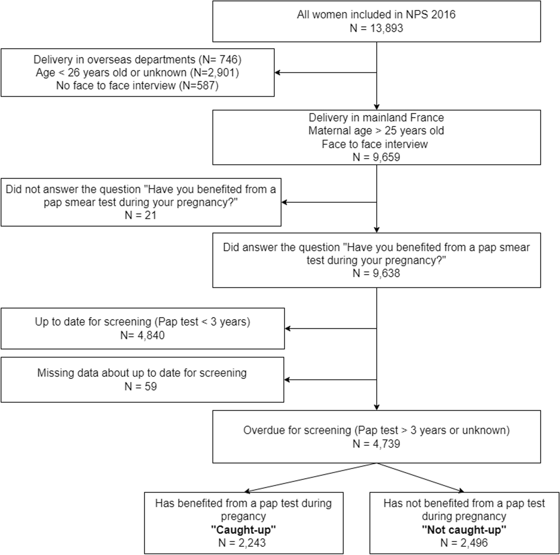
A total of 215 patients with OSCC met the inclusion criteria after carefully screening medical record files. All patients agreed to participate in the investigation; however, five patients were lost to follow-up, and the pathologic materials of ten potential participants were inadequate to perform immunohistochemistry (IHC). Finally, 200 patients with OSCC were enrolled in this clinicopathological correlation study. The study flowchart based on the STROBE (Strengthening the Reporting of Observational Studies in Epidemiology) statement [19] is shown in Fig. 1.
Fig. 1
The flow diagram describing the subjects’ enrollment as well as the working plan
Clinical data acquisitionThe items collected included age, sex, alcohol consumption, tobacco smoking, diet, oral hygiene habits, behavioral swallowing, periodontal condition, anatomical distribution of the tumor, TNM staging of OSCC, tumor differentiation, recurrence, treatment regime (surgery with sequential chemotherapy and/or radiotherapy, postoperative adjuvant immunotherapy or radiotherapy or chemotherapy), and survival status.
P. gingivalis assessmentP. gingivalis DNA was detected using PCR methods as established before [20]. To verify P. gingivalis-positive samples, one pair of 16S rDNA fragments were amplified from OSCC tissue and sequenced, confirmed by BLAST homology comparison (http://www.ncbi.nlm.nih.gov/BLAST) [12, 20].
Bioinformatics analysesTo ascertain the appropriate target transcript, gene expression omnibus (GEO), a public functional genomics data repository (https://www.ncbi.nlm.nih.gov/geo/), was searched. Retrieval of terms was combined in the following search string to identify relevant array- and/or sequence-based data: “Homo sapiens” (organism) AND “Oral squamous cell carcinoma” OR “Macrophage” OR “Porphyromonas gingivalis” (study keyword) AND “Expression profiling by array” (experiment type). After a systematic review, gene expression sequencing datasets of GSE24897 [20] and GSE138206 [13] were collected for further analyses. Specifically, the GSE24897 dataset contained nine samples of P. gingivalis-infected macrophages (GSM612265-73) and three samples of uninfected macrophages (GSM612262-4); the GSE138206 dataset contained six samples of OSCC tissue (GSM4101925-30), six samples of tissue adjacent to cancer (GSM4101937-42), and six samples of contralateral normal tissue (GSM4101931-6). The probes were converted into corresponding gene symbols based on the annotation information in the platform.
The differentially expressed genes (DEGs) between the two screened datasets were analyzed using the Limma package in R software (version 4.2.2; R Foundation for Statistical Computing, Vienna, Austria). Probes with > 1 gene symbol or without corresponding gene symbols were considered as intersections or removed. For analyzing and heat-mapping DEGs, adjusted P-value (adj. P) < 0.01 and |log2FC (fold-change)|> 1 were considered to have statistically significant difference [13, 20]. Furthermore, to explore the pan-cancer landscape of macrophage infiltration, TIMER 2.0, an online tool (http://timer.cistrome.org/ or http://timer.comp-genomics.org/) was utilized to analyze the immune module [20].
Histopathologic assessmentTissues specimens were provided by Biobank of Oral Medicine and Pathology, Central Scientific and Research Institute of Stomatology, Xinjiang, China, and representative tissue specimens from 200 OSCC patients were obtained from archival formalin-fixed paraffin-embedded (FFPE) tumor blocks to construct tissue microarrays, as described in our previous work [13, 20]. OSCC tissue microarrays were consecutively cut into 4 μm sections and dried on IHC microscope slides (BC075, Biosharp, Beijing, China). The sections were deparaffinized using standard xylene and hydrated using a gradient of ethanol in water. Antigen repair was performed by heating the sections with EDTA antigenic retrieval buffer (pH 8.0). IHC staining of P. gingivalis, downstream of kinase 3 (DOK3), and M2-TAM was consistent as follows: anti-P. gingivalis monoclonal antibody (#ab225982, Abcam, Cambridge, UK) at 1:100 dilution [21]; anti-DOK3 monoclonal antibody (#ab236609, Abcam, Cambridge, UK) at 1:500 dilution [20]; and anti-M2-TAM monoclonal antibody (CD206+; #MA5-44,409, ThermoFisher Scientific, Waltham. MA, USA) at 1:200 dilution of incubation. The DAB chromogenic agent (#D5905, Sigma-Aldrich, Saint Louis. Missouri, USA) was used as the substrate for P. gingivalis, DOK3, and M2-TAM expression.
Each slice was independently assessed by two professional pathologists who were blinded to clinical data. The immunoreactivity of P. gingivalis, DOK3, and M2-TAM was measured according to a score that added the intensity of staining to the proportion of positive cells using ImageJ software (version 1.8.0; National Institutes of Health, Bethesda, Maryland, USA) [22].
Statistical analysisData analysis was performed using R software (version 4.2.2; R Foundation for Statistical Computing, Vienna, Austria). Clinicopathologic characteristics of included patients were described as absolute frequency (percentage), and bivariate analysis to evaluate the association between clinicopathologic variables and P. gingivalis, DOK3, and M2-TAM immunoexpression levels in the tumor microenvironment (TME) of OSCC was determined using the Chi-square or Fisher’s exact test. The correlation between the levels of P. gingivalis, DOK3, and M2-TAM in specimens from patients with OSCC by immunohistochemical staining assays was analyzed using Pearson’s correlation. The Kaplan–Meier method was used to estimate the cumulative survival rate (CSR) probability over 10 years, and the log-rank test was used to compare prognosis among patients. Univariate and multivariate Cox proportional hazards regression models were employed to calculate the relevant hazard ratios (HR) with their 95% confidence intervals (CI). All tests were two-sided and P values less than 0.05 were considered statistically significant.
留言 (0)