Smith TA (1998) FDG uptake, tumour characteristics and response to therapy: a review. Nuclear Med Commun 19:97–105. https://doi.org/10.1097/00006231-199802000-00002
Article
CAS
Google Scholar
Coleman RE (2002) Value of FDG-PET scanning in management of lung cancer. Lancet 359:1361–1362. https://doi.org/10.1016/s0140-6736(02)08388-5
Article
PubMed
Google Scholar
UyBico SJ, Wu CC, Suh RD, Le NH, Brown K, Krishnam MS (2010) Lung cancer staging essentials: the new tnm staging system and potential imaging pitfalls. Radiographics 30:1163–1181
Article
PubMed
Google Scholar
Pommier P, Touboul E, Chabaud S et al (2010) Impact of (18)F-FDG PET on treatment strategy and 3D radiotherapy planning in non-small cell lung cancer: a prospective multicenter study. AJR Am J Roentgenol 195:350–355
Article
PubMed
Google Scholar
Postmus PE, Kerr KM, Oudkerk M et al (2017) Early and locally advanced non-small-cell lung cancer (nsclc): ESMO clinical practice guidelines for diagnosis, treatment and follow-up. Ann Oncol 28:iv1–iv21
Article
CAS
PubMed
Google Scholar
Manafi-Farid R, Askari E, Shiri I et al (2022) [(18)F]FDG-PET/CT radiomics and artificial intelligence in lung cancer: technical aspects and potential clinical applications. Semin Nucl Med 52:759–780. https://doi.org/10.1053/j.semnuclmed.2022.04.004
Article
PubMed
Google Scholar
Carles M, Fechter T, Radicioni G et al (2021) FDG-PET radiomics for response monitoring in non-small-cell lung cancer treated with radiation therapy. Cancers (Basel) 13:814. https://doi.org/10.3390/cancers13040814
Kong F, Hu C, Machta M et al (2021) OA02.04 randomized phase II trial (RTOG1106) on midtreatment PET/CT guided adaptive radiotherapy in locally advanced non-small cell lung cancer. J Thoracic Oncol 16:S104–S105
Article
Google Scholar
Vinod SK, Hau E (2020) Radiotherapy treatment for lung cancer: current status and future directions. Respirology 25:61–71
Article
PubMed
Google Scholar
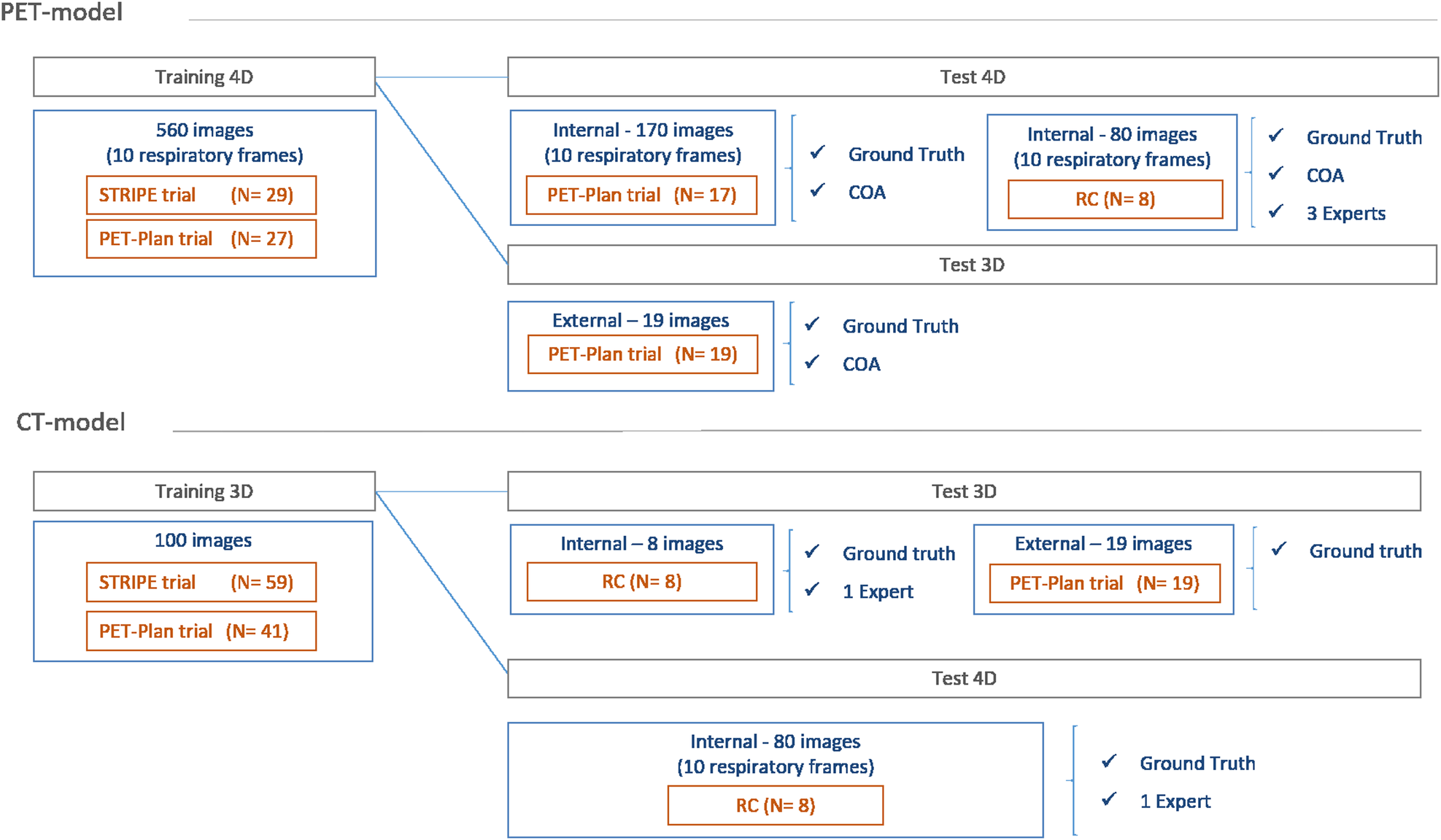
Nestle U, Schimek-Jasch T, Kremp S et al (2020) Imaging-based target volume reduction in chemoradiotherapy for locally advanced non-small-cell lung cancer (PET-plan): a multicentre, open-label, randomised, controlled trial. Lancet Oncol 21:581–592
Article
CAS
PubMed
Google Scholar
Cooke SA, de Ruysscher D, Reymen B et al (2023) [18F]FDG-PET guided vs whole tumour radiotherapy dose escalation in patients with locally advanced non-small cell lung cancer (PET-Boost): results from a randomised clinical trial. Radiother Oncol 181:109492. https://doi.org/10.1016/j.radonc.2023.109492
Article
PubMed
Google Scholar
Gkika E, Grosu AL, Nestle U (2023) The use of 18 F-FDG PET/CT for radiotherapy treatment planning in non-small cell lung cancer: a mini-review. Precision Cancer Med 6:1–7.
Article
Google Scholar
Bettinardi V, Picchio M, Di Muzio N, Gianolli L, Gilardi MC, Messa C (2010) Detection and compensation of organ/lesion motion using 4D-PET/CT respiratory gated acquisition techniques. Radiother Oncol 96:311–316. https://doi.org/10.1016/j.radonc.2010.07.014
Article
PubMed
Google Scholar
Büther F, Vehren T, Schäfers KP, Schäfers M (2016) Impact of data-driven respiratory gating in clinical PET. Radiology 281:229–238
Article
PubMed
Google Scholar
Huang TC, Chou KT, Wang YC, Zhang G (2014) Motion freeze for respiration motion correction in PET/CT: a preliminary investigation with lung cancer patient data. BioMed Res Int https://doi.org/10.1155/2014/167491
Chang G, Chang T, Pan T, Clark JW, Mawlawi OR (2010) Joint correction of respiratory motion artifact and partial volume effect in lung/thoracic PET/CT imaging. Med Phys 37:6221–6232
Article
PubMed
PubMed Central
Google Scholar
Dawood M, Büther F, Lang N, Schober O, Schäfers KP (2007) Respiratory gating in positron emission tomography: a quantitative comparison of different gating schemes. Med Phys 34:3067–3076
Article
PubMed
Google Scholar
Nehmeh SA, Erdi YE, Rosenzweig KE et al (2003) Reduction of respiratory motion artifacts in PET imaging of lung cancer by respiratory correlated dynamic PET: methodology and comparison with respiratory gated PET. J Nucl Med 44:1644–1648
PubMed
Google Scholar
Park SJ, Ionascu D, Killoran JJ et al (2008) Evaluation of the combined effects of target size, respiratory motion and background activity on 3D and 4D PET/CT images. Phys Med Biol 53:3661–3679
Article
PubMed
Google Scholar
Chirindel A, Adebahr S, Schuster D et al (2015) Impact of 4D- 18FDG- PET/CT imaging on target volume delineation in SBRT patients with central versus peripheral lung tumors. Multi-reader comparative study. Radiother Oncol 115:335–341
Article
PubMed
Google Scholar
Heath E, Unkelbach J, Oelfke U (2009) Incorporating uncertainties in respiratory motion into 4D treatment plan optimization. Med Phys 36:3059–3071. https://doi.org/10.1118/1.3148582
Article
PubMed
Google Scholar
Bittermann G, Scheifele C, Prokic V et al (2013) Description of a method: computer generated virtual model for accurate localisation of tumour margins, standardised resection, and planning of radiation treatment in head & neck cancer surgery. J Craniomaxillofac Surg 41:279–281. https://doi.org/10.1016/j.jcms.2012.10.011
Article
PubMed
Google Scholar
Carles M, Torres-Espallardo I, Alberich-Bayarri A et al (2016) Evaluation of PET texture features with heterogeneous phantoms: complementarity and effect of motion and segmentation method. Phys Med Biol 62:652–9251
Article
PubMed
Google Scholar
Carles M, Bach T, Torres-Espallardo I, Baltas D, Nestle U, Martí-Bonmatí L (2018) Significance of the impact of motion compensation on the variability of pet image features. Phys Med Biol 63:065013
Article
CAS
PubMed
Google Scholar
Zaidi H, El Naqa I (2010) PET-guided delineation of radiation therapy treatment volumes: a survey of image segmentation techniques. Eur J Nucl Med Mol Imaging 37:2165–2187. https://doi.org/10.1007/s00259-010-1423-3
Morra J, Tu Z, Toga A, Thomson P (2010) Machine learning for brain image segmentation. Biomedical image analysis and machine learning technologies: applications and techniques, edited by Gonzalez FA and Romero E, IGI Global, 102–126. https://doi.org/10.4018/978-1-60566-956-4.ch005
Kao D, Cheze Le Rest C, Jaouen V, Hatt M (2019) Artificial intelligence, machine (deep) learning and radio(geno)mics: definitions and nuclear medicine imaging applications. Eur J Nucl Med Mol Imaging 46:2630–2637. https://doi.org/10.1007/s00259-019-04373-w
Litjens G, Kooi T, Bejnordi BE et al (2017) A survey on deep learning in medical image analysis. Med Image Anal 42:60–88
Article
PubMed
Google Scholar
Halder A, Dey D, Sadhu AK (2020) Lung nodule detection from feature engineering to deep learning in Thoracic CT images: a comprehensive review. J Digit Imaging 33:655–677. https://doi.org/10.1007/s10278-020-00320-6
Article
PubMed
PubMed Central
Google Scholar
Liu X, Li KW, Yang R, Geng LS (2021) Review of deep learning based automatic segmentation for lung cancer radiotherapy. Front Oncol 11:717039. https://doi.org/10.3389/fonc.2021.717039
Article
PubMed
PubMed Central
Google Scholar
Wang S, Mahon R, Weiss E et al (2022) Automated lung cancer segmentation using a PET and CT dual-modality deep learning neural network. Int J Radiat Oncol Biol Phys 115:529–539. https://doi.org/10.1016/j.ijrobp.2022.07.2312
Article
PubMed
Google Scholar
Xiang D, Zhang B, Lu Y, Deng S (2023) Modality-specific segmentation network for lung tumor segmentation in PET-CT images. IEEE J Biomed Health Inform 27:1237–1248. https://doi.org/10.1109/JBHI.2022.3186275
Nestle U, Adebahr S, Kaier K et al (2020) Quality of life after pulmonary stereotactic fractionated radiotherapy (SBRT): results of the phase II STRIPE trial. Radiother Oncol 148:82–88
Article
PubMed
Google Scholar
Martinez-Movilla A, Mix M, Torres-Espallardo I et al (2022) Comparison of protocols with respiratory-gated (4D) motion compensation in PET/CT: open-source package for quantification of phantom image quality. EJNMMI Phys 9:80. https://doi.org/10.1186/s40658-022-00509-4
Article
PubMed
PubMed Central
Google Scholar
Nestle U, De Ruysscher D, Ricardi U et al (2018) ESTRO ACROP guidelines for target volume definition in the treatment of locally advanced non-small cell lung cancer. Radiother Oncol 127:1–5. https://doi.org/10.1016/j.radonc.2018.02.023
Article
PubMed
Google Scholar
Carles M, Fechter T, Nemmer U et al (2015) Feasibility of a semi-automated contrast-oriented algorithm for tumor segmentation in retrospectively gated PET images: phantom and clinical validation. Phys Med Biol https://doi.org/10.1088/0031-9155/60/24/9227
Isensee F, Jaeger PF, Kohl SAA, Petersen J, Maier-Hein KH (2021) nnU-Net: a self-configuring method for deep learning-based biomedical image segmentation. Nat Methods 18:203–211. https://doi.org/10.1038/s41592-020-01008-z
Article
CAS
PubMed
Google Scholar
Zou KH, Warfield SK, Bharatha A et al (2004) Statistical validation of image segmentation quality based on a spatial overlap index. Acad Radiol 11:178–189
Article
PubMed
PubMed Central
Google Scholar
Huttenlocher DP, Klanderman GA, Rucklidge WJ (1993) Comparing images using the Hausdorff distance. IEEE Trans Pattern Anal Mach Intell 15:850–863. https://doi.org/10.1109/34.232073
Article
Google Scholar
Zhang F, Wang Q, Li H (2020) Automatic segmentation of the gross target volume in non-small cell lung cancer using a modified version of ResNet. Technol Cancer Res Treat 19:1533033820947484. https://doi.org/10.1177/1533033820947484
Article
CAS
PubMed Central
Google Scholar
Jiang J, Hu YC, Liu CJ et al (2019) Multiple resolution residually connected feature streams for automatic lung tumor segmentation from CT images. IEEE Trans Med Imaging 38:134–144. https://doi.org/10.1109/TMI.2018.2857800
Article
PubMed
Google Scholar
Bi N, Wang J, Zhang T et al (2019) Deep learning improved clinical target volume contouring quality and efficiency for postoperative radiation therapy in non-small cell lung cancer. Front Oncol 9:1192. https://doi.org/10.3389/fonc.2019.01192
Article
PubMed
PubMed Central
Google Scholar
Wang C, Tyagi N, Rimner A et al (2019) Segmenting lung tumors on longitudinal imaging studies via a patient- specific adaptive convolutional neural network. Radiother Oncol 131:101–107. https://doi.org/10.1016/j.radonc.2018.10.037


留言 (0)