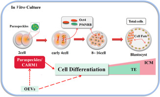
Paraspeckles are special nuclear structures that are involved in various physiological responses, such as nuclear chromatin recombination and histone modification. Paraspeckles regulate specific gene expression by retaining RNA in the nucleus and preventing certain RNA-binding proteins from limiting their biological function in the nucleus, thereby affecting physiological processes such as cell differentiation, foreign pathogen infection, and stress stimulation[1], [2]. Paraspeckles are often identified by the immune marker of paraspeckle protein 1 (PSPC1), and their number and size have been determined. Preliminary reports have stated that paraspeckles bind to determine cell fate in the early embryonic development of mice, but the physiological effects on the embryonic development of other mammals have not been studied in detail[3]. As an important biological event in early embryonic development, cell fate determination promotes the formation of the inner cell mass (ICM) and trophoblast cells (TE), and the development into extraembryonic tissues, such as the fetus and placenta, after implantation. Although the distinct grouping of embryonic cells occurs at the blastocyst stage, the regulation of cell fate is initiated at an earlier stage. Overexpression of H3R26 methyltransferase, CARM1, in the 4-cell stage of early preimplantation development in mice leads to increased expression of Sox2 and nuclear Nanog, which promotes the formation of ICM cells and affects the fate of early embryonic cells[3], [4]. During the early development of mouse embryos from 2-cell to 4-cell transition, CARM1 accumulates in the nucleus. The detection of paraspeckles using p54NRB and PSPC1 markers indicated that several paraspeckles that formed at the 2-cell stage of in mice corresponded to CARM1. It was also confirmed for the first time that there were paraspeckles in the nucleus of the 2-cell mammalian embryonic. Paraspeckle's main component, Neat1, and its cofactor, p54nrb, contribute to the binding between CARM1 and paraspeckles, leading to differences in H3R26 methylation. CARM1 also affects the formation of paraspeckles. Knockout of Neat1 and p54NRB leads to the arrest of embryonic development at the 16–32 cell stage, which is mainly due to the increased transcription of Cdx2, which promotes the transformation of cell fate to TE cells. Since this arrest occurs before CARM1 is depleted, paraspeckle, as an upstream signal of CARM1, can regulate cell fate at an earlier stage[3], [4]. CARM1 plays an important role in the regulation of autophagy[5]. Autophagy not only acts as a resource allocator during embryonic development but also improves embryonic quality and reduces embryonic apoptosis[6]. Whether this regulatory mechanism exists in other species is unknown. Therefore, during mammalian embryonic development, whether the first differentiation of cells and the first difference determin the trend of cell specificity during mammalian embryonic development is still a key open question.
Extracellular vesicles (EVs), also known as exosomes (40–100 nm) and microvesicles (100–1000 nm), are membrane-encapsulated vesicles released in an evolutionarily conserved manner from yeast to most mammalian cells. They exist in body fluids and have become a new way of intercellular communication. EVs contain a series of bioactive molecules, such as proteins, mRNAs, ncRNAs, lipids, and genomic DNA[7]. This molecular cargo is protected from extracellular degradation or modification[8]. The importance of EVs lies in their ability to transfer molecular goods (proteins, RNA, genomic DNA, lipids, and metabolites) to other cells while functioning in target cells. Cell-derived exosomes give cells the ability to protect their information transport and can choose to deliver multiple different signal messengers simultaneously at the proximal or even distal ends of the organism[9]. EVs are one of the main components of oviduct fluid in different species and are potential mediators for the interaction of oviduct components with gametes and embryos[10]. Oviduct exosomes (OEVs) act as unique multi-signal messengers in gamete/embryo-tubal interactions. To date, exosomes have been isolated from the oviduct fluid of mice[11], [12], cows[13], [14], humans[15], chickens[16], dogs[17], and turtles[18]. At the fertilization stage, studies have shown that OEVs play a key role in mouse sperm storage, promoting capacitation and regulating acrosome reaction and hyperactive movement. The molecular cargo carried by OEVs can be mediated by integrin αvβ3 and α5β to fuse into the sperm membrane. The plasma membrane Ca2+ -ATPase 4 protein (PMCA4) carried by OEVs can participate in maintaining Ca2+ homeostasis, preventing premature sperm capacitation, and stimulating sperm fertilization potential[11]. Moreover, during early embryonic development, OEVs added during in vitro culture (IVC) can be absorbed by in vitro produced (IVP) embryos, and can improve embryo blastocyst rate, hatching rate, in vitro survival rate, and blastocyst quality, and affect birth rate by increasing the efficiency of embryo transfer[12]. Although OEVs play such an important role in embryonic development, the effects of OEVs on embryonic paraspeckle formation and cell differentiation remain unexplored.
The occurrence time of zygotic genomic activation (ZGA) in bovine early embryonic development is similar to that of other mammals, including humans, macaques, sheep, and rabbits, as evidenced in the study of early embryos of multiple mammalian species. It is proposed that the early embryonic development model of cattle may be more representative of mammalian characteristics than mice[19], [20], [21]. The yak is unique species of cattle living in an alpine and hypoxic environment. Studies have shown that the development of yak embryos or germ cells in vitro is regulated by hypoxia-inducible factor (HIF-1α)[22], heat shock proteins (HSPs)[23], cold-inducible RNA-binding protein (CIRP) and other alpine hypoxia-related factors[24]. Previous studies have found that under the action of epidermal growth factor (EGF) and insulin-like growth factor (IGF), the development ability of yak embryos in vitro can be improved through apoptosis but also affect the quality of blastocysts and the distribution of different cell fates in blastocysts is affected by regulating factors related to the alpine environment[24], [25]. The in vitro regulation mechanism of early cell differentiation, blastocyst quality, and blastocyst attachment ability play a unique role in the in vitro development of mammalian embryos in an alpine hypoxic environment. Therefore, in this study, we aimed to analyze the effects of OEVs on paraspeckle formation and cell differentiation in IVF yak embryos. We also investigated the effects of OEVs on embryo autophagy and blastocyst apoptosis. Additionally, we discussed the relationship between CARM1 and paraspeckle formation and autophagy during yak embryonic development. These results elucidate the uniqueness of yak embryonic development and the importance of OEVs, paraspeckles, and autophagy in yak embryonic development.


留言 (0)