Lactational mastitis is a painful and debilitating inflammation of breast tissue that has an important impact upon breastfeeding duration and frequency in many lactating mothers [1]. Affected individuals usually develop a swollen, tender, and painful breast with a focal area of erythema, and flu-like symptoms including fever and chills [2]. Yet, if the condition is managed poorly or left untreated, a breast abscess can form along with bacteremia and sepsis [1,2,3].
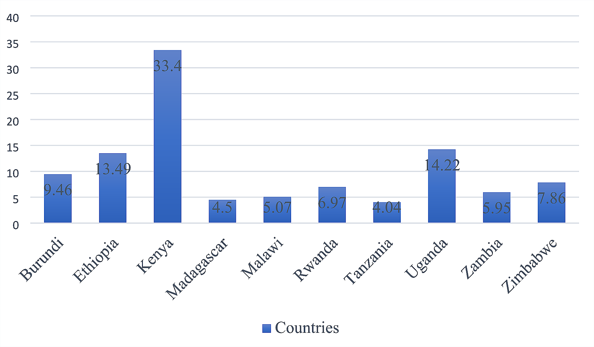
Whereas mastitis is a recognized cause of fever during the postpartum period, its precise incidence and epidemiology in the breastfeeding population is not well defined and can vary tremendously, depending on numerous factors such as geographic location, cultural practice, and breastfeeding preferences [1]. It is estimated that around 10% to up to 33% of breastfeeding women will experience mastitis at some point during their lactation period [2, 3]. The incidence of lactational mastitis tends to be highest within the first 6 to 12 weeks postpartum and gradually decreases over time [1,2,3]. The high incidence in the initial period is often related to women’s early adjustments to breastfeeding, as well as issues related to skin injuries, and ineffective milk removal [2, 3]. Furthermore, certain determinants have been identified as risk factors for lactational mastitis, including previous history of mastitis, poor breastfeeding techniques, cracked and sore nipples, incomplete breast emptying, infrequent or missed breastfeeding sessions, lower maternal immune status, and fatigue secondary to stress and sleep deprivation [1, 2]. Although the exact etiology in each patient is difficult to determine, hyperlactation causing milk duct narrowing and consequent breast engorgement or intrusion of baby’s mouth bacteria through sore and cracked nipples into the milk ducts are believed to be the most common causes of the disease [2]. A blend of different pathogens is involved in the bacterial form of lactational mastitis, with S. aureus being the most common pathogen [3].
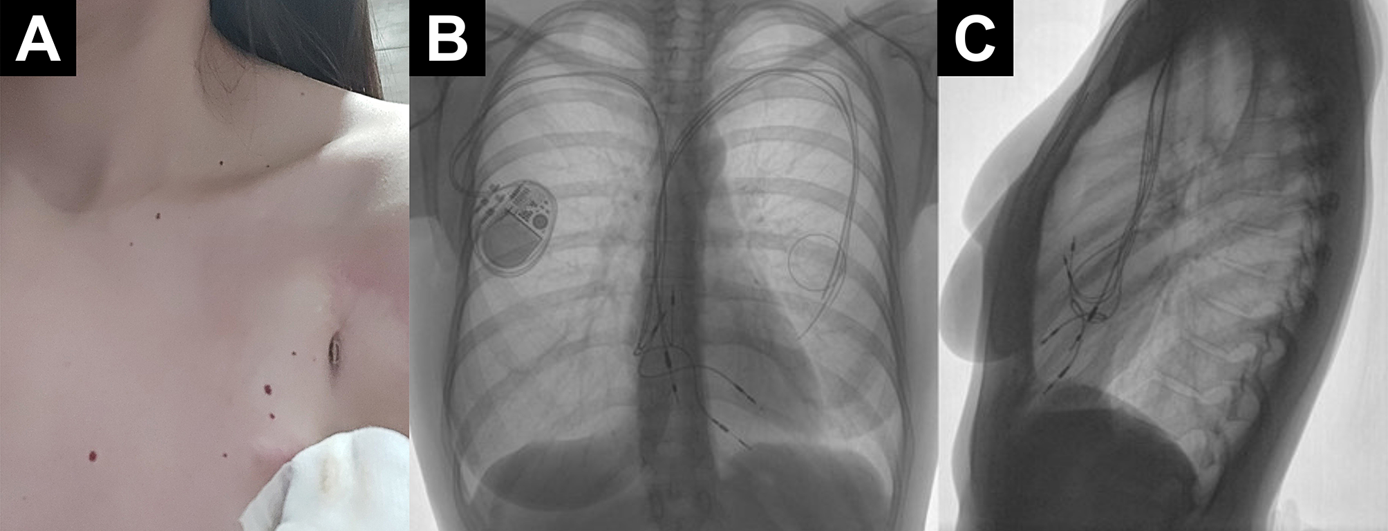
In the majority of patients with lactational mastitis, management of the disease is rather straightforward, including continuation of breastfeeding, cooling of the affected breast area, reverse pressure softening, and usage of nonsteroidal anti-inflammatory drugs and antibiotics in cases of severe pain and signs of systemic inflammation. If mastitis progresses to abscess formation, pus puncture using fine-needle aspiration technique or surgical incision and pus drainage is indicated as soon as possible [2, 3]. However, treatment of lactational mastitis and mastitis related complications in patients with implantable material in the breast or close to the breast region (i.e., silicone breast implants, CIED or implantable loop recorders) can be extremely challenging [2, 3], requiring patient-specific treatment strategies. This is especially so in cases of surgical implantable material infection which necessitates complete removal from the body [2,3,4,5,6,7,8,9,10].
Evidence indicates that lactational mastitis is fortunately only rarely complicated with secondary infection of implantable surgical material that lies in or near the affected breast [2,3,4,5,6,7,8,9,10], however such a clinical course is the most plausible scenario in the breastfeeding mother described above. We strongly believe that severe lactational mastitis in this woman was complicated with secondary pacemaker pocket infection through direct bacterial contamination. Other causes of pacemaker pocket infection, such as infection through the hematogenous route during an episode of bacteremia or late clinical presentation of bacterial contamination during previous surgeries seem highly unlikely. This is mainly due to the absence of retained leads endocarditis and the long and completely uneventful postoperative course [4,5,6].
According to current American Heart Rhythm Society and European Heart Rhythm Association guidelines for the treatment of CIED infection (including device pocket infection), prompt extraction of the entire electronic system, followed by targeted antibiotic treatment and reimplantation of a new device after infection cessation is indicated in virtually all affected individuals mainly due to high mortality (i.e., up to 35%) following untreated CIED infection [5,6,7,8,9,10,11,12].
Over the last decade, TLE has greatly advanced and is currently regarded as the premier surgical method when CIED removal is indicated: it provides a complete, safe and highly effective mode of implanted leads extraction with reported success rates ranging from 83.3 to 97.6% and major complication rates ranging from 1.5 to 2.4% [11,12,13]. Although the reported TLE complication rate is relatively low, especially in dedicated, high-volume centres, the nature of potential complications (i.e., anaesthesia-related severe adverse events, sepsis, respiratory distress, cardiac or vascular avulsion or tear requiring pericardiocentesis or open-heart surgery, and even death in 0.5% of patients [13, 14]), is non-negligible and may direct the patient’s decision-making process. Regardless of the clear recommendations from the scientific community it is sometimes difficult for some patients to decide whether to undergo a potentially risky surgical procedure of complete lead extraction or to leave infected electrodes in-situ and combine a less risky but incomplete surgical procedure with long-term antibiotic therapy, as was the case in this young breastfeeding mother [5, 6, 12].
Every TLE is regarded as a complex surgical procedure, yet, several clinical factors, such as very long dwell time (> 10 years); young age at primary implantation; preoperative anemia; and damage to remnant leads during previous surgery; have been reported as risk factors for even more demanding, technically more challenging, and potentially unsuccessful procedures [7, 13,14,15,16]. This was also the case in the young mother described above– she was only 18 years old when she underwent the first pacemaker implantation; the dwell time was 15 years (combined dwell time of all electrodes was 74 years); and all three leads were at least partially damaged during previous surgery. Of note, a fully functional pacemaker implanted from the contralateral side represented an additional risk factor for clinical failure of the TLE procedure [6]. Performing a TLE procedure by leaving the contralateral pacemaker system intact is technically even more demanding and raises the odds of an unsuccessful treatment outcome [12,13,14,15,16].
Since the current patient-orientated medical culture appreciates and encourages shared-decision-making and values each individual’s autonomy, especially among women in the antenatal and postpartum time-periods, our team respected the young mother’s requests and complied with her wish not to undergo a complete pacemaker system removal, despite having clinically evident pacemaker pocket infection. However, in everyday clinical practice we usually try to discourage our patients from declining such surgical strategies [6, 12]. Patients should be strongly advised and encouraged by all members of the multidisciplinary team to undergo a complete extraction to maximize the treatment success and to minimize the possible short- and long-term complications [6, 12]. By delaying consent for the indicated surgical procedure due to — according to her understanding — unacceptably high surgery-related risks, this young breastfeeding mother has unfortunately increased the likelihood of an unsuccessful treatment outcome and risks for potentially severe and life-threatening complications following a non-optimal treatment scenario. Even though our stepwise surgical therapy has resulted in an overall successful treatment outcome, such treatment strategies should be used only as a bailout clinical scenario if consent to recommended treatment is refused for any reason.


留言 (0)