Oral cancer is a type of head and neck cancer that can develop in any part of the mouth, including the lips, tongue, gums, and inner lining of the cheeks, and is the most prevalent type of head and neck cancer (Ali, 2022, D'Silva et al., 2023). Treatment options currently available include a combination of surgical resection, chemotherapy, radiation therapy, immunotherapy, or a combination of these approaches. Despite significant research efforts to improve treatment outcomes, the emergence of drug resistance remains a persistent challenge, leading to ineffective therapeutic outcomes and the development of local, regional, and distant recurrence. This underscores the need to develop innovative therapeutic strategies to overcome drug resistance and improve the efficacy of existing therapies (Atashi et al., 2021). One of the emerging approaches to enhance the sensitivity of cancer cells towards chemotherapy is to combine chemotherapy with hyperthermia therapy.
Hyperthermia therapy for cancer involves heating the tumor in the temperature range of 42 – 48 °C to induce apoptotic cell death (Amin et al., 2020). A rise in the temperature at the tumor site leads to an increase in the blood flow and permeability of the blood vessels, permitting the penetration of chemotherapeutic drugs deeper into the tumor. In addition, increased blood flow also increases the tumor pO2 and tumor pH. Hyperthermia also possesses the potential to regulate immune function and constrain tumor growth by eliciting immunogenic cell death. All these changes cumulatively contribute to an increase in the sensitivity of cancer cells toward chemotherapy (Dunne et al., 2020).
Among the various techniques available for inducing a hyperthermic rise in temperature, photothermal therapy has come to the forefront as a result of the recent clinical trials using AuroShell®, a pegylated silica gold nanoshell sponsored by Nanospectra Biosciences (Yao and Bojic, 2023). The reduced form of graphene oxide is another agent that produces a temperature rise on irradiation with a near-infrared laser. Our recent publication reports its potential application as an effective photothermal agent for implant development (Anup et al., 2023). Our investigation observed the potential of combined chemo-photothermal therapy in cancer therapy. The promising results that we observed prompted a more detailed investigation of the effectiveness of chemo-photothermal therapy in increasing the sensitivity of resistant cancer cells toward chemotherapy.
There has been a surge in reports using 3D-printing technologies to develop drug-loaded implants in recent years. One such report involves developing a cylindrical 3D-printed implant loaded with olanzapine by Picco et al. (Picco et al., 2023). The implant was prepared using polycaprolactone alone or in combination with poly(ethylene)glycol. The major objective behind the development of this implant was to deliver the drug in a single administration to eliminate the need for multiple administration at specific intervals. This helps reduce the pill burden and eliminate missing doses from forgetfulness. They achieved 60 % drug release in 200 days. Treating diseases in the posterior segment of the eye involves intravitreal injection of drugs. Frequent injection at this site can result in serious complications. However, the development of implants with sustained release properties will be highly beneficial in the treatment of diseases in the posterior segment of the eye.
Annuryanti et al. developed triamcinolone acetonide (TA) loaded 3D-printed implants of different shapes (filament, rectangular, and circle). They achieved the highest cumulative drug release of over 180 days with the filament-shaped implant. The implants showed good biocompatibility as well (Annuryanti et al., 2023). Drug-eluting 3D-printed personalized intrauterine implants incorporating paclitaxel and carboplatin were developed by Varan et al. They achieved controlled release for 10 days and a 60 % reduction in cell viability in in vitro cell culture studies (Varan et al., 2023). 3D printing offers flexibility in the fabrication of implants in terms of their size and shape to achieve the goal of personalized therapy and meet patient-specific needs. The CT image of the tumor bed left behind after surgical excision of the tumor can be converted to.stl format suitable for 3D printing. However, the conventional solvent casting approach, which can be used to prepare patches, lacks the virtue of personalized fabrication.
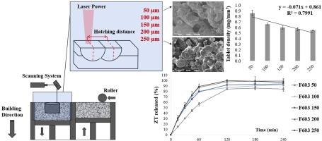
This investigation reports the development of a 3D-printed, biodegradable, laser-responsive, drug-loaded implant for treating resistant cancer cells. The 3D printer allows implant fabrication with varying sizes, shapes, doses, and drug types to meet patient-centric therapeutic needs. We employed cisplatin (CIS) as a model anticancer drug and plasmonic graphene oxide (PGO) as the photothermal active agent. The in vitro analysis of the implant was carried out in the tumor spheroid model as it has more resemblance to in vivo tumor setting.


留言 (0)