
記住我


Hepatic stellate cells (HSCs) are the major site of vitamin A (retinol) esterification and subsequent storage as retinyl esters within lipid droplets. However, retinyl esters become depleted in many pathophysiological states, including acute and chronic liver injuries. Recently, using a liver slice culture system as a model of acute liver injury and fibrogenesis, a time-dependent increase and decrease in the apparent formation of the bioactive retinoid all-trans-retinoic acid (atRA) and retinyl palmitate was measured, respectively. This coincided with temporal changes in the gene expression of retinoid-metabolizing enzymes and binding proteins, that preceded HSC activation. However, the underlying mechanisms that promote early changes in retinoid metabolism remain unresolved. We hypothesized that LX-2 cells could be applied to investigate differences in quiescent and activated HSC retinoid metabolism. We demonstrate that the hypermetabolic state of activated stellate cells relative to quiescent stellate cells may be attributed to induction of STRA6, RBP4, and CYP26A1, thereby reducing intracellular concentrations of atRA. We further hypothesized that paracrine and autocrine cytokine signaling regulates HSC vitamin A metabolism in both quiescent and activated cells. In quiescent cells, tumor necrosis factor α dose-dependently downregulated LRAT and CRBP1 mRNA, with EC50 values of 30–50 pg/mL. Likewise, interleukin-1β decreased LRAT and CRBP1 gene expression but with less potency. In activated stellate cells, multiple enzymes were downregulated, suggesting that the full effects of altered hepatic vitamin A metabolism in chronic conditions require both paracrine and autocrine signaling events. Further, this study suggests the potential for cell type–specific autocrine effects in hepatic retinoid signaling.
SIGNIFICANCE STATEMENT HSCs are the major site of vitamin A storage and important determinants of retinol metabolism during liver fibrogenesis. Here, two LX-2 culture methods were applied as models of hepatic retinoid metabolism to demonstrate the effects of activation status and dose-dependent cytokine exposure on the expression of genes involved in retinoid metabolism. This study suggests that compared to quiescent cells, activated HSCs are hypermetabolic and have reduced apparent formation of retinoic acid, which may alter downstream retinoic acid signaling.
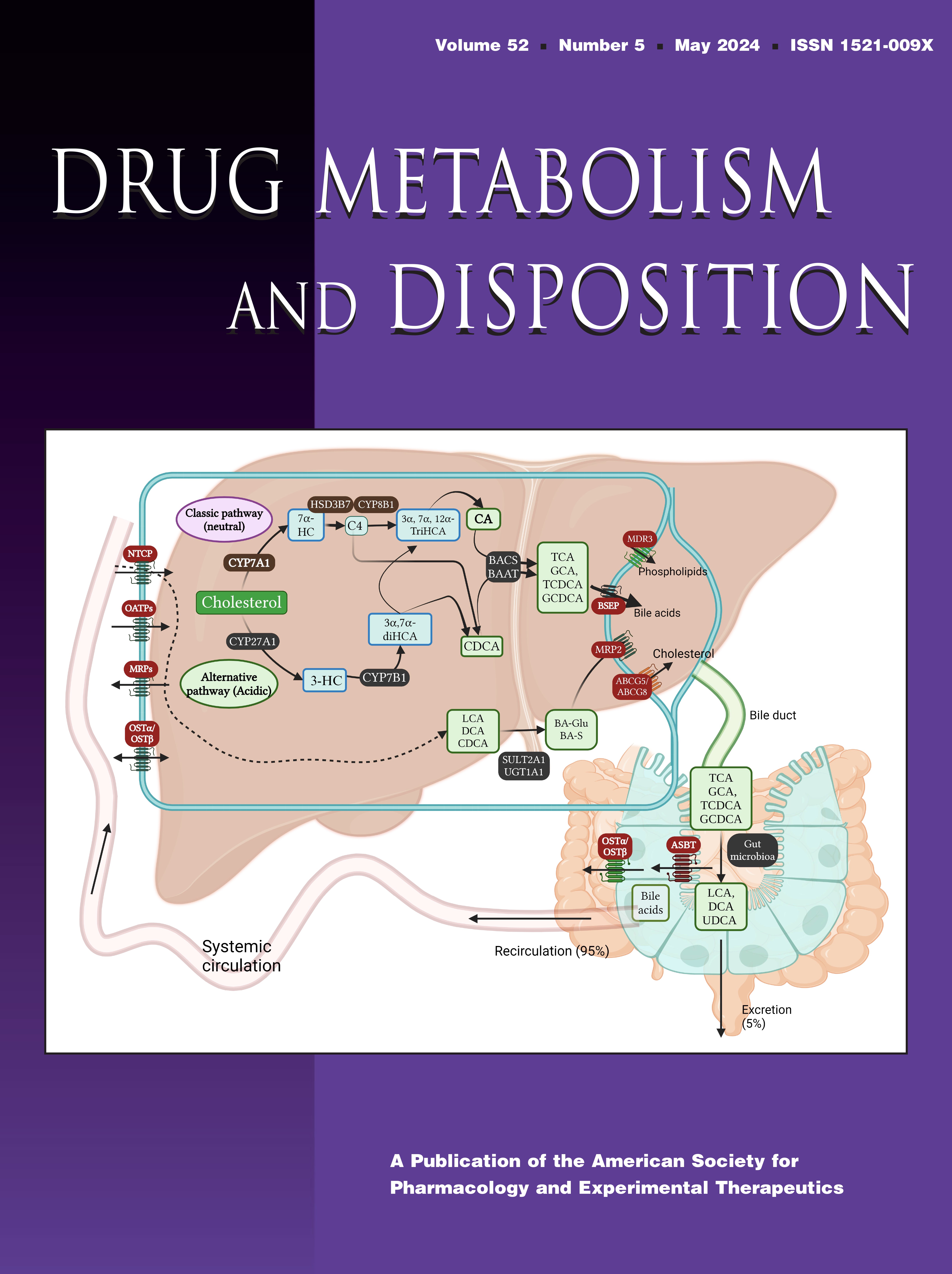
IntroductionVitamin A is an essential fat-soluble vitamin obtained from the diet and stored in the liver as retinyl esters. Vitamin A signaling is attributed to the transcriptionally active metabolite of retinol, all-trans-retinoic acid (atRA). atRA is an integral regulator of lipid metabolism (Chen and Chen, 2014), fatty acid oxidation (Zhao et al., 2012), gluconeogenesis (Obrochta et al., 2015), and extracellular matrix remodeling (Ye and Dan, 2010). atRA exerts its transcriptional effects through activation of the retinoic acid receptors (RAR)-α, RARβ, and RARγ (Huang et al., 2014). The transcriptional activity is dependent on atRA concentrations, which are regulated by a complement of enzymes and binding proteins (Fig. 1). Hepatic stellate cells (HSCs) esterify retinol to retinyl esters (REs) by lecithin retinol acyltransferase (LRAT) and store REs in lipid droplets. HSCs also support circulating retinol concentrations through the mobilization of REs via retinyl ester hydrolases. Retinol is secreted into systemic circulation from the hepatocytes bound to retinol-binding protein (RBP)-4 (Blaner et al., 2016). In cells, retinol oxidation to retinaldehyde is reversible and involves retinol dehydrogenases (RDHs), short-chain dehydrogenase reductases (DHRS) (Kedishvili, 2016; Belyaeva et al., 2020), and CYP1B1 (Chen et al., 2000). Retinaldehyde oxidation to atRA is irreversible and in human liver is mediated mainly by aldehyde dehydrogenases (ALDHs; ALDH1A1) and, to a lesser extent, aldehyde oxidase (AOX) (Zhong et al., 2021). Clearance of atRA is primarily attributed to the CYP26 family of enzymes, but other cytochrome P450s can also oxidize atRA in human liver (Isoherranen and Zhong, 2019).
 Fig. 1.
 Vitamin A metabolic pathway and the enzymes involved in retinoid metabolic flux in human liver. The scheme shows the major enzymes and retinoid-binding proteins involved in modulating the metabolic steps. The ligand-free apoCRBP has been shown to inhibit retinol esterification and stimulate ester hydrolysis in addition to having ALDH1A enzyme–specific effects on atRA formation. The CRABPs have been suggested to modulate RA oxidation by the apoCRABPs inhibiting CYP26 activity and the liganded holoCRABPs channeling their ligand to CYP26 for oxidation.
Vitamin A metabolome is altered in acute and chronic liver injury (Zhong et al., 2019b; Czuba et al., 2021). Yet, the exact mechanisms perpetrating vitamin A dysregulation in humans are unknown, and the consequences of altered atRA concentrations to retinoid signaling in the liver are not well characterized. In rodent models, activation of HSCs is associated with downregulation of LRAT mRNA and protein, mobilization of REs, and depletion of hepatic vitamin A stores (D’Ambrosio et al., 2011; Kida et al., 2011). In a human liver slice model, altered vitamin A metabolic activity preceded HSC activation and collagen deposition (Wu et al., 2018; Czuba et al., 2021). Within 12–24 hours of tissue slicing, LRAT, cellular retinol-binding protein (CRBP)-1, RBP4, and the retinol transporter STRA6 mRNAs were significantly downregulated in all donors tested. This coincided with a transient peak in tissue atRA concentrations. Kinetic modeling suggested that a combination of changes in retinoid-metabolizing enzyme activity was responsible for the peak (Czuba et al., 2021). Consistent with these findings, treatment of primary mouse HSCs with lipopolysaccharide (LPS) reduced RA cell concentrations, altered RAR signaling, and downregulated CRBP1 gene expression (Chen et al., 2017). Taken together, these data point to the critical role of vitamin A metabolic machinery in HSCs and to the importance of retinoid signaling in fibrosis. Yet, better model systems are needed to explore the mechanisms regulating vitamin A metabolism in human HSCs.
Stellate cells, including the immortalized stellate cell line LX-2, retain cytokine signaling and express the major receptors for interleukin (IL)-6, tumor necrosis factor (TNF)-α and IL-1β in culture (Xu et al., 2005; Tarrats et al., 2011; Harvey et al., 2013). TNF-α, IL-1β, and IL-6 are implicated in the early stages of liver injury, wound response and regeneration, and initiation of fibrogenesis (Tanaka and Miyajima, 2016). The secretion of soluble cytokines and signaling is elevated in many human conditions where vitamin A metabolome is also altered, including obesity (Trasino et al., 2015), nonalcoholic fatty liver disease (Saeed et al., 2017; Zhong et al., 2019b), alcoholic hepatitis (Ferdouse et al., 2022), and chronic kidney disease (Jing et al., 2016). In the human liver slice model, early changes to hepatic vitamin A metabolism parallel the time course of notable paracrine signals, including cytokines (Wu et al., 2018). Treatment of rat HSCs with IL-1 and TNF-α led to downregulation of LRAT mRNA expression (Kida et al., 2011). However, it is unknown if exposure to soluble cytokines promotes dysregulation at multiple steps of the vitamin A pathway as implicated by kinetic modeling. Here, we hypothesized that TNF-α, IL-1β, and IL-6 regulate the mRNA expression of hepatic retinoid-metabolizing enzymes. We used the immortalized stellate cell line LX-2 as a model system to investigate the expression of vitamin A–metabolizing enzymes in “quiescent” and “activated” stellate cells. We then tested whether TNF-α, IL-1β, and IL-6 regulate the mRNA expression of key genes in the vitamin A metabolic pathway.
Materials and MethodsChemicals and ReagentsHuman-derived (HumanKine) TNF-α, IL-1β, and IL-6 were purchased from MilliporeSigma (Burlington, MA). Human serum albumin, all-trans-retinoic acid, all-trans-retinol, retinyl acetate, acitretin, LPS, and TRI Reagent were purchased from MilliporeSigma. Retinyl palmitate and 13-cis-retinoic acid were from Toronto Research Chemicals (Toronto, Ontario). atRA-d6, retinol-d6, and retinyl palmitate-d4 were purchased from Cambridge Isotope Laboratories (Tewksbury, MA). Ethanol and Optima liquid chromatography/mass spectrometry (LC/MS)-grade water, acetonitrile (ACN), and formic acid were purchased from ThermoFisher Scientific (Waltham, MA). Gibco brand Dulbecco’s modified Eagle’s medium (DMEM; high glucose), charcoal-stripped FBS, penicillin-streptomycin (10,000 U/mL), Dulbecco’s phosphate-buffered saline (DPBS), trypsin, and GlutaMAX were from ThermoFisher Scientific.
Cell Culture Conditions for Quiescent and Activated Stellate CellsThe immortalized hepatic stellate cell line LX-2 (MilliporeSigma), derived from isolated stellate cells from a single donor and cultured under low serum conditions, was used (Xu et al., 2005). Cells were cultured in DMEM with 2% charcoal stripped FBS, 1% penicillin-streptomycin, and 1% GlutaMAX. Passages 3–10 were used for experiments, and during the experimental phase of research, the LX-2 cell line was authenticated by submitting cell samples to the American Type Culture Collection for short-tandem repeat analysis.
To promote an activated hepatic stellate cell phenotype, LX-2 cells were seeded at a density of 2 to 3 × 10⁵ cells per well to a 12-well tissue culture–treated plastic plate. These cells are referred to as activated cells due to the previously established phenotype based on the expression of α smooth muscle actin (αSMA), platelet-derived growth factor receptor β (βPDGF-R), and cell morphology when cultured on plastic plates (Xu et al., 2005; Olsen et al., 2011). To promote a quiescent HSC phenotype, LX-2 cells were seeded at a density of 3 to 4 × 10⁵ cells per well to 12-well plates precoated with Growth Factor Reduced Matrigel from Corning (Corning, NY) (“Matrigel culture”) as previously described (Sohara et al., 2002; Gaça et al., 2003; Olsen et al., 2011). Although these cells may represent a mixture of quiescent and senescent stellate cells, they are referred to as quiescent cells here for clarity and exhibit a nonfibroblast-like morphology similar to quiescent cells (Gaça et al., 2003; Olsen et al., 2011). Matrigel coating was performed according to manufacturer recommendations for the “Thin Gel Method” (190 μL Matrigel per well), gelled at 37°C for at least 30 minutes, and rinsed with warm DPBS prior to seeding cells (Sohara et al., 2002). The phenotype of the cultures was assessed based on the expression of αSMA mRNA and cell morphology as previously described (Olsen et al., 2011). Although not directly measured, the phenotype of the quiescent cells was apparent based on a notable reduction in cell proliferation once plated on the Matrigel matrix as previously observed (Gaça et al., 2003).
The expression of the enzymes regulating retinoid homeostasis is typically regulated via presence of retinoids through various feedback loops. We hypothesized that maintenance of baseline vitamin A levels in the culture similar to retinol concentrations observed in vivo is necessary to maintain the network of enzymes in HSC responsible for vitamin A metabolism. The culture of HepG2 cells in the presence of atRA was previously shown to be essential for constitutive CYP26A1 expression in vitro (Tay et al., 2010). To test the impact of retinol supplementation, cells seeded on plastic (activated cells) were cultured in the presence or absence of 2 μM retinol for 48 hours prior to measuring the expression of retinoid-metabolizing enzymes. Subsequently, for all other experiments, cells were seeded, and culture media (1 mL) was refreshed daily (every 24 hours) during experiments to maintain 2-μM-retinol concentrations in the media in both activated and quiescent cell systems, mirroring the approximated concentration of retinol in human circulation.
To test whether the metabolic pathways of retinoids (retinol esterification and oxidation to atRA) are present in the quiescent and activated LX-2 cells, cells were seeded on Matrigel or plastic, as described above, in the presence of 2 μM retinol. The following morning, the media was changed, and cells were cultured in the presence of 2 μM retinol and 100 μM palmitic acid for 96 hours (1 mL media was replenished every 24 hours). Palmitic acid was added to the media to support the formation of retinyl palmitate during activity assays. This concentration (100 μM) of palmitate was previously used in LX-2 cells to facilitate ester formation (Lee et al., 2010). To confirm the impact of palmitate on retinyl ester formation, an experiment was also conducted without palmitate addition, supplementing the cells only with retinol. The persistence of cell morphology in the presence of palmitate supplementation was confirmed through visual inspection. The concentrations of retinol, retinyl palmitate, and retinoic acids were measured in the cells following a culture to steady state to assess the presence of vitamin A metabolic machinery. The metabolic activity of LX-2 cells toward retinoids was then measured using deuterium-labeled retinoids. Following the initial 24-hour culture with 2 μM retinol, culture media was replaced with 1 mL media supplemented with 2 μM retinol-d6 and 100 μM palmitic acid, and the cells were incubated at 37°C for 8 hours, 24 hours, or 48 hours. At each time point, media was collected, light protected, and stored at −80°C. Cells were briefly rinsed with ice-cold DPBS followed by complete aspiration of residual liquid. Plates were wrapped in parafilm, light protected with foil, and stored at −20°C pending retinoid extractions.
Quantification of Cellular RetinoidsTo extract retinoic acids, retinol, and retinyl esters, cell monolayers from Matrigel coatings or from plastic were scraped in DPBS buffer, transferred to borosilicate tubes, and spiked with 20 µL of an internal standard mixture of 1 μM acitretin and 10 μM retinyl acetate. A two-round liquid-liquid extraction was performed as previously described (Czuba et al., 2020). Two milliliters of 25 mM potassium hydroxide in ethanol were added to each tube and extensively mixed on a vortex mixer, and retinol and retinyl esters were extracted using 10 ml of hexanes. Following centrifugation, the top organic layer was transferred to a new glass tube, and 120 μl of 4N hydrochloric acid was added to the bottom aqueous layer. The acidified aqueous layer was vigorously vortexed, and retinoic acids were extracted with 10 mL of hexanes. Hexane extracts were dried under N2 gas. Residues containing retinol and retinyl esters were reconstituted in 100 μl of ACN and aliquoted to two separate amber vials. One vial was diluted threefold for the analysis of retinyl palmitate-d6, and the other vial was diluted 30-fold for the analysis of retinol, retinol-d6, and retinyl esters. The extracted residue containing retinoic acids (RA) was resuspended in 100 μl of 60% ACN. Liquid chromatography tandem mass spectrometry (LC-MS/MS) was performed using an AB Sciex 5500 QTRAP mass spectrometer coupled with an Agilent 1290 UHPLC (Agilent Technologies; Santa Clara, CA) according to validated published methods (Czuba et al., 2020, 2021). Peaks were integrated with MultiQuant (AB Sciex).
Cytokine TreatmentsTo determine the effect of cytokines on the gene expression of hepatic vitamin A–metabolizing enzymes, LX-2 cells were cultured on either plastic or Matrigel in DMEM with 2 μM retinol and treated with 10 ng/mL of TNF-α, IL-1β, IL-6, or vehicle control for 48 hours. As a positive control, LX-2 cells were treated with 100 ng/mL of LPS. The treatment media (1 mL) was refreshed after 24 hours. To confirm that cytokine treatment alone did not alter the activation phenotype of LX-2 cells, αSMA (ACTA) gene expression was also quantified. To determine the dose dependency of treatments on the mRNA expression of retinoid-metabolizing enzyme gene expression, LX-2 cells were treated as above with 0, 0.001, 0.01, 0.1, 1, and 10 ng/mL of TNF-α, IL-1β, or IL-6. The cells were harvested at 48 hours for mRNA extraction and quantification.
Quantitative Real-Time Polymerase Chain ReactionTotal mRNA was extracted using TRI Reagent according to the manufacturer’s recommendations. mRNA (0.5–1 μg) was converted to cDNA using a TaqMan Reverse Transcription kit with oligo (dT)16 reagent (Thermo Fisher, St. Louis, MO). Changes to the relative mRNA expression of genes were measured using a StepOnePlus real-time polymerase chain reaction (PCR) instrument (Applied Biosystems, Waltham, MA). TaqMan gene expression master mix and TaqMan primer probes used were as follows: LRAT (Hs00428109), RDH10 (Hs00416907), RDH11 (Hs00211283), CYP1B1 (Hs00164383), ALDH1A1 (Hs00946916), ALDH1A2 (Hs00180254), CYP26A1 (Hs00175627), CYP26B1 (Hs01011223), STRA6 (Hs00980261), RARA (Hs00940446), RARB (Hs00977140), ACTA (cat. Hs00426835), RBP4 (Hs00924047), cellular retinoic acid-binding protein (CRABP)-1 (Hs00171635), CRABP2 (Hs00275636), and CRBP1 (Hs01011512). The expression of GusB (Hs00428126) was selected as the housekeeping gene based on a literature precedent describing its use as a stable housekeeping gene in LX-2 cell experiments involving cell activation stimuli (McVicker et al., 2020; Kataoka et al., 2021).
For plates involving technical replicates of PCR measurements, the average cycle threshold (CT) of the replicates within a plate for each gene was used. Undetermined CT values were set to 40, the maximum number of cycles run for real-time PCR. The relative change in mRNA expression following cytokine treatment in comparison with vehicle controls was measured using the 2-ΔCT method and normalized to the 2-ΔCT from treated samples to the arithmetic mean of the 2-ΔCT measurements from the untreated samples. Individual data points are shown as the geometric mean of the fraction of expression remaining from technical replicates from one experiment. Each experiment was repeated on 3 separate days unless otherwise indicated.
Data AnalysisFor activity assays, data are represented as the mean (± S.D.) pmol of retinoids extracted per well from n = 3 experiments. Each data point represents the average amount extracted from three separate wells within a single experiment.
For gene expression analysis, data are normalized to the expression of housekeeping gene GusB and presented as “% of expression remaining” relative to vehicle-treated control measurements. Undetermined CT values were defined as 40, which is the maximum number of cycles run during PCR cycling. Experiments were performed on 3 separate days in triplicate measurements except where indicated. For experiments testing a single concentration of cytokine treatments, the data from each experimental replicate were averaged to a single representative value, and the data bars are shown as the mean ± standard deviation of the average expression. No statistical test was performed for treatments using supraphysiological concentrations (single concentration) of cytokines. A significant effect of individual cytokine treatments on vitamin A–metabolizing enzyme gene expression was inferred if there was a measurable exposure-response relationship, and EC50 values were calculated. Each experiment was repeated on 3 separate days (n = 3) and performed as technical triplicates at each dose. Gene expression was normalized to the measured average CT values at 0 ng/mL treatment conditions, which was designated as 100% expression. For each experiment, the [inhibitor] versus response (three parameters) curve was fit to the average gene expression level at the specific concentrations to obtain an EC50 using GraphPad Prism. Data points are plotted as the mean ± standard deviation of the expression level (% response). For each experiment, the EC50 was fit and constrained to a bottom >0% and top <120%. The EC50 values are reported as mean ± standard deviation of the experiments conducted on 3 separate days.
ResultsLX-2 Cells Are a Model System for Vitamin A Metabolism in Hepatic Stellate CellsHepatic stellate cells isolated from human or rodent livers express the complement of enzymes required for regulation of hepatic vitamin A homeostasis and retinol metabolic flux (Bruschi et al., 2017; Belyaeva et al., 2018; Shajari et al., 2019; Shmarakov et al., 2019; Czuba et al., 2021) (Fig. 1). However, isolated cells can represent a mixed population of HSCs present in vivo with different states of activation and quiescence. This is due to the fact that activated cells arise from any paracrine or endocrine stimuli that lead to tissue injury. Activating stimuli promote the resting, retinoid, and lipid-laden quiescent cells to proliferate and transdifferentiate. Stellate cell activation is a progressive process whereby gene expression and phenotypic changes occur along a continuum until full differentiation into a fibrogenic myofibroblast (Friedman, 2008). We hypothesized that LX-2 cells could be used as a model system to study hepatic vitamin A homeostasis specifically in activated and/or quiescent cell states. LX-2 cell phenotype and retinoid metabolic activity were evaluated, and the mRNA expression of a panel of retinoid-metabolizing enzymes, binding proteins, and receptors was measured in LX-2 cells cultured under different conditions. Based on prior studies (Xu et al., 2005; Olsen et al., 2011; Zhang et al., 2019), modification of cell culture conditions, such as changing the serum percentage, retinol concentration, or incorporation of a basement membrane matrix, allows for LX-2 cells to adopt either an “activated” fibroblast-like phenotype or a reverted, quiescent-like (hereby called quiescent) phenotype. Indeed, cells grown on Matrigel appeared quiescent and had a star- or dendrite-like morphology with long cellular processes (Olsen et al., 2011; Zhang et al., 2019) (Fig. 2). Reproducing previous reports, growing LX-2 cells on plastic resulted in the activated phenotype (Fig. 3). The cells had a flattened, fibroblast-like morphology and expressed the gene encoding α-SMA, ACTA.
 Fig. 2.
 Phenotype of LX-2 cells grown on a thin gel of Matrigel and retinoid metabolism in the quiescent LX-2 cells. (A) Representative image of LX-2 cells grown on Matrigel. Cells have a quiescent HSC morphology with extending cellular processes and compact cell body. (B–D) LC-MS/MS chromatograms of retinol (B), retinyl esters (C), and retinoic acids (D) extracted from LX-2 cells grown on Matrigel. (B) Quiescent LX-2 cells take up retinol and (C) esterify retinol to retinyl palmitate and other retinyl esters. (D) All-trans-retinoic acid and 13-cis-RA are formed in quiescent cells after retinol media supplementation.
 Fig. 3.
 Phenotype of LX-2 cells grown on cell culture–treated plastic plates and retinoid metabolism in the activated LX-2 cells. (A) Representative image of LX-2 cells grown on plastic. Cells have an activated HSC morphology similar to myofibroblasts. (B–D) LC-MS/MS chromatograms of unlabeled and labeled retinol (B), retinyl esters (C), and retinoic acids (D) extracted from LX-2 cells grown on plastic to characterize metabolic activity of the cells. Activated LX-2 cells take up (B) retinol and esterify retinol to (C) retinyl palmitate and other retinyl esters. Activated cells do not accumulate (D) all-trans-retinoic acid and 13-cis-RA. Black traces depict unlabeled retinoid chromatograms. Blue traces depict the -d6-labeled retinoids following an 8-hour incubation with stable-labeled retinol-d6. Data in (E) represent the average cellular amounts (pmol per well) of retinol-d6, retinyl palmitate-d6, all-trans-retinoic acid-d6, and 13-cis-retinoic acid-d6 quantified from LX-2 cells cultured on plastic (n = 2 experiments), where each data point is the average amount extracted and quantified from three separate wells within a single experiment. N.D., not detected.
To test whether LX-2 cells have the complete enzymatic machinery for vitamin A metabolic flux that is found in human liver (Fig. 1), the formation of retinyl esters and atRA from retinol was tested. Hepatic stellate cells store the majority of the body’s vitamin A as retinyl esters in vivo, and the esterification enzyme LRAT is considered a marker enzyme for hepatic stellate cells. Hence, esterification of retinol is a key reaction to monitor to ensure functionality of stellate cells in culture. Retinyl esters were detected in LX-2 cells grown on Matrigel in the presence of retinol (2 μM) and excess palmitate (100 μM to support transesterification by LRAT) in the culture media (Fig. 2). Retinyl palmitate was the main retinyl ester formed under these conditions (Fig. 2), although other retinyl esters were also detected. Retinoic acid formation was also measured in cultures supplemented with retinol. The quiescent LX-2 cells cultured in the presence of retinol and palmitate produced atRA and 13-cis-retinoic acid (13cisRA) (Fig. 2). This supports the hypothesis that quiescent stellate cells have the complete enzymatic machinery of vitamin A metabolic flux.
In the activated LX-2 cells, retinyl ester formation was also detected when the cells were cultured on plastic and media supplemented with retinol and palmitate (Fig. 3). Surprisingly, no atRA nor the isomer 13cisRA were detected in activated LX-2 cells (cells grown on plastic) in the presence of supplemented retinol (Fig. 3). This suggests that activated stellate cells lose the ability to synthesize the bioactive retinoid atRA and its isomer 13cisRA from retinol. To confirm these findings of the metabolic activity in the activated cells, the LX-2 cells were treated with retinol-d6, and the formation of retinyl palmitate-d6 and RA-d6 isomers was measured. Consistent with the findings with steady-state retinoid concentrations, neither atRA-d6 nor 13cisRA-d6 formation was detected. This is despite the fact that the overall cell number was higher in these experiments than in the experiments with the quiescent cells. Robust retinol esterification to retinyl palmitate-d6 was still observed in the activated cells (Fig. 3). Taken together, the differences in retinoid metabolite detection between the culture conditions suggest that there are distinct differences in the expression and/or activity of the enzymes responsible for retinol metabolism between the two culture conditions.
To assess the expression of retinoid-metabolizing enzymes and retinoid-binding proteins in the quiescent and activated LX-2 cells, mRNA was extracted from quiescent and activated cells, and the mRNA expression of genes encoding retinoid-metabolizing enzymes, receptors, and binding proteins was measured (Table 1). When LX-2 cells were cultured on Matrigel in the presence of 2 μM retinol, the LX-2 cells expressed a complement of retinoid-metabolizing enzymes (Fig. 1). The mRNA for enzymes for retinyl ester synthesis and hydrolysis, LRAT and PNPLA3, were detected together with the mRNAs of enzymes responsible for RA synthesis, RDH10, RDH11, and ALDH1A2, and for RA catabolism, CYP26B1. The mRNAs of the retinol uptake transporter STRA6 and the plasma retinol-binding protein RBP4 were not expressed in the quiescent cells (Table 1), suggesting that these cells do not take up RBP4-bound retinol nor secrete RBP4-retinol. The mRNAs for the enzymes associated with atRA synthesis and clearance in human hepatocytes, ALDH1A1 and CYP26A1, were also not detected in the quiescent LX-2 cells cultured with retinol, differentiating these cells from hepatocyte-like cell lines such as HepG2s. The mRNAs for retinoic acid receptors RARα and RARβ were detected in the quiescent LX-2 cells similar to prior observations in HepG2 cells (Tay et al., 2010). Of the cellular retinoid-binding proteins, mRNAs for CRBP1 and CRABP2 were consistently detected, whereas CRABP1 was only occasionally detected with a high CT value.
TABLE 1Retinoid-metabolizing enzyme, binding protein, and nuclear receptor expression patterns in different LX-2 cell culture conditions
The activated plastic-cultured cells expressed an extended complement of mRNAs for the enzymes and proteins for vitamin A homeostasis when compared with the quiescent cells. In addition to the mRNAs detected in the quiescent cells, activated cells cultured with equal concentrations of retinol in the media expressed the mRNAs for the retinol uptake transporter STRA6, the plasma retinol-binding protein RBP4, and the atRA hydroxylase CYP26A1. The mRNA for ALDH1A1 was not detected, and CRABP1 was only occasionally detected (Table 1). Surprisingly, STRA6 mRNA expression was only detectable following culture of LX-2 cells in the presence of retinol, whereas other tested mRNAs were not sensitive to the presence of retinol and were detectable in the presence and absence of retinol in the LX-2 cells grown on plastic (data not shown).
The mRNA data suggested that quiescent LX-2 cells support the complete vitamin A metabolic flux. To test this hypothesis, the kinetics of retinol metabolism were assessed in the quiescent LX-2 cells. The cells were treated with stable-labeled retinol-d6 and palmitate, and the formation of the retinoid metabolome was measured. Within 8 hours, retinol-d6 accumulated in the cells, and the cell concentrations of retinol-d6 remained constant through the 48 hours of treatment (Fig. 4). Formation of retinyl palmitate-d6 and atRA-d6 was observed within the first 8 hours, and the concentrations of both metabolic products increased between 8 and 24 hours, reaching apparent steady state by 24 hours and remaining stable until 48 hours. The net formation of retinyl esters was about 100-fold higher than the net formation of RA, confirming the predominant metabolic fate of retinol being the formation of retinyl esters rather than oxidation to RA in the quiescent cells. These findings demonstrate that, as predicted from the mRNA expression data, quiescent LX-2 cells have the complete complement of vitamin A metabolic machinery, and they can carry out the full vitamin A metabolic flux, including retinol esterification and formation of atRA and 13cisRA. The in vitro activity in the quiescent LX-2 cells recapitulates the predominant role of hepatic stellate cells in storing vitamin A as retinyl esters and human liver–like formation of RA.
 Fig. 4.
 Metabolism of vitamin A in quiescent LX-2 cells. Average cellular amounts and representative chromatograms of retinol-d6 (A and B), retinyl palmitate-d6 (C and D), all-trans-retinoic acid-d6 (E and F), and 13-cis-RA-d6 (F and G) after 8-, 24-, and 48-hour incubation with 2 µM retinol-d6 and 100 µM palmitate. Data are represented as the average pmol per well (mean ± S.D.) from n = 3 experiments, where each data point is the average amount extracted from three separate wells within a single experiment.
LPS, TNF-α, IL-1β, and IL-6 Alter the mRNA Expression of Retinoid-Metabolizing Enzymes in LX2 cellsStimulating cultured rat stellate cells with LPS induces the expression of genes involved in cytokine synthesis and signaling and increases the secretion of TNF-α, IL-1β, and IL-6 (Harvey et al., 2013). To test the impact of LPS on the mRNA expression of retinoid-metabolizing enzymes in stellate cells, quiescent and activated LX-2 cells were cultured with a supraphysiological concentration of LPS (100 ng/mL) in the media. LPS treatment decreased LRAT mRNA expression in both quiescent and activated LX-2 cells (Fig. 5). This demonstrates the cellular response to exogenous treatments, which stimulate cytokine signaling cascades in both quiescent and activated cultures. LPS also reduced CRBP1 and CRABP2 mRNA expression to ∼50% of the control expression in both culture types. In the activated cells, the single-concentration LPS treatment also showed a trend toward a decrease of CYP1B1, ALDH1A2, CYP26A1, and RARB mRNA, suggesting that cytokine signaling pathways also affect these mRNAs. Surprisingly, LPS treatment appeared to increase the mRNA expression of STRA6, RBP4, and RARA in the activated cells.
 Fig. 5.
 Modulation of retinoid-metabolizing enzyme gene expression by cytokines in quiescent LX-2 cell cultures (left) and activated LX-2 cultures (right). LX-2 cells grown on a thin gel of Matrigel in the presence of 2 μM retinol were treated with (A) 100 ng/mL of LPS or 10 ng/mL of (B) TNF-α, (C) IL-1β, or (D) IL-6. All data are normalized to the expression of housekeeping gene GusB and presented as the percentage of expression remaining relative to vehicle-treated control measurements. Experiments were performed on 3 separate days (quiescent) or 2 separate days (activated), with each experiment having triplicate measurements. The data from each experimental replicate was averaged to a single representative value. Data bars are shown as the mean and S.D. (quiescent) or mean (activated) of the average expression from each experiment. n/a, not available.
We hypothesized that the LPS-stimulated changes in the transcription of vitamin A–metabolizing enzymes were due to the activation of canonical cytokine signaling pathways. To test this hypothesis, quiescent and activated LX-2 cells were treated with 10 ng/mL TNF-α, IL-1β, or IL-6. The concentration of 10 ng/mL was chosen to mimic liver concentrations following acute or local liver injury and/or inflammation. Systemic concentrations of cytokines are generally lower than 10 ng/mL in humans, but specific cytokines within the liver, such as TNF-α, IL-1β, and IL-6, are part of the acute phase response after injury, and therefore, local concentrations are likely to be higher in the liver than in systemic circulation. LPS treatment of quiescent rat stellate cells in vitro alters the expression of numerous cytokines, chemokines, and receptors, including those essential to both the sensitivity and secretion of TNF-α, IL-1β, and IL-6 (Harvey et al., 2013). Notably, prior work has shown that 24-hour treatment with LPS promotes the secretion of TNF-α into culture media (Harvey et al., 2013), although the 10 ng/mL concentration used here likely exceeds the expression levels of these cytokines following the LPS treatment in vitro. To confirm that the 10 ng/mL concentration of cytokines does not alter the activation status of the LX-2 cells, α-SMA (ACTA) mRNA expression was measured in both systems. It was not affected by treatment with LPS (100 ng/mL) or supraphysiological concentrations (10 ng/mL) of specific cytokines (TNF-α, IL-1β, IL-6) (Fig. 6).
 Fig. 6.
 α smooth muscle actin (ACTA) gene expression was not induced following exogenous treatment with the known stellate cell activators LPS (100 ng/mL) or cytokines (10 ng/mL). Data are normalized to the housekeeping gene GusB and presented as the percentage of expression remaining relative to vehicle-treated control measurements. Experiments were performed on (A) 3 separate days (quiescent) or (B) 2 separate days (activated), with each experiment performed as triplicates. The data from each experimental replicate was averaged to a single representative value. Data bars are shown as the mean and S.D. (quiescent) or mean (activated) of the average expression from each experiment.
Similar to what was observed with LPS treatment and consistent with the hypothesis that LPS treatment induces cytokine secretion and gene expression via cytokine signaling, TNF-α and IL-1β decreased LRAT, CRBP1, and CRABP2 mRNA expression by more than 50% in quiescent and activated cells, reproducing the effects observed with LPS (Fig. 5). In contrast, IL-6 (10 ng/mL) did not alter the expression of any of the retinoid-related genes in quiescent LX-2 cells (Fig. 5). IL-6 did, however, decrease the expression of LRAT, CRBP1, and CRABP2 mRNA in the activated cells (Fig. 5), demonstrating differences in the sensitivity of the quiescent and activated cells to cytokine signaling. Similar to the changes observed with LPS treatment, in the activated cells all three cytokines caused a decrease in mRNA expression of RARB and CYP26A1, suggesting that the cytokines influence the mRNA of the classic retinoid responsive (RA-regulated) genes.
In addition to the observations of the mRNAs altered following LPS treatment, TNF-α also reduced the mRNA expression of PNPLA3 and CYP1B1 by more than 50% in the quiescent cells (Fig. 5). Of the tested cytokines, TNF-α was the only one that affected the mRNA expression of PNPLA3 in either culture condition and the mRNA expression of CYP1B1 in the quiescent cells. Both IL-1β and IL-6 decreased CYP1B1 mRNA expression in the activated LX-2 cells, similar to LPS, but not in the quiescent cells. In the activated cells, TNF-α and IL-1β also appeared to decrease the mRNA expression of ALDH1A2. Surprisingly, the mRNA expression of RDH10, RDH11, and CRABP1 was increased in cells treated with TNF-α compared with untreated cells. The mRNAs of RARα and STRA6 were increased in the activated LX-2 cells treated with IL-6. RBP4 mRNA was increased in the activated LX-2 cells treated with IL-1β. Taken together, different patterns of mRNA changes were observed with individual cytokine treatments, but the individual cytokine effects together capture the full complement of mRNA changes observed following LPS treatment. These findings indicate different regulatory pathways for the different genes involved in modulating retinoid homeostasis.
To further define the impact and biologic significance of TNF-α and IL-1β on vitamin A metabolism in quiescent stellate cells, dose-response studies were conducted for genes that were impacted by 10-ng/mL cytokine treatment using the quiescent model, which is the most relevant for constitutive regulation of vitamin A homeostasis in the liver. IL-6 was not investigated due to the limited effects of this cytokine in the quiescent cultures at 10 ng/mL (Fig. 5). TNF-α exhibited a dose-dependent downregulation of LRAT (Fig. 7A), CRBP1 (Fig. 7B), and CYP1B1 (Fig. 7C) mRNA expression in quiescent LX-2 cells (Table 2), with the largest maximum decrease in gene expression observed for LRAT and CRBP1. Prior work using primary rat HSC cultures has demonstrated the capacity of LPS-stimulated cells to secrete ∼300 pg/mL of TNF-α (Harvey et al., 2013), a concentration 6–10 times higher than the EC50 values indicated here. Likewise, the estimated EC50 values (Table 2) are lower than the serum TNF-α concentrations measured in patients with chronic hepatitis C with advancing liver fibrosis (Hammam et al., 2013). Despite the decrease in PNPLA3 mRNA observed in initial experiments done at a single concentration of TNF-α, no full dose response was observed in the follow-up experiments. This suggests that the TNF-α EC50 value for PNPLA3 downregulation is >10 ng/mL or that PNPLA3 mRNA half-life is longer than 48 hours, preventing observations of complete downregulation in these experiments. The impact of mRNA and protein half-life on the magnitude of observed downregulation has been previously illustrated with the observed magnitude at a given time point depending on the maximum downregulation at steady state and the half-life of the mRNA and protein (Stevison et
留言 (0)