
記住我


The animal experiments in this study were approved by the Peking University Institutional Review Board (PUIRB) with the registration number (PUIRB-LA2023295). Female SD rats (Vitalriver, Beijing, China) aged 8 to 9 months were housed in controlled environmental conditions, with a temperature of 25 °C, humidity of 55%, and a 12-hour light-dark cycle.
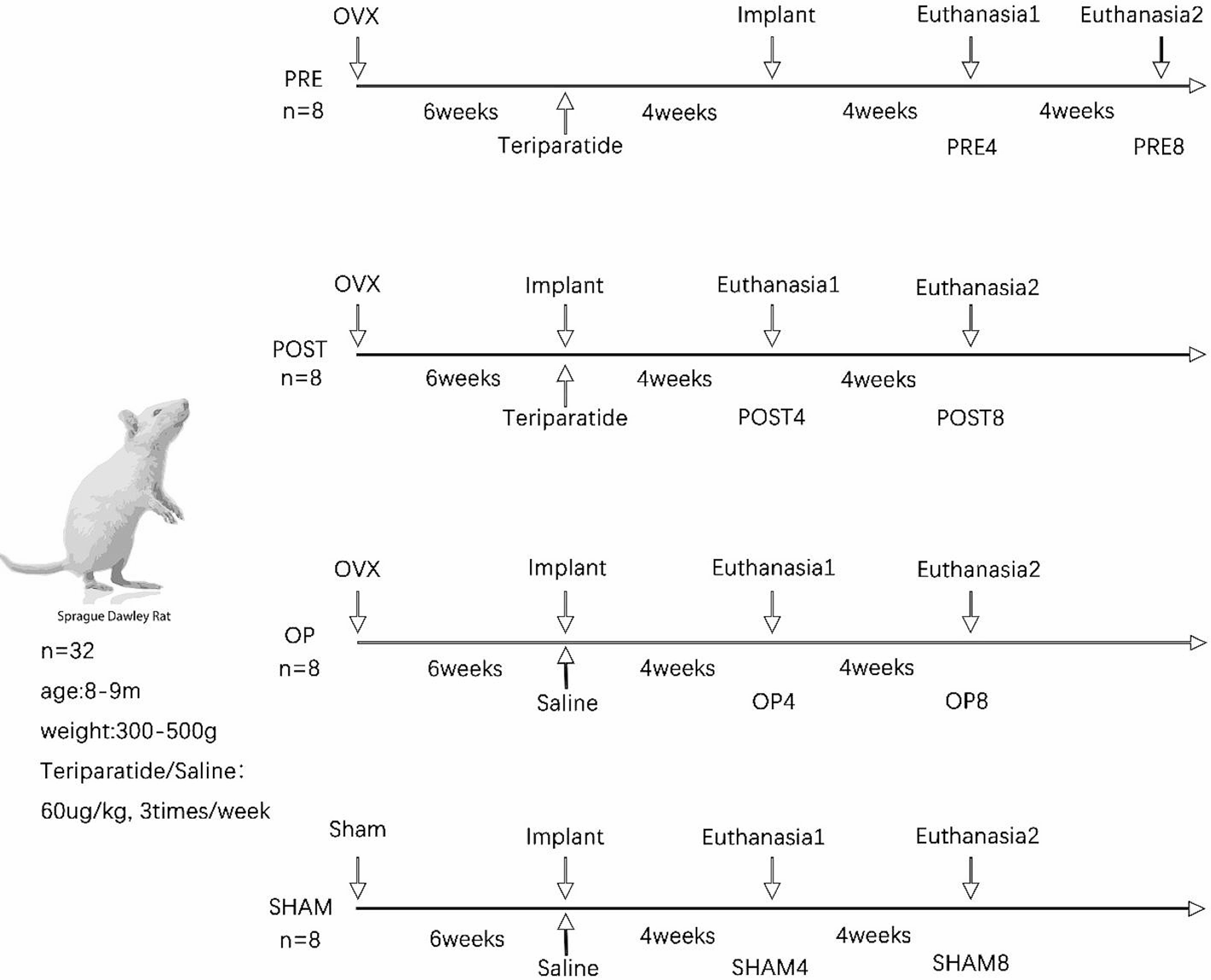
As shown in Fig. 1, a total of 32 rats were randomly divided into 4 major groups: PRE, POST, OP and SHAM. After bilateral ovariectomy (OVX) or sham surgery, rats were fed with maintain feed which containing 1.4% Ca, 0.8% P and water for 42 days to induce osteoporosis according to a previous research [14]. In PRE group, the administration of teriparatide (XINFUTAI, Shenzhen, Guangdong, China) commenced at 4 weeks prior to the implantation. In POST group, the administration started with the implantation. As a blank control group, rats in OP group received an injection of normal saline (NS) after implantation. And in SHAM group, the implants were placed 42 days after undergoing a sham surgery and then NS was injected. Half of rats were euthanized at 4 or 8 weeks after implantation surgery and four major groups were divided into eight subgroups (n = 4). Rats in PRE4-SHAM4 subgroups were euthanized at 4 weeks, and rats in PRE8-SHAM8 subgroups were euthanized at 8 weeks.
Previous research suggests that daily PTH treatment may raise the concentration of the bone resorption marker [15], whereas teriparatide administered three times a week promotes bone formation on the endocortical surface without improving bone turnover markers, resulting in an increase in cortical thickness and bone density [16]. Consequently, we administered 60 µg/kg of teriparatide or NS subcutaneously three times a week, which might promote implant osseointegration effectively.
Fig. 1
Experimental design is shown above. A total of 32 female SD rats (8–9 months old) were divided into 4 major groups: PRE, POST, OP and SHAM. Titanium implants were placed in the bilateral proximal tibiae. Rats in each group (n = 8) were euthanized at 4 weeks (n = 4) or 8 weeks (n = 4) after implantation and the four major groups were divided into eight subgroups (PRE4, PRE8, POST4, POST8, OP4, OP8, SHAM4 and SHAM8). After euthanasia, 8 tibiae were collected in each subgroup
Validation of osteoporosisThe serum concentration of osteocalcin (OCN) and type I collagen C-terminal telopeptide (CTX-I) were measured to verify the successful establishment of osteoporosis models [17, 18]. Blood samples were randomly collected from five osteoporosis rats (in PRE, POST and OP groups) and five normal rats (in SHAM group), and centrifuged at 1000 x g for 20 min using a high-speed freeze centrifuge (Thermo Fisher, Waltham, MA, USA). The serum concentrations of OCN and CTX-I was tested using the OCN Elisa serum test kit (Sabbiotech, College Park, MD, USA) and the CTX-I Elisa protein test kit (Cloud-Clone, Wuhan, Hubei, China) separately. The absorption intensity of each well at 450 nm was analyzed using a multimode microplate detector (PerkinElmer, Waltham, MA, USA) to establish a standard sample function curve, and the concentrations of OCN and CTX-I were calculated based on this curve.
Implantation procedureSixty-four grade IV pure titanium screws (BioMaterials, Beijing, China), with 2 mm in diameter and 5 mm in length, were chosen to simulate the dental implants. The self-tapping implant had four continuous threads, and the special design of the tip facilitated the removal of bone debris during the implantation process.
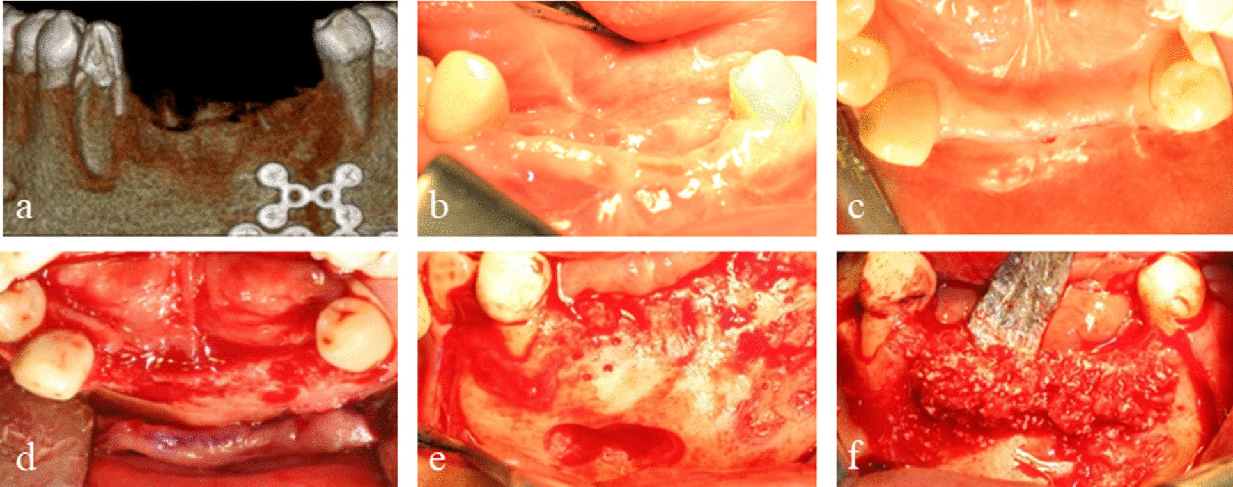
The abdominal injection of Zoletil@50 (Aladdin, Shanghai, China) was used for anesthesia with the dosage of 307 mg/kg. After anesthesia, the skin was incised at the proximal tibia to expose the underlying tibial surface, and each rat then received an implant in the right and left tibial metaphysis respectively. Using a bone drill (DISHENGDE, Jinhua, Zhejiang, China), we bored a pilot hole 2–3 mm away from the tibial growth plate and perpendicular to the medial surface of tibial metaphyseal under NS flow. The hole measured 1 mm in diameter and 3 mm in length. Then with the help of a torque screwdriver (AIGU, Dongguan, Guangdong, China), the implant was progressively inserted in the pilot hole until the point where every thread was fully inside the bone. The maximal torque encountered in PRE, OP and SHAM groups were recorded during the insertion process, indicating the insertion torque. Finally, the skin and mucous membranes were sutured to close the incision site.
The animals in PRE and POST groups were continually treated with teriparatide after implant operation, while rats in OP and SHAM groups were injected NS. Rats were euthanized by excessive CO2 inhalation following euthanasia principles. Eight tibiae from four rats in each subgroup were collected 4 or 8 weeks after implantation. Then the specimens were fixed in a 10% formalin neutrophilic fixation (Yulu, Nanchang, Jiangxi, China) for one week. Three tibiae were randomly collected from different rats preparing for undecalcified sections analysis, while the rest five tibiae were subjected to micro-CT scanning and torque test.
Micro-CT analysisAfter fixation, 5 tibiae were scanned by micro-CT (Bruker Skyscan 1172, Aatselaar, Antuérpia, Belgium), with parameters included a voltage of 90 kV, a current of 80 µA, an exposure time of 1050 ms, a rotation angle of 180°, a rotational step length of 0.4°, a filter of 0.5 mm AL, and a scan resolution of 13.75 μm. Then the images were reconstructed three dimensionally and analyzed using the Data Viewer software (SkyScan, version 1.5.6.2) and the CTAn software (SkyScan, version 1.18.8.0).
The Region of interest (ROI) was defined as the area around implants and the bone tissues (Fig. 2a, 2.5 mm in diameter and 1 mm in height). The CTAn software was used to quantify various metrics within the ROI, including bone volume percentage (BV/TV), trabecular thickness (Tb.Th), trabecular number (Tb.N) and trabecular separation (Tb.Sp). For the CTAn analysis, a grayscale option ranging from 40 to 80 was selected.
Biomechanical testingImmediately after micro-CT scanning, the five tibiae with implants in each subgroup were fixed by a torque horizontal clamp and connected to a digital torque meter (HBO, Beijing, China), in anticipation of the torque test. A screwdriver was used to remove the implant from the bone and the maximal removal torque needed to remove the implant was recorded during this process.
Undecalcificated bone-implant sectionsAfter fixation, the three tibiae underwent a dehydration process using a graded series of ethanol concentrations: 70%, 80%, 90%, and finally 100%. Following dehydration, the tibiae were soaked in a solution consisting of 50% ethanol and 50% light-curing resin (Technovit 7200VLC, Hanau, Germany) for a period of 2 weeks. Subsequently, the tibiae were embedded in resin (Technovit 7200VLC, Hanau, Germany) and photopolymerized for 10 h. A special fixed glue (Tech 4000, Hanau, Germany) was used to fix the specimens block to the cover glass, and the EXAKT hard tissue cutting system (EXAKT 300CP, Apparatebau, Gmbh, Hamburg, Germany) was used to cut vertically along the long axis of the implant at a thickness of 100 μm, and grinded in the polishing machine (EXAKT 400CS, Apparatebau, Gmbh, Hamburg, Germany). Then the hard tissue sections were dyed using methylene-blue/acid fuchsin stain to visualize trabecular bone growth around the implants. Finally, the multi-mode intelligent living cells imaging analysis system (MICA) (Leica, Wetzlar, Hessian, Germany) was used to observe the growth patterns of trabeculae. Thread’s BIC was defined as the square of the newborn trabeculae in the thread pit (Fig. 2b) divided by the overall square of the thread pit. And the overall BIC of the section was defined as the average of the two consecutive threads’ BIC values in the cancellous bone area. After locally enlarged images of two consecutive threads (in cancellous bone area) were transported to software ImageJ (Image Processing and Analysis Software, Bethesda, MD, USA), the values of overall BIC of different subgroups were calculated and compared.
Statistical analysisThe SPSS 24.0 software (SPSS, Chicago, IL, USA) was used throughout for statistical analysis. One-way analysis of variance (one-way ANOVA) was used to compare the radiological, biomechanical and histological parameters among groups or subgroups. When ANOVA indicated significant differences, post-hoc multiple comparisons were conducted using either the Bonferroni method (in cases of homogeneity of variance) or the Tamheini test (in cases of heterogeneity of variance). To evaluate differences in serum concentrations of OCN and CTX-I between osteoporotic and sham rats, and to analyze the difference of parameters in PRE4 and POST8, a two-independent sample t-test was employed. A significance level of p < 0.05 was used to determine statistical significance. All values are shown as the average and the standard difference.
Fig. 2
(a) The area surrounding the implant, measuring 2.5 mm in diameter and 1 mm in height, was designated as the region of interest. (b) The trabeculae within the orange dashed line were identified as newborn trabeculae in thread pits (marked by white arrows). Thread’s BIC was defined as the square of the newborn trabeculae in the thread pit divided by the overall square of the thread pit
留言 (0)