
記住我


PWS represents a complex challenge for spine surgeons given the high prevalence [6,7,8,9] of scoliosis. Scoliosis, alongside with muscle weakness, muscle hypotonia and obesity, is considered an important risk factor for cardiopulmonary impairment due to the chest deformities associated with PWS [18]. Therefore, a prompt diagnosis of spinal deformities is mandatory in these patients, in order to plan the most appropriate therapeutic interventions. For what may concern scoliosis prevalence in patients affected by PWS, the available literature reports a variable rate between 15 and 86% [7,8,9]. In our cohort of PWS patients affected by scoliosis, most of the diagnoses were made at a juvenile age, in contrast with the trend of adolescent patients reported in the literature [10]. In this current work, we divided subjects with scoliosis according to the degree of severity, as established by the SRS [17], showing a major prevalence of moderate and severe forms of scoliosis with a Cobb angle included between 20 and 40° and above 40°. Few studies are available in the literature regarding the partition of patients with PWS according to the degree of severity of scoliosis divulgated by the SRS, and therefore, a more extensive evaluation of this matter should be considered in the future [19]. In our study, we had similar data to the work of Crinò et al. [19] concerning the gender distribution of patients with scoliosis affected by PWS, with a prevalence of female patients. Apart from the overall coronal plane curve assessment, there are few differences between idiopathic scoliosis and the PWS type. The idiopathic deformity is characterized by a thoracic lordoscoliosis, while in PWS most curves are kyphoscoliotic [14]. In our experience, most of the patients we evaluated had normal kyphosis curve values, while only a few numbers had a kyphoscoliotic asset.
Conservative treatment, with different types of braces, for scoliosis in PWS patients seems safer than spinal fusion surgery in this specific population due to high rate of associated complications, even though surgery remains the gold standard choice in this particular cohort of scoliotic patients. There is a lack of literature regarding brace treatment for PWS patients, and this therapeutic option still remains controversial. A difficult brace molding due to the obesity of these patients, the necessity of frequent remodeling due to weight fluctuations and poor compliance of these patients, considering the intellectual deficits associated, are all risk factors for inadequate results (Fig. 4). In our experience, conservative treatment showed poor results with six patients, among the 11 treated with a brace, presenting a coronal progression of the curve, maintaining however a good sagittal balance as almost all patients had good kyphotic curve values. Compliance to treatment remains the main challenge for spine surgeons, and we believe that a more intensive brace program, with a more frequent follow-up and strict collaboration with the family, should be considered for these patients in order to prevent the necessity for a corrective surgery, with all the associated risks. Surgery remains the gold standard for patients affected by PWS and scoliosis. In our experience, we reserved surgery for patients with scoliotic curves above 45° Cobb angles at diagnosis with similar indications as patients affected by idiopathic scoliosis, with posterior hybrid arthrodesis in two cases and growing rods positioning in two patients where diagnosis was made at a younger age. Surgery in patients affected by PWS represent a difficult challenge because of the frequently associated complications. In our experience, complication rate was high, with a prevalence of 75% of the operated cases. In our series, one patient (25%) developed a PJK after 3 years from index surgery that required reoperation. This complication could be related to several features of PWS, such as muscle weakness, ligamentous laxity and osteoporosis, and therefore, an accurate planning before operative treatment should always be considered by expert surgeons. Preoperative lateral radiographs must be carefully analyzed to choose the right upper instrumented level and an accurate contouring of the rods, and the preservation of the posterior ligaments in the upper vertebrae has to be of primary importance when surgery is planned. Spinal cord injury represents the most fearsome complication during spine surgery, and in our case, one patient developed paraplegia and signs of medullary suffering at MRI after pedicle screws positioning that required removal of the instrumentation after 24 h after first surgery without any neurological recovery. We recommend performing a total spine MRI examination before surgery and spinal cord monitoring in order to identify any spinal abnormalities and check neurological status during the surgical procedures. Wound infection and deep surgical site infection were seen in one patient after 10 years from index surgery which required a DAIR procedure with instrumentation removal, after a TC scan demonstrated a good arthrodesis site. Deep infection in PWS patients is a dangerous complication due to the high risk of repeated surgeries and the high anesthesiologic risks related to these clinically complex patients; therefore, a strict control of a correct wound healing, a prompt evaluation by the infectious disease specialist at any sign of infections, in order to start the correct antibiotic therapy, even after years from index surgery must be mandatory in the follow-up of these patients. Rod breakage was seen in one case, requiring revision surgery. This could be related to obesity and hypotonia with increased loads on the instrumentation. Correction of the scoliotic deformity after surgery was satisfactory in two patients where we obtained a halving of the curve while the remaining two patients did not have any improvement of the scoliotic curves, on the contrary a worsening, due to the occurrence of the previously mentioned complications; thus, prevention of complications must be of main importance for the spine surgeon in order to avoid the loss of correction achieved with surgery.
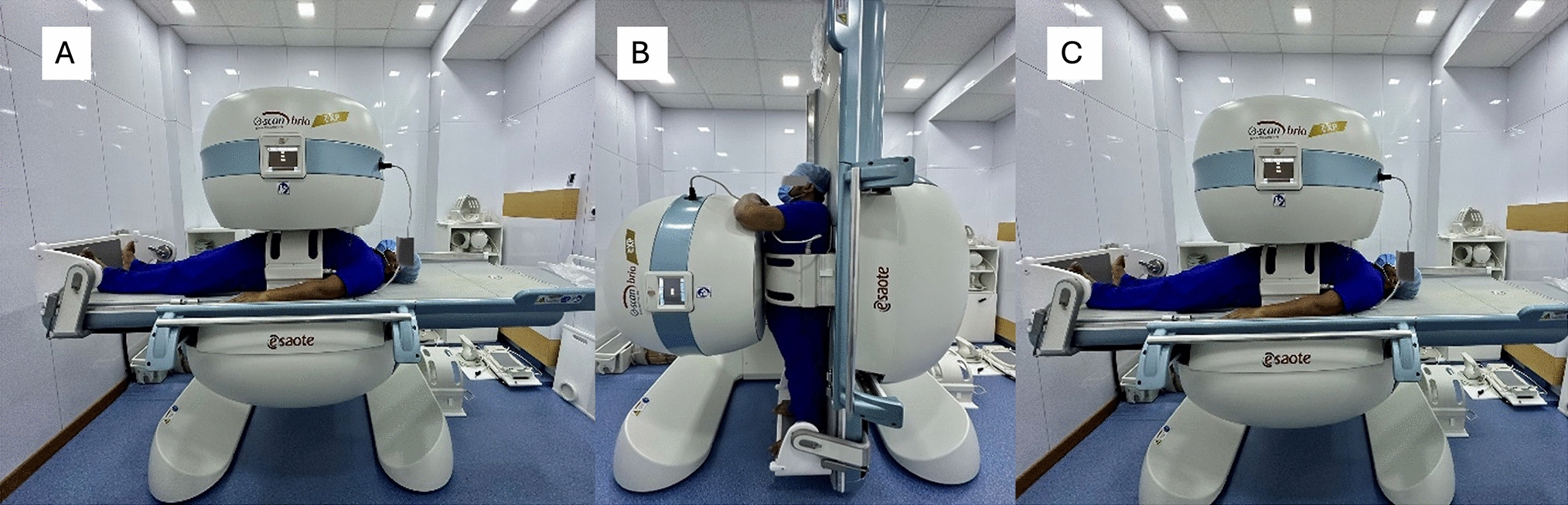
Fig. 4
Brace molding is difficult in patients affected by Prader–Willi Syndrome due to the severe obesity associated with this condition
留言 (0)