Patient cohort
The study was conducted in accordance to local ethical guidelines. Patient consent has been obtained from each patient after full explanation of the purpose and nature of the all procedures used, and approval by the ethical board was granted (number 21-1025_1, institutional review board of the University Hospital of Cologne).
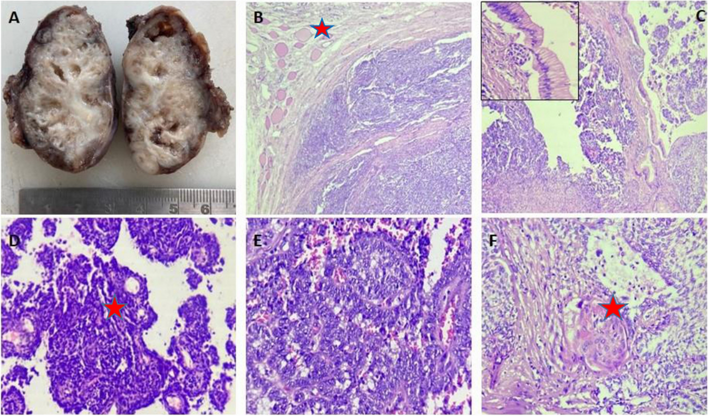
Twenty-two specimens of papillary thyroid carcinoma with a tumor size ≤ 1 cm (traditionally classified as papillary thyroid microcarcinoma) and corresponding lymph node metastases diagnosed between 2012 and 2022 were retrieved from the surgical pathological archives. No prophylactic lymphadenectomy is performed in case of tumors ≤ 1 cm at our institution, according to the current German Guidelines. Accordingly, all patients had either preoperative sonographic or biopsy diagnosis of suspicious lymph nodes. In four patients papillary carcinoma lymph node metastases were detected on lymph node dissection for head and neck squamous cell carcinoma, followed by subsequent thyroidectomy. Lymph node metastasis removed later than 6 months after diagnosis were considered metachronous and were not included in the present study, as well as all with concomitant carcinoma > 1 cm.
Histopathological evaluation
Whole slide images of thyroid resection specimens and lymph node dissection samples were evaluated by two experienced pathologists (AMS, MLE). For establishment of diagnosis of lymph node metastasis of papillary thyroid carcinoma TTF1 and PAX8 immunohistochemistry was performed in routine diagnostic workflow. Positivity of TTF1 and PAX8, indicating thyroid origin along with morphologic features, lead to the diagnosis of lymph node metastasis from papillary thyroid carcinoma. The concomitant lymph node metastasis from head and neck squamous cell carcinoma were negative for these two markers.
Both primary tumors and lymph node metastases were examined with regard to presence of stromal desmoplasia, histologic variant of papillary thyroid carcinoma (infiltrative follicular, classic, and others), presence of inflammation, psammoma bodies, and extracapsular extension. Lymphovascular invasion in primary tumors was also examined, as well as the presence of underlying Hashimoto’s thyroiditis.
Immunohistochemical assessment
Whole slide sections of primary tumor and the largest lymph node metastasis were used to perform immunohistochemistry for p53 (clone: DO-7. 1:1800, Dako / CE), Ki-67 (clone: SP6. 1:100. Cellmarque / CE) and BRAF V600E (clone: VE1, ready to use, Roche / CE) following the manufacturer’s protocol. Detection of immunolabeling was performed using anti-mouse or anti-rabbit horseradish peroxidase–conjugated secondary antibodies and developed using 3,3′-diaminobenzidine.
For BRAF V600E diffuse cytoplasmic staining of tumor cells was considered positive and used as a surrogate marker for an underlying mutation. P53 was considered abnormal, if a strong nuclear expression in a majority of nuclei was present, a strong cytoplasmic staining or the absence of staining. A varying staining pattern across tumor nuclei was considered wild type staining pattern.
For evaluation of proliferation index via Ki-67, whole slide images were digitized with a Hamamatsu S360 scanner. Representative tumor areas were annotated in QuPath 0.3.2. Positive cells were detected running the built-in positive cell detection command using following parameters: detection image optical density sum, requested pixel size 1 µm; nucleus parameters: background radius 8 µm, median filter radius 0 µm, sigma 2 µm, minimum area 12 µm2, maximum area 400 µm2; intensity parameters: threshold 0.1, max background intensity 2; and cell parameters: cell expansion 5 µm, cell nucleus included.
TERT RNAscope®
The largest lymph node metastasis of each case was selected for analysis. RNAscope® detection for TERT was performed manually using the Hs-TERT-01 probe. Staining was done a per manufacturer’s protocol (ACDBio). Appropriate accompanying positive and negative controls (universal negative control dapB probe) were used to evaluate the staining procedure. After pre-treatment, probe hybridization and detection, the slides were counterstained with 50% hematoxylin, dehydrated, and mounted.
Expression quantification was carried out as described by Momeni-Boroujeni et al. [13] TERT signals in 100 cells were counted in hotspot regions.


留言 (0)