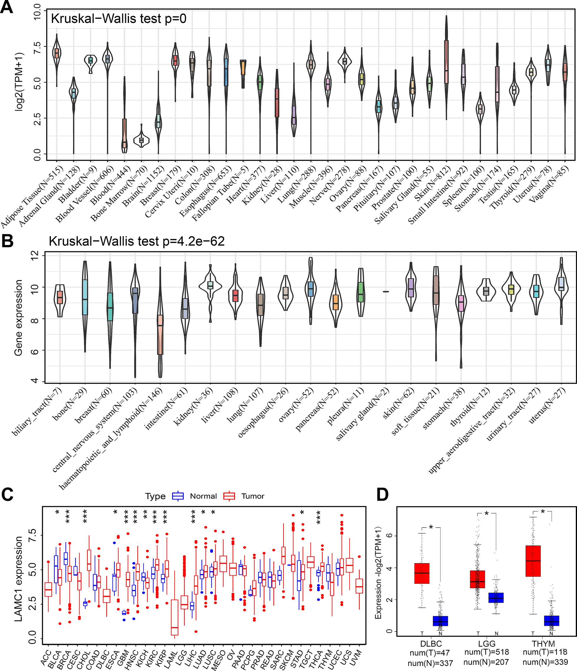
SKOV3 (ATCC, HTB-77), NIH/3T3 (ATCC, CRL-1658), OAW42 (Sigma, 85073102), and ID8 (Merck, SCC145) cell lines were purchased and cultured in mediums recommended by the manufacturers. Breast cancer-associated fibroblasts (CAF) were generated by Prof. Orimo, Juntendo University, Japan [26]. To prepare stable monoclonal cells, COMP expressing (COMP) and an empty pcDNA3 vector (mock) were transfected to CAF and NIH/3T3 cells (9 × 10⁵) using lipofectamine 3000 transfection reagent (Cat: L300008, Invitrogen, USA). Cells were then treated with 0.7 mg/ml G418. Stable colonies were then picked up and cultured for a month under G418 exposure. ELISA further confirmed the secretion of COMP by COMP-expressing CAFs. All cell lines were maintained at 37 °C in a humidified incubator with 5% CO2 and were checked monthly for mycoplasma contamination using the VenorGeM classic kit (Minerva Biolabs). The passage number of cells in all experiments was below 10.
Cell proliferation assay
In brief, 3 × 10⁴ cells were seeded in 96-well cell-binding black plates (Cat: 3340, Corning, USA) for 24 h. Cells were then treated with increasing concentrations of COMP for another 24 h. After that, cells were incubated with the detection reagent for one h at 37 °C, and fluorescence intensities were then measured by Cytation 5 Cell Imaging Multimode Reader (BioTek, USA) at 480/535 nm. The percentage of cell survival was calculated and compared to untreated cells as a control.
A coculture assay was performed to evaluate the effect of secreted COMP by COMP-expressing CAFs on the proliferation of SKOV3 cells. In brief, SKOV3 cells were first stained using a CellTrace CFSE cell proliferation kit (Cat: C34554, Invitrogen), according to the manufacturer’s instructions. An equal number (1 × 10⁵) of CFSE-labeled SKOV3 cells and COMP-expressing CAFs or the counterparts mock control CAFs were mixed and seeded in 24-well plates. Unstained SKOV3, COMP-expressing CAFs and the mock control CAFs were used as negative controls to gate out CFSE negative cells. Cells were collected, resuspended in PBS at each time point and analyzed by flow cytometry (Beckman Coulter CytoFLEX). The shift of the CFSE positive peak to the left was evaluated as a measure of the SKOV3 cells proliferation, as CFSE dye was lost with each cell division.
COMP binding assay
SKOV3 and OAW42 cells (3 × 10⁵) were resuspended in FACS binding buffer (10 mM HEPES, 140 mM NaCl, 5 mM KCl, 1 mM MgCl2, 2 mM CaCl2, and 0.02% NaN3, (pH:7.2)) and incubated with increasing concentrations of recombinant COMP for 1 h. Cells were then incubated with an anti-human COMP primary antibody (homemade) and Alexa-flour 488 conjugated secondary antibody (Invitrogen, USA), respectively, for 1 h at 37 °C. Untreated cells were considered as a control. COMP binding to the cells’ surface was finally evaluated by flow cytometer (Beckman Coulter CytoFLEX, CA, USA), and data were analyzed using the FlowJo software (BD Life Sciences).
Migration & invasion assay
SKOV3 (4 × 10⁴) and OAW42 (3 × 10⁴) cells were suspended in a serum-free medium with increasing COMP concentrations and then seeded in 24-well plate transwell inserts (PET, 8 μm, Falcon, USA) for a 24-h migration assay or Biocoat Matrigel invasion chambers (Corning, USA) for a 48-h invasion assay. Untreated cells served as a control, and the medium in the lower chamber was supplemented with 10% FBS as a chemoattractant. After steps of washing with PBS, fixing by 3.7% formaldehyde, and staining the cells with a 0.5% crystal violet solution, respectively, the chamber's inner cells were removed using cotton swabs.
In the co-culture assay, mock or COMP-expressing 3T3 or CAF cells (1.5 × 10⁵) were seeded in 24-well plates with 10% FBS and incubated for 3 days. Subsequently, ID8 or ovarian cancer cells (SKOV3 and OAW42) (3 × 10⁴) were seeded in the upper chamber of transwell inserts for 24 h. Staining was performed as described earlier. Images from different areas of each chamber were captured at 4 X or 10 X magnification, using the EVOS XL Core Cell Imaging System (Thermo Scientific, MA, USA) and analyzed with ImageJ software.
ALDH activity assay
SKOV3 and OAW42 cells were seeded in 6-well plates and treated with 20 μg/ml COMP or PBS as a control for 48 h. Cells were then trypsinized, and 5 × 10⁵ cells per sample were collected to determine the ALDH activity using the ALDEFLUOR assay kit (STEMCELL Technologies, Canada) according to the manufacturer’s instructions. DEAB, a selective inhibitor of ALDH, was used as a control of background fluorescence. Cells were analyzed by flow cytometer (Beckman Coulter CytoFLEX, CA, USA), and data analysis was performed using the FlowJo software (BD Life Sciences).
Apoptosis assay
Cells (1 × 10⁵) were initially seeded in 6-well plates for 24 h, followed by treatment with COMP (20 μg/ml), BSA (20 μg/ml) (negative control), and cisplatin (positive apoptosis inducer control) either individually or in combination with COMP for 48 h. SKOV3 and OAW42 cells were treated with 10 μM and 20 μM of cisplatin, respectively. Subsequently, the cells were trypsinized and washed with FACS binding buffer (10 mM HEPES, 140 mM NaCl, 5 mM KCl, 1 mM MgCl2, 2 mM CaCl2, and 0.02% NaN3, (pH:7.2)). Incubation with 5 μl Annexin V and 0.4 μl Zombie aqua was carried out for 30 min at room temperature. After washing with FACS binding buffer, the cells were subjected to flow cytometry (Beckman Coulter CytoFLEX, CA, USA), and data was analyzed using the FlowJo software (BD Life Sciences).
RNA isolation, cDNA synthesis, and RT-qPCR
For recombinant COMP treatment, 80% confluent cells in 6-well plates were treated with recombinant COMP (20 μg and 50 μg/ml) or BSA (50 μg/ml) as a control for 24 h. For cell treatment with TGFβ proteins, 80% confluent cells in 12-well plates were incubated with 10 ng/ml TGFβ1, TGFβ2, TGFβ3, BSA, and the same volume of PBS as a control for 48 h. Then, the medium was refreshed, and cells were incubated for 48 h again. Subsequently, total RNA was isolated from cells using RNeasy Plus Mini Kit (Qiagen, Germany), according to the manufacturer’s instructions. The purity of isolated RNA was checked by measuring the 260/280 absorbance ratio via Nanodrop. The RNA integrity number (RIN) of isolated RNA samples was also checked using an Agilent 2100 Bioanalyzer instrument with an Agilent RNA 6000 Nano kit (Agilent Technologies). Total RNA samples were then quantified and used for cDNA synthesis immediately. Otherwise, they were stored at −80 °C. Total RNA (500 ng) was reverse transcribed to cDNA using SuperScript™ IV First Strand Synthesis System (Cat: 18091050, Thermo Scientific, USA), employing Oligo (dT)20, according to the manufacturer’s instructions and stored at −20 °C. RT- qPCR was performed using TaqMan probes (Thermo Scientific, MA, USA) for each gene. GAPDH and HPRT1 were considered internal controls, and relative mRNA expression for each gene was calculated using 2−ΔΔCt or 2−ΔCt as indicated in the figure legend. The array of epithelial to mesenchymal transition genes was performed using PrimePCR Custom Plate (Cat: 10034487, Bio-Rad) according to the manufacturer’s protocol. Results were analyzed using GAPDH as an internal control, and were presented in volcano plots.
Western blotting
Cell lysates were prepared using cold 1X radioimmunoprecipitation assay lysis buffer (RIPA) (10 mM Tris–HCl (pH: 7.2), 150 mM NaCl, 0.1% SDS, 1% Triton X-100, 1% deoxycholate) supplemented with 1% (v/v) Halt Protease and Phosphatase inhibitor Cocktail (100X) (Thermo Scientific, USA). Protein concentrations were quantified using the Pierce BCA Protein Assay Kit (Thermo Scientific, USA). After protein separation on SDS-PAGE gels and transferring to PVDF membranes, blots were incubated with respective primary antibodies, and HRP-conjugated secondary antibodies. The used antibodies are listed in Additional file 1: Table S1. Blots were then visualized under Chemidoc (Bio-Rad, USA) using Immobilon Western Chemiluminescent HRP substrate (Millipore, MA, USA). The bands’ intensities were measured by Image Lab software (Bio-Rad) and normalized to the expression of β-tubulin or GAPDH as a control.
Proximity ligation assay
In brief, 2 × 10⁴ cells were seeded in a 12-well removable chamber (ibidi, Germany) for 24 h, and then they were treated with 0.25 mg/ml recombinant COMP. After 24 h, cells were fixed with 4% paraformaldehyde, permeabilized with 0.1% triton-X100, blocked with 3% BSA blocking buffer, and then incubated with primary antibodies for 1 h at room temperature, respectively. Respective isotype control antibodies were included as a negative control. Subsequently, incubation with the goat-mouse probes and three steps of enzymatic reactions were performed based on the NaveniFlex GM instructions. Duolink in situ mounting medium with DAPI (Sigma, USA) was used for nuclear staining. At least three images from different areas of each chamber were captured at 63 X magnification under a Confocal microscope (Zeiss LSM800, Germany). Images were analyzed by the ImageJ software to count the spots per cell values.
Tumorsphere formation assay
Cells (5 × 10⁴) were suspended in the Mammocult medium (STEMCELL Technologies, Canada) supplemented with 4 μg/ml heparin (STEMCELL Technologies, Canada) and 0.48 μg/ml hydrocortisone (STEMCELL Technologies, Canada) to prepare a serum-free single cell suspension. Then, cells were seeded in 6-well Ultra-low binding plates (Corning, USA) with increasing concentrations of COMP and incubated for a week at 37 °C in a humidified incubator with 5% CO2. For the NOTCH inhibition studies, cells were incubated with either COMP alone (20 μg/ml) or in combination with NOTCH inhibitors, DAPT (1 μM, Sigma-Aldrich, USA), and anti-Jagged1 antibody (2 μg/ml, R&D systems, USA). A minimum of 10 images per well were captured using the EVOS XL Core Cell Imaging System (Thermo Scientific, MA, USA). The length of at least 10 tumor spheres per well was measured using the ImageJ software.
Luciferase reporter assay
In brief, 1.4 × 10⁵ SKOV3 cells were seeded in a 24-well plate and incubated overnight to reach 70% confluency on the day of transfection. Cells were then transfected with the following plasmids using lipofectamine 3000 (Invitrogen): the positive control: 2488 ng M50 Super 8 × TOPFlash [27], 2 ng pIS2 [28], and 10 ng pcDNA3-S33Y β-catenin [29]. The negative control: 2498 ng M51 Super 8 × FOPFlash (TOPFlash mutant) [27] and 2 ng pIS2. For the detection of β-catenin activation: 2498 ng M50 Super 8 × TOPFlash, 2 ng pIS2. The next day culture media was replaced, cells were treated with BSA (50 μg/ml), BSA in combination with DAPT (1 μM), COMP (50 μg/ml), and COMP in combination with DAPT (1 μM), and incubated for 24 h. Untreated cells were included as a control. Luciferase activity was detected using a Dual-luciferase reporter assay system (Promega) according to the manufacturer’s instructions. Luminescence was measured via Cytation 5 Cell Imaging Multimode Reader.
Plasmid pIS2 was a gift from David Bartel (Addgene plasmid # 12177), M50 Super 8 × TOPFlash and M51 Super 8xFOPFlash were a gift from Randall Moon (Addgene plasmid, # 12456 and #12457), and pcDNA3-S33Y Beta-catenin was a gift from Eric Fearon (Addgene plasmid # 19286).
Recombinant COMP purification
In brief, Freestyle 293-F cells (ThermoFisher Scientific, USA) were cultured in FreeStyle 293 Expression Medium (Thermo Scientific, MA USA) supplemented with 1% (v/v) penicillin–streptomycin. Cells were transfected with histidine tagged-COMP pCEP4 plasmid using Freestyle Max transfection reagent (Thermo Scientific, USA). Transfected cells were cultured for 10 days, and the supernatant was collected every 2 days and stored at −20 °C. A Ni–NTA affinity column (Ni–NTA Superflow, Qiagen) loaded in ÄKTAprime plus machine (GE Healthcare) was used for COMP purification. Fractions containing COMP protein were subsequently pooled and dialyzed against 1X PBS (pH = 7.4) and concentrated by 10 kDa ultra centrifugal filter units (Millipore, MA, USA). The purified recombinant COMP protein was finally stored at −80 °C for further experiments.
Statistical analysis
SPSS software (version 29) was used for survival analyses. GraphPad Prism was used for all other statistical analyses. The data were represented as mean ± standard deviation (SD). To calculate the p-value, the student’s T-test, one-way ANOVA, and two-way ANOVA were utilized. P value < 0.05 was considered statistically significant.


留言 (0)