
記住我


West Nile virus (WNV) is a single-stranded ribonucleic acid (RNA) flavivirus transmitted mainly by the bite of infected mosquito species of the genus Culex and Aedes spp [1].
Human infections show a seasonal trend, with most cases reported between July and October, with peaks in late August [2]. Nearly 80% of WNV infections in humans are asymptomatic, up to 20% of infections present with a mild flu-like syndrome, and less than 1% of WNV infections develop a neuroinvasive disorder. When the central nervous system is involved, the mortality rate is about 10% [3].
Multifocal chorioretinitis is a common ocular manifestation of WNV infection with neuroinvasive disease, which is frequently asymptomatic and self-limiting [4] However, involvement of the choroid and retina is rare in patients with asymptomatic systemic disease [5,6,7].
The present study aims to describe a case of bilateral multifocal chorioretinitis as the only presentation of acute WNV infection.
Case presentationA 78-year-old Caucasian woman was admitted to the ophthalmic emergency room of Sant’Orsola-Malpighi Hospital (Bologna, Italy) because she noticed blurry vision in both eyes. The patient lived in a small rural town in the district of Bologna in the Emilia Romagna region. She was not under medical treatment. She did not have diabetes mellitus and she was immunocompetent. In addition, her vital signs were normal, and she did not report fever, fatigue, or neurological symptoms in the last few days, except for the onset of blurred vision in both eyes, which started in the last week. Best-corrected visual acuity (BCVA) was 20/30 in both eyes, there were no relative afferent pupillary defects (RAPD), intraocular pressure was normal, and there was mild flare in the anterior chamber in both eyes. Fundoscopy examination revealed bilateral vitreitis (grade 1 + , according to NIH grading system [8]) and bilateral swelling of the optic disc. Computed Tomography (CT) scan of the brain was immediately performed, revealing no pathological findings. In accordance with the clinical scenario, a standard workup for uveitis was immediately requested. Waiting for the laboratory test results, empirical therapy with topical dexamethasone in both eyes twice daily was started. During the next few days, the diagnostic workup was completed. Visual field test was performed and revealed a mild physiological blind spot enlargement on both eyes; spectral-domain optical coherence tomography (SD-OCT) (Spectralis OCT, Heidelberg Engineering, Germany) showed the presence of oval hyperreflective deposits on the retinal surface in the left eye, focal alterations in the retinal pigment epithelium (RPE) and the ellipsoid zone (EZ), and granular hyperreflective specks located predominantly within the outer and inner nuclear layers in both eyes (Figs. 1A and B). Fluorescein angiography (FA) showed the presence of hyperfluorescent lesions in the late phase with a linear distribution, corresponding to the hypocyanescent spots on indocyanine angiography (ICGA) (Figs. 1C and D). FA was more sensitive in detecting lesions than ICGA. In both eyes, the OCT scan at the level of the lesion showed the presence of alterations in the RPE and EZ (Figs. 2A and B). Optical Coherence Tomography Angiography (OCTA) showed diffuse attenuation of the capillary network in the choriocapillaris at the level of the lesions, with no evidence of alterations at the level of the superficial and deep capillary plexus (Figs. 2C and D). Considering the characteristic linear pattern of the chorioretinal lesions, antibody serology and serum RNA for West Nile virus, Toscana virus, and Usutu virus were also requested.
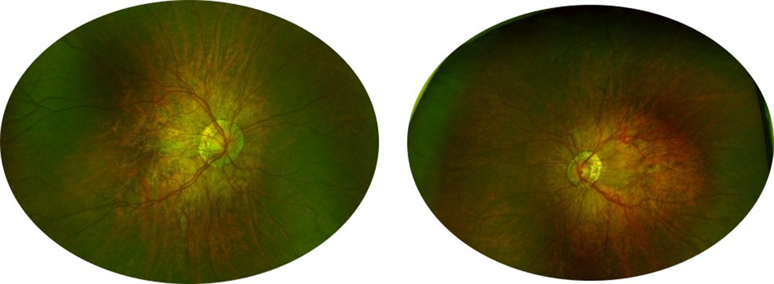
Fig. 1
Multimodal imaging. A In the right eye, fluorescein angiography (FA) showed the presence of hyperfluorescent lesions with a linear distribution, corresponding to the hypocyanescent lesions in indocyanine green angiography (ICGA). B In the left eye, FAF showed the presence of vitreitis, while OCT showed the presence of hyperreflective dots in the vitreous chamber, hyperreflective oval deposits on the retinal surface, focal alteration in the retinal pigmented epithelium (RPE) and the ellipsoid zone (EZ), and granular hyperreflective specks located predominantly in the outer and inner nuclear layers. C In the right eye, fundus autofluorescence (FAF) showed the presence of hyperautofluorescent lesions with a linear distribution, corresponding to focal alteration in the retinal pigmented epithelium (RPE) and the ellipsoid zone (EZ) at the OCT. D In the left eye, FA showed the presence of “target” lesions with a rim of hyperfluorescence and a central spot of hypofluorescence, corresponding to focal alteration of the RPE and the EZ
Fig. 2
Optical Coherence Tomography Angiography (OCTA) at the level of the lesions in the right eye. A OCT en face. B OCTA showed diffuse capillary network attenuation in the choriocapillaris at the level of the lesions. C and D OCT at the level of the lesions
Antibody results were available the following week. The Quantiferon-TB Gold test, Venereal Disease Research Laboratory, and Treponema pallidum particle agglutination test were negative. In addition, IgM antibodies were negative for Herpes simplex virus type-1, Herpes simplex virus type-2, Epstein-Barr virus, Cytomegalovirus, Varicella-zoster virus, and Toxoplasma. However, the antibody serology detected IgM antibodies, IgG antibodies, and RNA for West Nile virus.
Therefore, the patient was immediately contacted and referred to the infectious diseases department, where she underwent magnetic resonance imaging (MRI) of the brain, which ruled out central nervous system involvement.
Hence, asymptomatic West Nile Virus infection with bilateral multifocal chorioretinitis was diagnosed.
One month after the first ophthalmological examination, visual acuity in both eyes was 20/25, there was no involvement of the anterior segment and vitreitis was absent. OCT showed a reduction of the oval hyperreflective deposits on the retinal surface in the left eye. In addition, FA and ICGA scans in both eyes showed reduced hyperfluorescent spots in the late phase (Figs. 3A and B). OCT showed recovery of the EZ and RPE at the level of the lesions (Fig. 3C and D), and OCTA showed a resolution of the attenuation of the capillary network in the choriocapillaris at the level of the lesions. In accordance with the clinical scenario, topical dexamethasone was discontinued. No adverse effects occurred.
Fig. 3
One-month follow-up. A In the right eye, OCT at the level of the lesions showed recovery of the RPE and the EZ. B In the left eye, OCT showed a reduction in the hyperreflective oval deposit on the retinal surface in the left eye
Three months after the initial evaluation, the patient reported an improvement in her visual acuity. Visual acuity was stable at 20/25 in both eyes, and there was no evidence of active chorioretinitis on fundoscopic examination.
留言 (0)