Endoscopic ultrasound-guided fine needle biopsy diagnosis of circumferentially extraluminal mucosa-associated lymphoid tissue lymphoma in the transverse colon: a case report
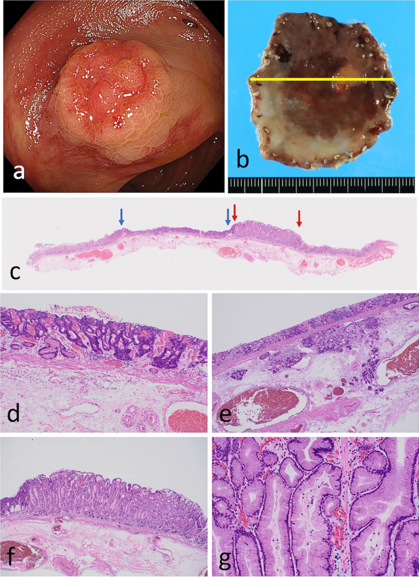
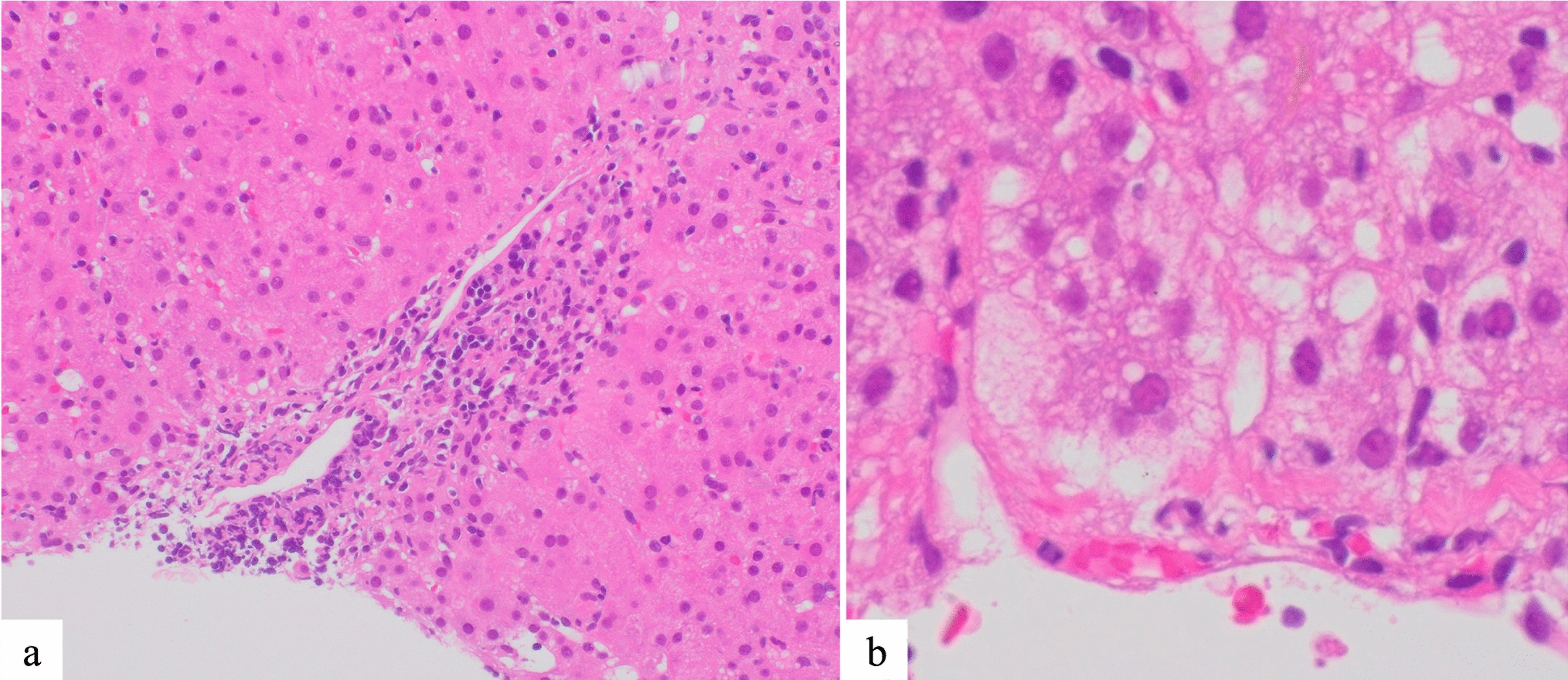
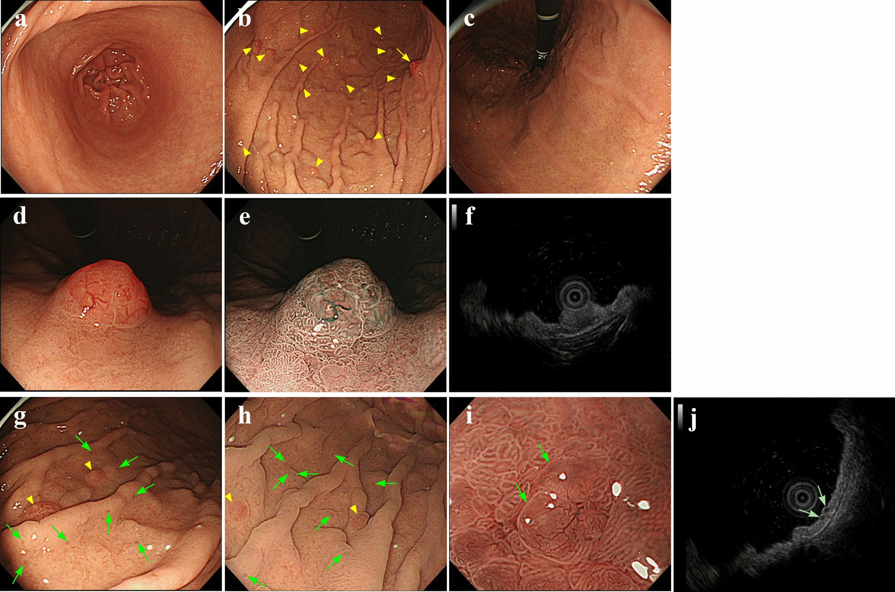
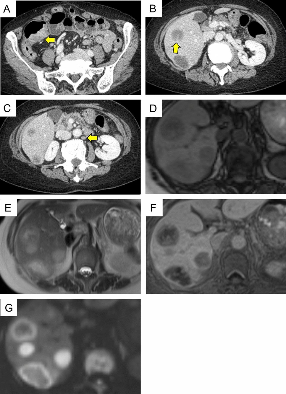
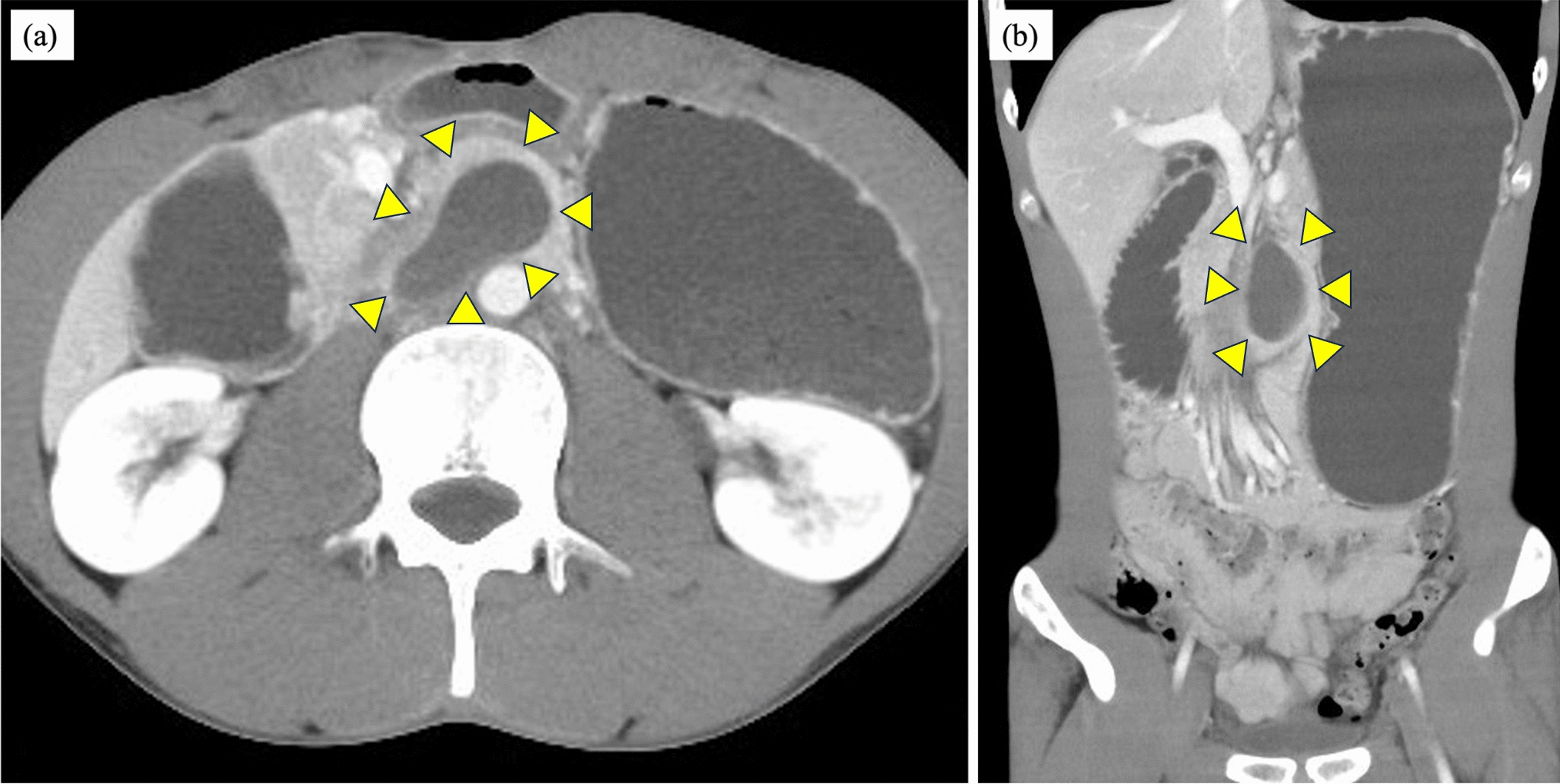
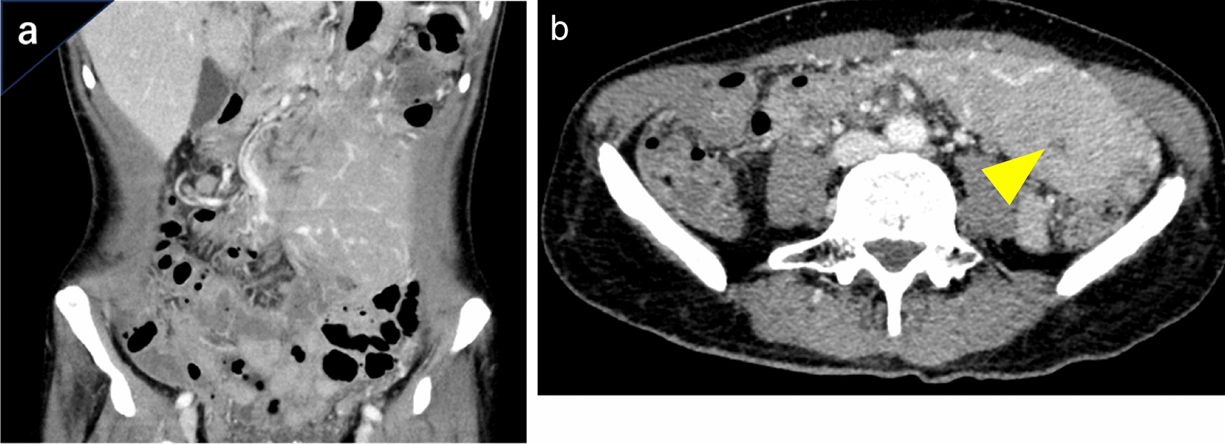
A 61-year-old man present to us with continued abdominal pain without abdominal tenderness for 1 month. Blood testing showed elevated biliary enzymes and inflammation. Contrast-enhanced computed tomography (CT) revealed thickening of the transverse colon with relatively strong enhancement but no bile duct dilatation. Colonoscopy revealed localized edema and granular mucosa in the transverse colon. Fluoroscopic endoscopy exhibited the absence of haustra. Multiple biopsies were performed, but differentiation between mild inflammation and mucosa-associated lymphoid tissue (MALT) lymphoma was inconclusive. To establish a definitive diagnosis, transgastric endoscopic ultrasound-guided fine needle biopsy of the hypoechoic mass was performed. Histopathological analysis exhibited the proliferation of small-sized lymphocytes. Fluorescence in situ hybridization revealed the characteristic API2-MALT1 translocation of MALT lymphoma. We performed liver biopsy to investigate biliary enzyme elevation. Histopathology confirmed lymphocytic infiltration within Glisson’s capsule. Immunohistochemistry showed positive for CD20 and negative for CD3 and CD5, signifying the infiltration of MALT lymphoma in the liver. Based on these findings, we diagnosed MALT lymphoma, Lugano classification Stage IV. We performed bendamustine–rituximab (BR)-combined therapy. After six courses of BR-combined therapy, colonoscopy revealed improvement in the lead pipe sign and CT revealed disappearance of the mass.


留言 (0)