
記住我


Available online 10 April 2024
 Author links open overlay panel, , , , , Highlights•
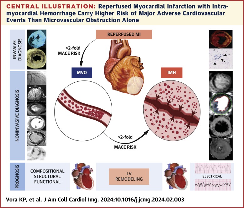
In patients reperfused for acute MI, microvascular injury following reperfusion therapy substantially increases the risk for MACE, with those that develop intramyocardial hemorrhage within the MI zone carrying the greatest risk for MACE in the post-MI period.
•Currently available advanced imaging techniques can noninvasively detect and accurately characterize the type of microvascular injury following reperfusion therapy as those with microvascular obstruction only or microvascular obstruction and intramyocardial hemorrhage.
•Therapies to mitigate reperfusion injury and improve outcomes need to take the type and extent of microvascular injury into account.
AbstractMicrovascular injury immediately following reperfusion therapy in acute myocardial infarction (MI) has emerged as a driving force behind major adverse cardiovascular events in the postinfarction period. Although postmortem investigations and animal models have aided in developing early understanding of microvascular injury following reperfusion, imaging, particularly serial noninvasive imaging, has played a central role in cultivating critical knowledge of progressive damage to the myocardium from the onset of microvascular injury to months and years after in acute MI patients. This review summarizes the pathophysiological features of microvascular injury and downstream consequences, and the contributions noninvasive imaging has imparted in the development of this understanding. It also highlights the interventional trials that aim to mitigate the adverse consequences of microvascular injury based on imaging, identifies potential future directions of investigations to enable improved detection of disease, and demonstrates how imaging stands to play a major role in the development of novel therapies for improved management of acute MI patients.
Section snippetsIschemia: From “Time Is Muscle” to “Time Is Vessel”The notion that “time is muscle,” borne out of the celebrated “wave front” hypothesis of Reimer and Jennings in 1973, captures the need to rapidly abolish ischemia following a sudden coronary obstruction to preserve cardiomyocyte necrosis within the area at risk.3 Closely associated with this phenomenon but not often placed in the same stature is “time is vessel”—ie, with prolonged ischemia not only does the cardiomyocyte necrosis progress, but so does the injury to the microvascular bed
Minimally Invasive Diagnostic Approaches for Characterizing MVO and IMHDespite their strengths, invasive methods typically only provide a single measurement, which limits access to time-dependent changes following acute MI in the same subject. Serial evaluation of experimental and clinical subjects post-MI, including assessment of therapy response, has been made possible by the technical advances in noninvasive imaging. This section outlines the key minimally invasive and noninvasive imaging (coronary angiography, echocardiography, nuclear medicine, and
Investigations Into Prognosis: Imaging-Guided Observational and Interventional Clinical TrialsIn the management of MI, particularly ST-segment elevation myocardial infarction (STEMI), clinical attention is acutely focused on the coronary arteries and timeliness of reperfusion.39 Despite advances in reperfusion therapy, patients surviving AMI are at risk of both near- and long-term complications whose risk can be traced back to the burden of myocardial damage at the time of the acute event. Large cohort studies have reported heart failure in 13% within 1 month of MI and in 20% to 30% by
Outlook: Building on What We KnowHistopathology studies grounded in experimental models and human cadavers have been instrumental in the establishment of microvascular injury, emanating as MVO and IMH, postreperfusion. However, it is not until the emergence and use of noninvasive imaging approaches in living MI patients, that we were able to establish the long-term clinical impact of reperfusion injury. Although, a number of imaging modalities can assess microvascular injury, as outlined in the previous text, some are better
Funding Support and Author DisclosuresThis work was funded in part by National Institutes of Health (HL133407, HL136578, and HL147133) to Dr Dharmakumar. Dr Dharmakumar has an ownership interest in Cardio-Theranostics, LLC. All other authors have reported that they have no relationships relevant to the contents of this paper to disclose.
View full text© 2024 by the American College of Cardiology Foundation. Published by Elsevier.
留言 (0)