Collection of human lung tissues
Fresh human fibrotic lung specimens were obtained from PF patients undergoing lung transplantation. Fresh human normal lung specimens were obtained at least 10 cm away from the lesion in patients undergoing surgery for pulmonary nodules. All human specimens were obtained from the Second Xiangya Hospital of Central South University. Protocols using human specimens were approved by the Ethics Committee of National Clinical Research Center of the Second Xiangya Hospital of Central South University (approved no. 2022-052). Informed consent was obtained from all subjects. The study conformed to the principles outlined in the Declaration of Helsinki.
Cell culture
MRC5 cells were cultured in minimal essential medium (Thermo Fisher) with 10% FBS, penicillin, streptomycin, and nonessential amino acids (NEAA) and maintained at 37 °C and 5% CO2.
Animal studies
C57BL/6J male mice (8 weeks old) were purchased from SJA Laboratory Animal Co., Ltd. (Hunan, China). Znf451-/- mice were constructed by Cyagen Biosciences Inc. (Suzhou, China). Mice were bred and housed under specific pathogen-free conditions at the Animal Care Center in the Second Xiangya Hospital of Central South University. All mouse experiments were approved by the Ethics Committee of the Second Xiangya Hospital of Central South University. Mice were randomly assigned to experimental groups. Treatment groups were blinded, and no outliers were excluded from the datasets.
Bleomycin-induced mouse PF model
The bleomycin (BLM)-induced mouse PF model was generated by repetitive intratracheal BLM spray as previously described [20]. Briefly, mice (C57Bl/6J, 9–10 weeks of age) were anesthetized with Avertin (Sigma), 40 mg/kg i.p., intubated, and given an intratracheal instillation of 1 U/kg BLM (Selleck) or an equivalent volume of saline for a total of 6 times, with 14-day intervals between each instillation. Mice were sacrificed by excessive anesthesia 10 days after the last BLM challenge.
Isolation of primary mouse lung fibroblasts
Primary lung fibroblasts were isolated from mice as previously reported [21]. After the fresh lung tissue of mice treated with or without BLM was chopped, the tissue was cut into 1 mm3 pieces in DMEM. After centrifugation at 1,500 rpm for 10 min, the tissue suspension was suspended in DMEM containing 15% FBS and spread evenly in a 10-cm dish. After 4–5 days of culture, the adherent fibroblasts were harvested for passage or for assays. Primary lung fibroblasts were cultured for no more than 3 passages.
Immunofluorescence staining
Cells cultured on coverslips were fixed with 4% (v/v) formaldehyde at room temperature for 15 min and permeabilized with 0.5% Triton X-100 at room temperature for 20 min. After blocking with 3% BSA at room temperature for 30 min, the cells were incubated with antibodies against alpha smooth muscle actin (α-SMA) at 4 °C overnight, followed by staining with Alexa Fluor 594-conjugated anti-mouse antibodies at room temperature. For visualization of F-actin, cells were stained with Alexa Fluor™ 488-phalloidin for 15 min at room temperature. The sections were mounted with 4,6-diamidino-2-phenylindole (DAPI)-containing mounting medium and imaged using a Zeiss LSM 780 Laser Scanning Confocal Microscope.
Immunoblotting
Mouse lung tissues or cells were lysed with RIPA Lysis Extraction Buffer (Beyotime Technology) along with protease inhibitor cocktail (Selleck). The total protein concentration was determined by a BCA protein assay reagent kit (Applygen Technologies Inc.) according to the manufacturer’s protocol. Total protein (20 µg) was loaded and separated by 10% SDS‒PAGE and then transferred to a PVDF membrane. Membranes were blocked with 5% skim milk in TBST buffer for 1 h and then incubated with primary antibodies overnight at 4 °C, followed by incubation with horseradish peroxidase-conjugated secondary antibodies. The following antibodies were used: anti-ZNF451 (Proteintech), anti-α-SMA (BOSTER), anti-Col1 (Abcam), and anti-GAPDH (ZSGB BIO). The signaling was visualized using a ChemiDocTM XRS + with Image LabTM Software (Bio-Rad, Hercules, California, USA) with an ECL kit (Tanon).
RNA isolation and quantitative RT‒PCR
Total RNA from cells was prepared using the EastepTM Super Total RNA Extraction Kit (Promega, Beijing, China), and cDNA was synthesized using EasyScript one-step gDNA Removal and cDNA Synthesis SuperMix (TransGen, Beijing, China). qRT‒PCR analyses were performed using 2X qPCR Master Mix (KAPA BIOSYSTEMS, Wilmington, Massachusetts, USA) and Applied Biosystems® ViiA™ Real-Time PCR System (Applied Biosystems, Carlsbad, CA, USA). All reactions were carried out in triplicate. PCR primer sequences for qRT‒PCR are listed in Table S1.
Cell migration assay
Fibroblasts were cultured in serum-free DMEM for 24 h prior to cell migration assays. This assay was performed using transwell inserts (8 μm pore size) in 24-well culture plates. Cells were seeded (5 × 10⁵ cells/mL) into the upper chamber in FBS-free medium, while the lower chamber contained medium with 10% FBS. Normal IgG or PDGFB antibody was added to the lower chamber. After 12 h, the medium was removed, and the migrated cells in the lower chamber were stained with crystal violet. Invasive cells from 3 nonoverlapping fields of each membrane were imaged and counted using a brightfield microscope (Olympus IX71, Olympus Optical, Tokyo, Japan) with a ×10 objective.
Oris™ pro cell migration assay
This assay was performed with an Oris cell migration assay kit (Platypus Technologies, Fitchburg, WI, USA) according to the manufacturer’s instructions. Briefly, primary mouse lung fibroblasts were seeded (2.5 × 10⁴ cells/well) in an Oris Pro Cell Migration Assay 96-well tissue culture-treated plate. Two hours later, once the cells were adhered to the plates, the plate was incubated for an additional 12 h to allow cells to migrate. The wells were then washed with PBS and fixed with 4% paraformaldehyde (PF) for 15 min. After being washed 3 times, the wells were incubated with CoraLit® 594-phalloidin for 2 h at room temperature. Images were captured with a confocal microscope (TCS SP2, Leica Microsystems, Heidelberg, GmbH) utilizing a 4× objective.
Hydroxyproline assay
The collagen content was measured using a hydroxyproline assay kit (NanJing JianCheng Bioengineering Institute) according to the manufacturer’s instructions. Briefly, mouse lung tissues were hydrolyzed by adding alkaline acid at 95 °C for 20 min. Then, the pH was adjusted to 6.0-6.8 using the reagent provided. Finally, active carbon was added to each sample, and the supernatant was carefully taken for measurement using an HTS 7000 Plus Bio Assay Reader (Perkin Elmer). Hydroxyproline content in each sample was calculated as ‘ug per right lung’.
Lung function measurement
Mice were placed on a flexiVent pulmonary system (SCIREQ Inc., Montreal, Canada) under anesthesia. Mice were mechanically ventilated with a tidal volume of 10 ml/kg, a positive end expiratory pressure (PEEP) of 3 cmH2O, and a ventilatory frequency of 150 breaths/min. Static compliance (Cst) was recorded for endpoint measurements.
Lentivirus transduction
For lentivirus administration, lentiviruses (5 × 10⁷ I.U.) overexpressing Znf451 in 50 µl of PBS were administered to mice via intratracheal instillation for a total of two treatments at 2-week intervals beginning on day 10 after the last BLM administration.
Generation of stable cell lines
To generate MRC5 cells stably expressing ZNF451-shRNA or the Ctrl-shRNA, lentivirus were infected into MRC5 cells in the presence of 5 µg/mL polybrene. After 48 h of infection, cells were selected in medium containing 1 µg/ml puromycin (Gibco, CA, USA).
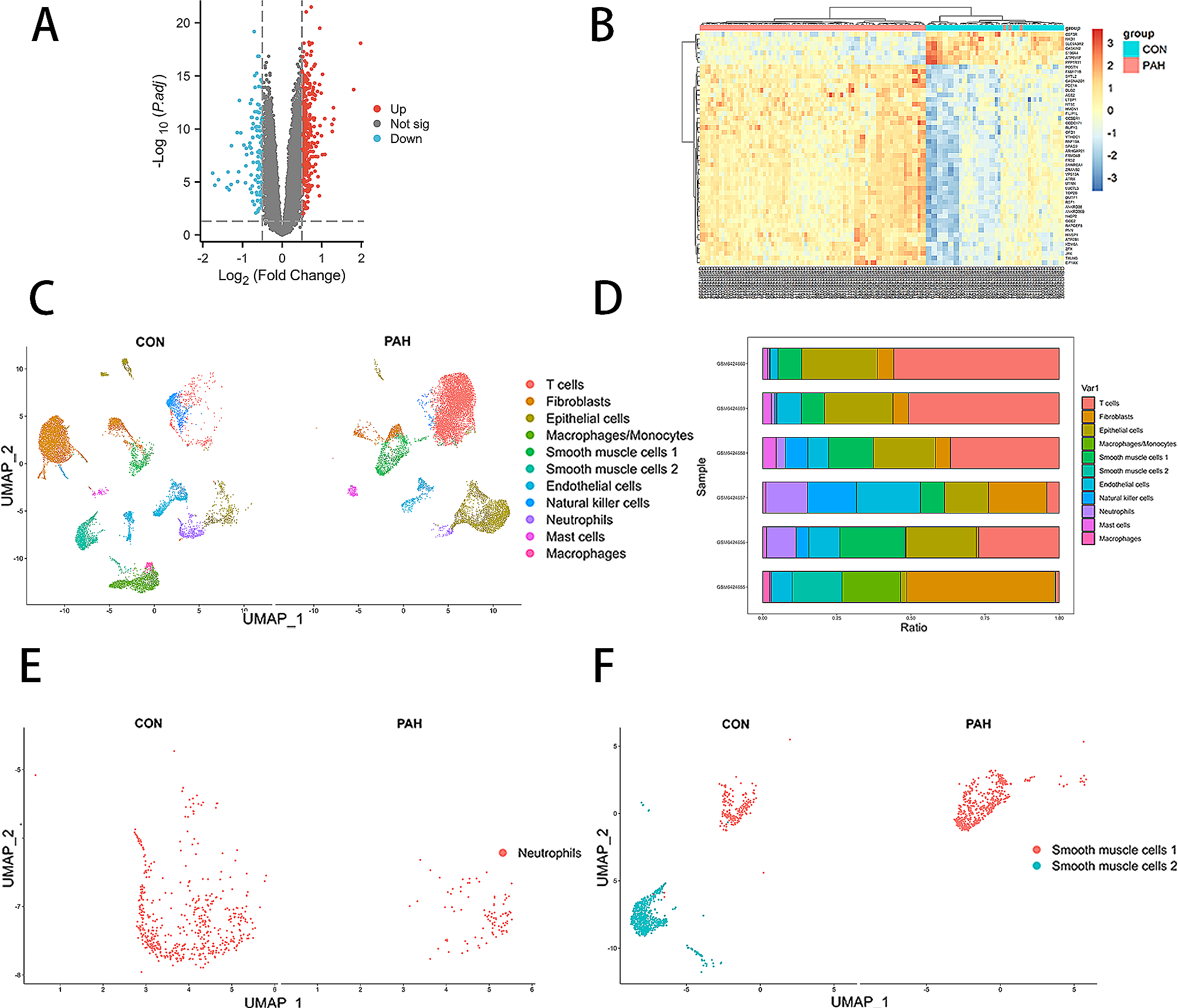
RNA-Seq
Total RNA from MRC5CTRL and MRC5ZNF451KD cells was extracted using TRIzol reagent (Invitrogen, CA, USA) according to the manufacturer’s protocol. RNA purity and quantification were evaluated using a NanoDrop 2000 spectrophotometer (Thermo Scientific, USA). RNA integrity was assessed using an Agilent 2100 Bioanalyzer (Agilent Technologies, Santa Clara, CA, USA). Then, the libraries were constructed using the VAHTS Universal V6 RNA-seq Library Prep Kit according to the manufacturer’s instructions. Transcriptome sequencing and analysis were conducted by OE Biotech Co., Ltd. (Shanghai, China). The SRA accession number is PRJNA963236 in this study.
Statistical analysis
Data are expressed as the mean ± standard error of the mean (SEM). Statistical significance was evaluated by Student’s t test or one-way ANOVA. Differences between groups were significant at a P value of < 0.05. Statistical analyses were performed with GraphPad Prism 9 (GraphPad Software, Inc., San Diego, CA).


留言 (0)