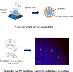
In the field of pharmaceuticals, the efficacy of certain drugs is persistently hindered by a set of recurring challenges. These obstacles encompass issues such as insolubility in water, limited stability, suboptimal distribution throughout the body, and a lack of specificity in targeting desired cells. To address these formidable challenges, drug carriers have emerged as pivotal solutions. Among the various carrier systems available, nanoparticles have garnered substantial recognition due to their unique advantages. Many studies comprehensively explored the myriad benefits associated with the utilization of nanoparticles in drug delivery systems, including the precise manipulation of nanoparticle size, surface characteristics, and the controlled release of active compounds. Previous investigation delved into how these nanoparticles, with their inherent capability to traverse blood vessels and gain access to cellular structures, have revolutionized drug delivery across a diverse spectrum of treatment targets, thereby preserving the stability and efficacy of therapeutic agents [1].
At the forefront of innovative cancer treatments, gene therapy has emerged as a groundbreaking approach [2]. Within the domain of genetic materials employed for gene therapy, microRNAs (miRNAs) play a pivotal role [3]. Gene therapy utilizing miRNA-mimicking (miRNA mimics) involves the administration of synthetic miRNAs designed to closely mimic the structural characteristics of endogenous miRNAs naturally occurring within the body [4]. This therapeutic strategy encompasses three primary objectives: (1) the replacement of essential but dwindling native miRNAs; (2) the suppression of target mRNA expression through gene silencing; and (3) the inhibition of protein translation for specific mRNA targets [5].
Within the domain of drug delivery strategies, a promising avenue involves the utilization of liposome nanoparticles as non-viral vectors [6]. Liposomes, spherical entities renowned for their ability to encapsulate a portion of the surrounding solvent and facilitate solvent diffusion to their core, have garnered attention due to their versatility in accommodating both hydrophilic and lipophilic compounds. Liposomes as drug delivery vectors are highly efficient at encapsulating hydrophilic substances within the inner core of the liposome [7].
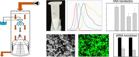
The utilization of liposome nanoparticles as drug delivery systems has gained significant attention in the field of pharmaceutical research. Despite their promising applications, liposomes do exhibit certain limitations, including challenges related to the large-scale production of nanoparticles and the high cost associated with commercial liposome products. To address these constraints, there is an urgent need to explore innovative materials possessing similar capabilities to liposomes while remaining cost-effective. Scientists have identified plant-derived nanoparticles as a promising option for drug delivery, potentially serving as an alternative to liposomes. Yang et al. discovered that nanovesicles from plants exhibit a similar structure to mammalian exosomes and conventional liposomes, characterized by a simple lipid bilayer [8]. Furthermore, Wang et al. demonstrated that nanovectors derived from lime lipids were not only non-toxic but also exceptionally efficient in delivering a wide range of therapeutic agents, including chemotherapeutic drugs, DNA expression vectors, siRNA, and proteins [9]. Zhang et al. further showed the potential of ginger-derived nanovectors (Dox-FA-GDNVs) in reducing cancer cell proliferation [10]. Furthermore, Yang et al. revealed the abundant availability of exosome-like nanoparticles in edible plants like lime, ginger, grape, and lemon [8]. Those research highlighted the potential of plant-based nanoparticles for drug delivery applications.
Black cumin (Nigella sativa L.), a prevalent plant in Indonesia, has garnered attention due to its reported phospholipid content, particularly phosphatidylcholine as much as 46.1 % − 48.5 % and phosatidylethanolalamine (PE) 25 % [11]. The phosphatidylcholine and phosatidylethanolalamine content in black cumin is greater than the phosphatidylcholine content in ginger (5 % − 6.5 %) and grapefruit (phosphatidylcholine 23 %; and phosphatidylethanolamine is 24 %) [10], [12], [8]. Notably, phospholipids, including phosphatidylethanolamine, phosphatidylglycerol, phosphatidylcholine, phosphatidylserine, and phosphatidylinositol, are essential components in the production of liposome nanoparticles [13]. Phosphatidylcholine will affect the fluidity of liposomes. Several studies also explained that liposomes containing phosphatidylcholine will be more stable and not easily bind to serum proteins [14], [15], thereby increasing the distribution efficiency of liposomes. Phosphatidylethanolamine was reported to play a role in membrane fusion [16] and liposome rigidity [17]. Therefore, the primary objective of this study is to isolate and characterize nanovesicles derived from black cumin (Nigella sativa) for their potential application as carriers for delivering miRNA.


留言 (0)