Glaucoma is a multifaceted optic nerve disease with characteristic nerve damage and visual field defects [1]. Glaucoma is the second most common blinding eye disease and the most common irreversible disease worldwide [2], and glaucoma drainage valve implantation is an important treatment method of advanced glaucoma [1]. Pressure-reducing valve implantation uses a drainage valve with a restrictive device (e.g., Ahmed valve) to drain the intraocular aqueous humor around the eyeball for diffusion and absorption, reducing intraocular pressure [3]. The current short-term postoperative success rate of valve implantation is 74–90%; however, the long-term success rate (> 2 years) is as low as 45–56% [4]. Fibrous encapsulation of the GVs outlet due to overgrowth of peripheral cells (e.g., fibroblasts) is the main cause of long-term postoperative failure [5]. 5-fluorouracil [6], mitomycin C [7], and other drugs have often been used as anti-fibroproliferative agents to prevent the silica rubber (SR) GV from forming fiber capsule after implantation. These medications can inhibit fibroblast proliferation at the initial implantation stage; however, they must be repeatedly used after surgery. However, in the long term, medication does not overcome the stimulation of the drainage valve as a foreign body, leading to a high long-term blockage rate after medication discontinuation [8].
Fiber coating formation on GVs is closely related to poor material biocompatibility [9]. SR is the primary material used for GVs with the advantages of good flexibility and promotes the formation of postoperative functional filtration blebs [10]. However, SR has some disadvantages as a typical inert material, such as poor hydrophilicity, no antifouling ability, and no biological activity [11]. Its poor hydrophilicity and lack of resistance to fouling lead to the adhesion of large amounts of proteins that have lost their natural structure, leading to the aggregation of inflammatory cells[12]. In addition, SR lacks bioactivity and cannot regulate the oxidative stress microenvironment[13], which is both an initiator and a driver in the core of the fibrotic cascade reaction[14] . Thus, the unfavorable factors due to defects in SR biocompatibility, such as inflammation, oxidative stress, and denatured adsorbed proteins, combine to promote the overproliferation of tissue cells around GVs, which ultimately causes fibrotic encapsulation.
The interaction of materials with living organisms often depends on their physical, chemical, and biological properties, which range from several nanometers to micrometers [14]. Therefore, a critical development path for biomaterials has been used in surface modification technology to design and create the surfaces of materials necessary to satisfy clinical needs. Recently, researchers have begun experimenting with surface-modified GVs to improve GV surface hydrophilicity. Poly (2-methacryloxyethyl phosphocholine) (pMPC) was introduced onto the surface of GVs and implanted into rabbit eyes. pMPC decreased the protein adsorption on the valve surface, reducing the intensity of foreign body reactions [15]. Hydrophilic coatings were used on intravascular catheters to improve fluidity, prevent friction, and minimize particle formation to reduce inflammatory responses [15], [16], [17]
To alter the GV surface topological morphology, Hu et al. prepared micropatterns comprising bacterial cellulose and arginine-glycine-aspartic acid-serine on hydrogel plates using CO2 laser lithography to guide and control the growth orientation of the cells to reduce the fibroblasts on the hydrogel surface [18]. Compared to ordinary GVs, surface-topologically structured GVs had lower outflow and better fibrosis resistance. The fibrosis degree was not considerably different from that of ordinary GVs; however, hydrophilic GVs can effectively lower the outflow resistance [19]. This study suggests that GV surfaces with topology and hydrophilic molecules, which are expected to have low outflow resistance and improved resistance to fibrillation, may be a superior strategy for hydrophilic surface construction. However, no research has been done on improving the biocompatibility of SR valves from two perspectives: stabilizing antifouling ability and regulating peripheral inflammation and the oxidative stress microenvironment.
Mussel-inspired polydopamine (PDA) coatings are promising materials for surface modification because of their ability to form coatings on various materials and complex surfaces [20]. Similarly, a superhydrophilic polydopamine (SPD) coating with a surface micro-nanometer topology was developed by adding a two-electron oxidant to accelerate the oxidative cross-linking of dopamine during PDA film formation[21,22], which has a significant short-term antifouling effect [23]. In addition, introducing SPD also helped positively modulate the inflammatory and oxidative stress microenvironments surrounding GV-implanted tissues. SPD contains a rich catechol group, which effectively regulates the inflammatory and redox microenvironment of nodes [24]. Phenolic hydroxyl groups reduce oxidative stress by scavenging reactive oxygen species(ROS), interrupting ROS chain reactions, and chelating metal ions [25]. Moreover, the catechol group can modulate the inflammatory response of implanted materials by inhibiting M1 macrophage polarization [26].
As a classical long-chain hydrophilic antifouling molecule, pMPC has been widely used for the surface modification of intraocular lenses [27], contact lenses [28], intravascular stents [29], and acetabular liners [30] to improve the hydrophilicity and biocompatibility of implant materials [20]. Subsequently, photocatalytic radical polymerization was used to introduce pMPC onto the surface of the GV materials [31]. However, pMPC polymerization on the surface of the material often requires additional chemical reagents and complex processing, which easily leads to pMPC-modified surface instability. PDA generates ROS and polymerizes C=C bond-containing monomers via free-radical polymerization under UV photocatalysis [32].
Compared with existing GV surface modification strategies, amphiphilic surfaces comprising SPD and 2-methacryloxyethylphosphocholine (MPC) may have the following advantages: the hydrophilicity is more stable and long-chain MPC can provide long-range hydrophilicity, whereas SPD with a topology can provide short-range hydrophilicity. This multistage hydrophilic structure might offer long-lasting superhydrophilicity and enhance the in vivo anti-contamination action of GVs, decreasing fibrous cyst formation.
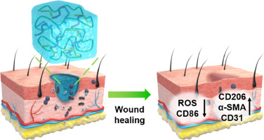
To address the biocompatibility issues of SR valves, we propose a synergistic surface engineering strategy that optimizes antifouling and microenvironment control, which can realize a comprehensive improvement in the biocompatibility of GVs by constructing a dual-hydrophilic antifouling coating with nano topology that regulates the microenvironment (Figure 1). Furthermore, using photocatalytic radical polymerization, we propose a simple and green method to introduce pMPC onto an SPD surface, which can reduce instability due to complex processes. First, using polyphenol chemistry, we introduced SPD coatings with a micro-nano topology on the surface of SR coated with microenvironment-controlled SPD-MPC dual-hydrophilic coatings. Two SPD+MPC coatings were created by modulating the feed concentration of MPC during polymerization. In addition, the surface physicochemical characteristics, resistance to protein adhesion, and anti-oxidant properties of the coatings were thoroughly examined. The anti-inflammatory and fibro-proliferative properties of the coatings were assessed in vitro. Finally, the ability of the coating to resist fibrous encapsulation in vivo was investigated via subcutaneous implantation in rats and normal rabbit eyes.


留言 (0)