
記住我

This cross-sectional, observational study adhered to the STROBE cross-sectional reporting guidelines [21]. The study protocol was approved by the Ethics Committee of Tokyo University Hospital. All participants provided written informed consent in accordance with the Declaration of Helsinki.
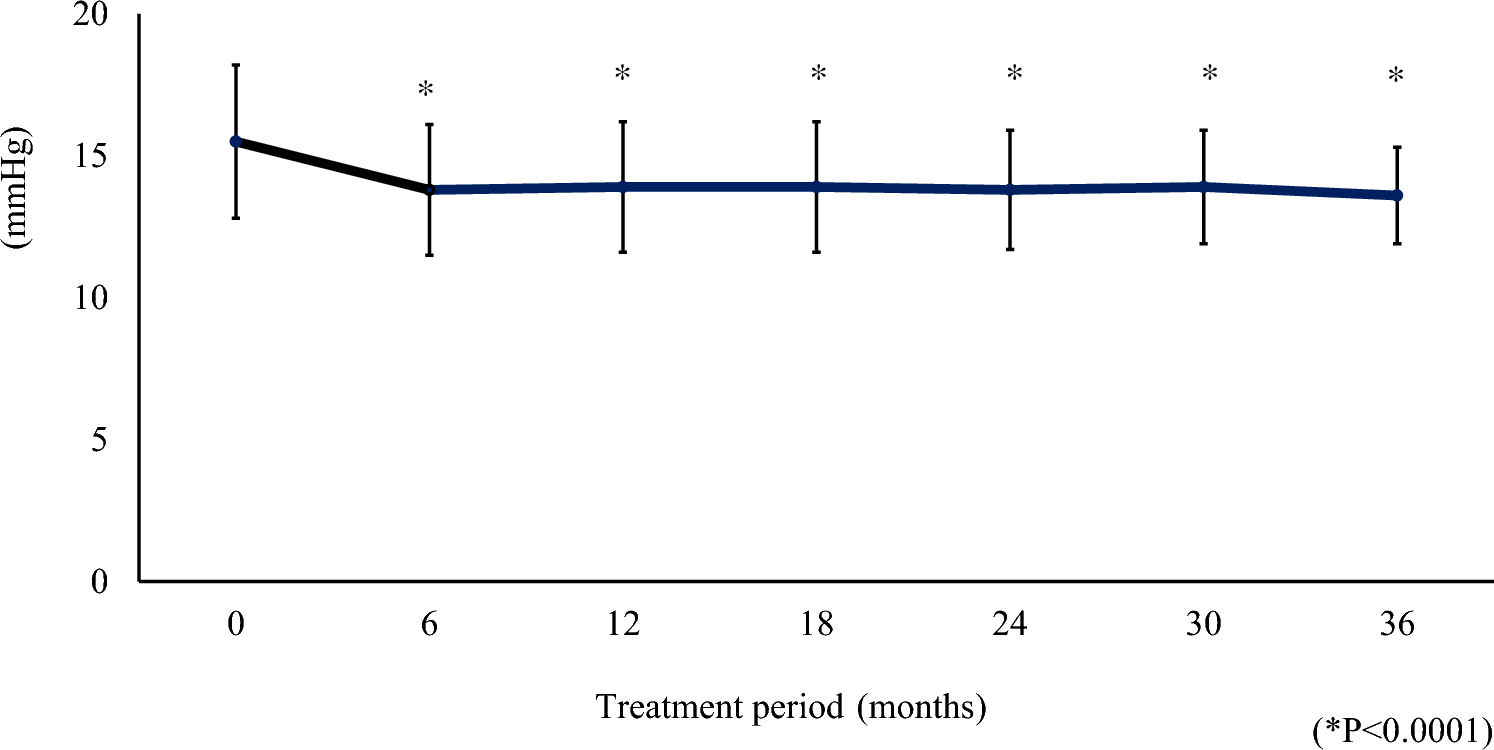
Between November 2018 and February 2020, 20 patients with glaucoma (mean ± SD age: 56.7 ± 13.6 years; 7 men and 13 women) were enrolled; in addition, 20 age-matched healthy volunteers (mean ± SD age: 56.0 ± 9.6 years, 10 men and 10 women) were enrolled as normal controls. The eligibility criteria for the glaucoma patients included a diagnosis of normal-tension glaucoma, receiving treatment at Tokyo University Hospital for > 1 year with topical agents or surgical intervention, experiencing a visual-field scotoma in the affected eye(s), and undergoing medical check-ups every 3 months.
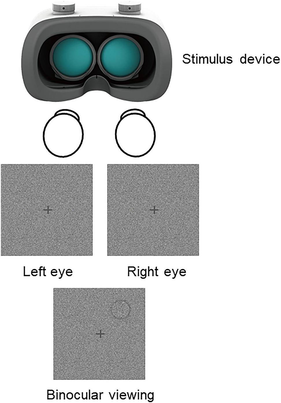
Behavioral proceduresThe visual stimuli and tasks performed in the present study have been described previously [16]. Briefly, the tasks investigated 3D shape perception and discrimination of 1D elementary features that serve as cues for 3D shape perception. The stimuli were presented on a 13-in LCD monitor (resolution: 1,920 × 1,080 pixels; refresh rate: 60 Hz) with the participants seated 40 cm away from the screen, like our previous study, and 30 cm from the Humphrey Field Analyzer (HFA). The head of each participant was constrained using a head and chin rest. Stimulus presentation and response registration were controlled by a personal computer using in-house software and Presentation 11.3 (Neurobehavioral Systems).
Task 1: Three-dimensional shape perceptionVisual stimuli employed in previous studies, created, and rendered in three modalities (shading, 3D-SfS; texture, 3D-SfT; motion, 3D-SfM), were used [16, 20]. These stimuli depicted 11 randomly generated complex and meaningless 3D objects including a variety of hills, ridges, valleys, and dimples [22,23,24,25]. Examples of a visual stimulus described under each modality are shown in Fig. 1a–c. A depth–color map of the stimulus is shown in Fig. 1d. Stimuli from each of the three modalities were presented in blocks of 11 trials, in which each 3D shape was shown once per block in a random order. In each trial, a single 3D shape (average size: 9° × 9°) was presented at the center of the screen. The participants were instructed to identify the foremost vertex on the convexity of the 3D surfaces (the highest convex point or global maximum of the surface, i.e., the point nearest to the observer) by superimposing a red cross on this point using a computer mouse under free-viewing and binocular conditions without any time limitation; these conditions were similar to those in daily life. Eye-tracking was not performed. The locations of the true global maximum of the 11 visual stimuli are shown in Fig. 1e. The depth difference between the global maximum of the surface, as identified by the participant, and the true global maximum was defined as the “error-in-depth” (cm).
Fig. 1
Display of a visual stimulus: a 3D shape defined by motion (3D-SfM), b shading (3D-SfS), and c texture (3D-SfT). d Depth–color map of the stimulus, with the depth difference (cm) from the base of the visual stimulus (highlighted with a blue background) represented by the color bar (blue, 0 cm; red, 5 cm). The white scale bar is 64 pixels. e The x-y distribution of the true global maximum for each stimulus (marked by yellow dots) is set against the blue backdrop of the visual stimulus. A white dotted line divides the four quadrants. The white scale bar is 64 pixels. f–h Elementary feature discrimination tasks for brightness of luminance, texture coarseness, and speed of motion, respectively
Task 2: simple feature discriminationTwo squares (5.7° × 5.7°) were presented simultaneously on the monitor, for 6 s, on either side (at 6.4°) of the fixation point, as described previously. Each square consisted of a single luminance value (Fig. 1f), texture coarseness (Fig. 1g), or speed of motion (Fig. 1h). Three levels of each cue were presented: a basic speed (1.9 °/s) and 20% faster or slower speeds, a basic luminance (60% greyscale) and 15% brighter or darker luminances, a basic texture (Fig. 1g, left stimuli) and 20% finer or 20% coarser textures. For each cue, three combinations of squares were derived from the different stimulus levels, and each combination was presented four times, yielding 12 trials. Moreover, an identical stimulus level was presented in the two squares in three trials. Thus, 15 trials for each cue were presented in random order and a block design was used. The participants selected the square with the faster motion, brighter luminance, or coarser texture by pressing the right or left shift key on the keyboard. If the two squares were considered identical, the participant was required to press the space key.
Visual-field sensitivity testingPatients underwent visual-field testing in each eye using the HFA 24-2 or 30-2 Swedish Interactive Threshold Algorithm (SITA) standard program semi-annually. Details on visual acuity, foveal sensitivity (dB), mean deviation value (dB), pattern standard deviation value (dB), and superior and inferior total deviation values (dB) captured using the HFA are summarized in Table 1. According to the glaucoma staging system of Mills et al., 2006, who modified the Hodapp-Parrish-Anderson criteria [26], eight, two, seven, and three participants were classified as stages 1, 2, 3, and 4, respectively, based on their visual-field scores for their worst eye [27]. The binocular visual fields of the glaucoma patients were assessed to gauge the influence of binocular-VFS on 3D shape perception. Due to the lack of available appropriate instruments to evaluate binocular-VFS, we used the method delineated by Matsuura et al. [28]. Briefly, the glaucoma patients were tested using the right 30-2 standard SITA program, but with both eyes open. During this binocular visual-field testing, the position of the chinrest was adjusted to its furthest left setting, allowing patients to position their chins over the right side of the chinrest. This configuration facilitated patients in aligning with the perimeter by adjusting both the vertical position of their head and the horizontal alignment relative to the bridge of their nose.
Table 1 Characteristics of visual acuity and index of visual fieldStatistical analysisThe accuracy for each cue in the 1D feature discrimination task, together with the error-in-depth values for each cue in the 3D shape perception task, were compared between the glaucoma patients and the healthy volunteers using Welch’s t-test. In the formula (t[df] = X, p = Y), t, df, and p indicate the t-value, degree of freedom, and p-value, respectively. The threshold for statistical significance was set at p < 0.05, with the adjusted threshold determined at p < 0.017 after applying Bonferroni correction.
As depicted in Fig. 1e, the positions of the true global maximum of the 3D visual stimuli within the visual field were evenly distributed around the center of the stimuli. The perception of 3D shape, as examined in this study, necessitates integration over a retinal area that surpasses single tested points in the HFA. Additionally, the specific region of the visual field responsible for 3D shape perception remained ambiguous due to the binocularly free-viewing conditions. The diminished monocular visual-field sensitivity in glaucoma patients manifested across various regions of the visual field because of the sporadic nature of visual-field defects. In a report that assessed temporal contrast sensitivity in the parvocellular and magnocellular pathways among healthy subjects, in subjects suspected of glaucoma and perimetric glaucoma patients, the stages of glaucoma were controlled [29]; whereas in the current study the glaucoma patients were considered together despite their wide range of visual-field defects and glaucoma stages [27]. Therefore, to evaluate the influence of binocular-VFS on 3D shape perception, we used visual-field index (VFI) values [30] of binocular-VFS. The VFI has more relevance to the central visual field, which largely covered the 3D-shape images. The correlations between VFI values (%) of binocular-VFS and error-in-depth values (cm) were evaluated using Spearman’s rank test. The glaucoma patients were divided into two subgroups based on their VFI values of binocular-VFS. The VFI values of binocular-VFS range from 0% (perimetrically blind) to 100% (normal visual field) [30]; patients with a VFI of 100% were classified as unimpaired, whereas all other patients were classified as impaired. The error-in-depth values of the healthy volunteers and the two subgroups were scrutinized using analysis of variance (ANOVA), taking the three groups as factors, and adjusting for significance using Bonferroni correction.
留言 (0)