Clinical presentation
A 67-year-old male patient had undergone excision of cutaneous epidermoid carcinoma on his anterior chest the year before and sought medical care at the surgical oncology outpatient clinic at Barretos Cancer Hospital, Brazil, with a persistent tumor in the left armpit (Supplementary Fig. 1). The patient had a history of acute myocardial infarction, coronary stent placement, and diabetes. A tumor biopsy at Barretos Cancer Hospital revealed a cutaneous squamous cell carcinoma (cSCC), and the patient underwent surgery and received postoperative carboplatin, taxane, and radiation therapy. Unfortunately, the patient experienced a recurrence shortly after treatment and was deemed no longer treatable, ultimately resulting in his death.
HCB-541 cell line establishment
Surgical specimens were collected for cell cultivation immediately after surgery and maintained in Dulbecco’s Phosphate Buffered Saline solution until processing begins as previously reported [13]. Biopsy tumor was fragmented into small pieces with a surgical scalpel in a petri dish. Then, it was incubated in 0.5 ml of accutase/trypsin enzymatic solution for total tissue dissociation, at 37 °C for 30 min. After dissociation, the cell solution was incubated in DMEM medium supplemented with 10% Fetal Bovine Serum (SBF) and 1% penicillin and streptomycin to complete enzyme inactivation. Throughout the study, experiments were consistently conducted using aliquots from this identical FBS batch (210625 K) to ensure quality and minimize variations in component composition. Cells were cultivated in DMEM medium supplemented with 1% penicillin/streptomycin, 10% FBS and maintained in culture flask at 37 °C, 5% CO2. Primary cell line was named HCB-541, and stock vials were frozen in different passage numbers to report the protein expression change during establishment processes, and for DNA and RNA analysis at the 15th passage.
Morphological and doubling time characterization
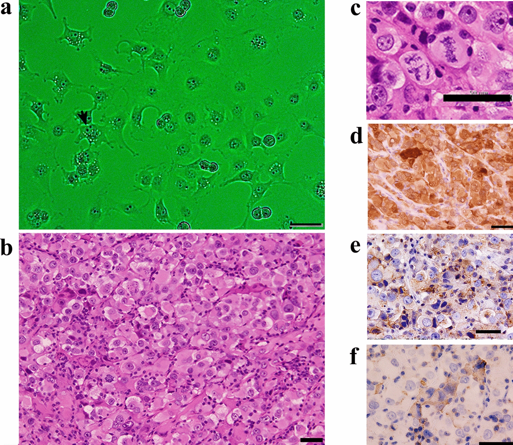
To assess the morphologic profile, after continuous passages (> 30), the HCB-541 cell line was seeded in plates and cultivated in DMEM supplement with 1% P/S and 10% SFB at 37 °C, 5% CO2. Cells were photographed under an optical microscope at × 100 magnification (Olympus XT01), until complete 90% of total confluence. Doubling time values were obtained using xCELLigence real-time cell analysis (Agilent Technologies, Inc), according to the manufacturer’s instructions. Initially, 3.5 × 10³ or 7 × 10³ cells were plated into E-plate (Agilent Technologies, Inc) and were cultivated in DMEM medium supplemented with 1% P/S and 10% for 72 h. The cell index was calculated using a specific RCCA® software analysis.
Tumorigenicity of HCB-541 cell line in NOG-SCID mice
To assess the tumorigenicity of HCB-541, subcutaneous injections of either 1 × 10⁶ or 3 × 10⁶ cells mixed with Matrigel® at a 1:1 (v/v) ratio were administered into the right flank of 8-week-old female NSG mice (NOD.Cg-Prkdcscid Il2rgtm1Wjl/SzJ, The Jackson Laboratory, USA), as previously described [14]. Tumor size was measured weekly with calypter, and volume was calculated using the formula: volume = length × width2/2. After two weeks of transplantation, tumors were excised surgically, measured, and subjected to morphological evaluation using hematoxylin and eosin staining and immnunohistochemistry (IHC) analysis. Tumors were fixed in a 4% paraformaldehyde, and paraffin-embedded sections were prepared using standard procedures for histological analysis. The mice used in this study were maintained under SPF conditions, on a 12-h light/dark cycle, and received sterile food and water ad libitum.
Immunophenotypic characterization
We conducted an immunocytochemistry (ICC) analysis of the HCB-541 cells and compared with controls A431 (human vulva epidermoid carcinoma cell line) (RRID:CVCL_0037), from ATCC (ID number: CRL-1555) and HACAT (immortalized normal human keratinocyte cell line)(RRID:CVCL_0038) from BCRJ (ID number: 0341). Cells were seeded in a chamber slide with 8 wells on a glass slide overnight and then fixed with paraformaldehyde in DPBS for 5 min. After the fixation step, cells were permeabilized with Triton-X 100 0.25% in DPBS for 10 min. Protein blocking was performed with Lab Vision™ UltraVision™ (Thermo Scientific), according to the manufacturer's protocol. Primary antibodies ready to use anti-vimentin (RRID:AB_2722716), anti-KI67 (RRID:AB_2250503), anti-cytokeratin 20 (RRID:AB_563800), anti-cytokeratin AE1/AE3 (RRID:AB_1587224)) anti-cytokeratin 8/18 (RRID:AB_563833), anti-cytokeratin 17 (RRID:AB_2133033)) were incubated overnight. After the incubation, the slides were in biotinylated goat polyvalent antibody for 10 min, followed by washing and subsequently incubated with streptavidin peroxidase, stained with DAB chromogen and counterstaining with hematoxylin eosin. Finally, the cells were photographed using an optical microscope Olympus XT01.
Immunohistochemistry expression of CK 5/6 (RRID:AB_563822), p63 (RRID:AB_10582857), p40 (RRID:AB_3073535) and p10 (RRID:AB_2549617) were performed in tumor tissue biopsy, HCB-541 cell line, both in formalin-fixed paraffin-embedded (FFPE) sections using BenchMark ULTRA IHC/ISH System (Roche) automated staining platform.
Cell line authentication by short tandem repeat (STR) profiling analysis and mycoplasma contamination test
STR analysis was performed using DNA from the cell line and FFPE tumor biopsy as previously described for authenticity confirmation [15]. The mycoplasma contamination test was carried out in DNA from the cell line using MycoAlert Mycoplasma Detection Kit (Lonza, Swiss), according to the manufacturer's protocol, in different moments throughout the time it was in culture.
DNA and RNA isolation
DNA was isolated from the patient's formalin-fixed and paraffin-embedded (FFPE) tumor biopsy, using QIAamp DNA Micro Kit (Qiagen) as previously reported [16]. DNA from the HCB-541 cell line was isolated using Biopur Mini Spin Plus 250 Extraction kit (Biopur), both according to the manufacturer's recommendations. All samples were quantified by NanoDrop 2000 System (Thermo Scientific) and stored at −20 °C. RNA was isolated from HCB-541 and HACAT (immortalized normal human keratinocyte) cells using RecoverAll Total Nucleic Acid Isolation kit (Invitrogen), according to the manufacturer's recommendations. Samples were quantified by NanoDrop 2000 System (Thermo Scientific) and Qubit 2.0 Fluorometer (Life Technologies), and stored at −80 °C.
Cellular viability assay (MTS)
Cell viability was determined 72 h after drug exposure, using the colorimetric CellTiter 96® AQueous One Solution Cell Proliferation Assay (Promega, Madison, WI), according to the manufacturer’s instructions and as previously reported [15]. To assess cytotoxicity, a total of 5 × 10³ were plated in 96 well plates in DMEM with 10% FBS and 1% allowed adherence overnight. Then, cells were treated with increased concentrations of pharmaceutical agents and reader (Thermo Scientific, Finland), at 490 nm. Results were normalized with DMSO control values. To calculate IC50 values the GraphPad Prism software (Version 9.0) was used to evaluate a nonlinear regression curve. All experiments were performed in triplicates at least three times.
Western blot and reverse phase protein arrays (RPPA)
Protein lysate of HCB-541 and A431 (epidermoid cancer cell line) cells were used to perform western blot analysis. Both cells were rinsed in DPBS and after lysed in lysis buffer following our group protocol [17] and were incubated overnight with primary antibody: pEGFR-Tyr1068 (D7A5), t44/42 MAPK (137F5); p.p44/42 MAPK-Thr202/Tyr204 (D13.14.4E); AKT(pan) (C67E7); pAKT-Ser473 (D9E); phosphoP53-Ser15 (82530), phosphor-P53-Ser46 (2521), ROR-2 (88639) FGFR1 (ab76464) and β- actin (3700) were used. Both primary antibodies from Cell signaling were diluted in TBS-T at 1:1000. Membranes were incubated with anti-rabbit secondary antibody Anti-rabbit (7074) at dilution 1:5000. Chemiluminescent signals were detected by ECL in automatic ImageQuant mini LAS4000 (GE Healthcare Protein array was Human Phospho-Mitogen-activated (ARY002B; R&D Systems, MN) and Human Phospho-RTK Array (ARY001B; R&D Systems, MN) were conducted according to the manufacturers' instructions). All the experiments were performed three times.
Pharmacological agents
Small-molecules inhibitors Afatinib (Cat.N° S1011); Allitinib (Cat.N° S2185); Lapatinib (Cat.N° S2111); Erlotinib (Cat.N° S1023) were purchased from Selleck Chemicals (Houston, TX). Carboplatin (Cat.N° C2538), Cisplatin (Cat.N° 15,663-27-1) and 5-Fluoracil (Cat.N° 51-21-8) were purchased from Sigma Aldrich (Sigma-Aldrich, USA) and Cetuximab was purchased from Merck and were diluted in DMSO at 10 mM and stored at 20 °C for posterior use. Carboplatin was dissolved in distilled water and Cetuximab is ready to use. DMSO or water were used as a negative control in all cytotoxicity experiments. Details of concentrations, doses and exposition time are explained in supplementary Table 1.
Table 1 Mutation profile of both tumor biopsy and HCB-541 cell lineChromosome preparation and GTG-banding
The karyotype analysis was performed as described previously [18]. HCB-541 cell line at the fifteenth passage was incubated with 120 ng/ml colcemid (Life Technologies-Gibco) for 125 min. The cells were harvested by treatment with 0.025% trypsin EDTA, suspended in KCl 0.075 M solution at room temperature for 5 min, and fixed with methanol/acetic acid (3:1) five times. To examine the chromosomal distribution of constitutive heterochromatin, GTG-banding was performed using the standard trypsin/Giemsa method [19]. The karyotype analysis of HCB-541 cells was done on twenty metaphases.
Mutational and fusion analysis and clinical significance variants classification
Mutations from the patient's FFPE tumor biopsy and HCB-541 cell line were evaluated by Next Generation Sequencing (NGS), using the Custom Cancer Solution cSTS kit (SOPHiA Genetics). This 48-gene panel plus microsatellite instability (MSI), includes all protein-coding exons for all transcripts of 8 genes, as well as a selection of protein-coding exons of 40 genes with ± 10 bp flanking into the intron and, if applicable, including 5’ UTR exons that contain coding sequence (Supplementary Table 2). No normal DNA tissue (blood or adjacent normal skin) was available for germline mutation analysis. Genetic variants were evaluated in both HCB-541 cell line and the primary tumor. Moreover, the presence of gene rearrangements was performed on the HCB-541 cell line, using also the Custom Cancer Solution (cSTS) kit (SOPHiA GENETICS, Switzerland) according to the manufacturer’s protocol. The somatic RNA panel for fusion research includes 16 genes: ALK, BRAF, EGFR, FGFR1, FGFR2, FGFR3, FGFR4, MET exon 14 skipping, NTRK1, NTRK2, NTRK3, RET, ROS, ERG, PPARG and NRG1.
Briefly, from 20 ng of DNA, fragments were generated using an enzymatic fragmentation step. The three subsequent enzymatic steps, end-repair, A-tailing, and ligation to Illumina adapters, were performed to produce NGS libraries. A capture-based target enrichment was carried out on the pooled libraries. The quantitation of the final pool of libraries was performed using Qubit dsDNA HS fluorimetric assays (ThermoFisher). Quality control of fragment size was assessed using DNA ScreenTape analysis 4150 TapeStation system (Agilent). Sequencing was achieved with the final library concentration of 10 pM onto a 600-cycle format V3 flow-cell, via Illumina MiSeq platform Illumina. Data analysis was performed to detect Single Nucleotide Variants (SNVs), insertions/deletions (INDELs), and Copy Number Alterations (CNAs). Sequencing FASTQ data were analyzed by the Sophia DDM® platform (SOPHiA Genetics). For gene fusions, 200 ng of RNA was used to convert it into cDNA, and library construction followed the RNA target Oncology solution (ROS) kit by SOPHiA GENETICS, Switzerland, as per the manufacturer protocol. DNA fragments were generated through enzymatic steps, including end-repair, A-tailing, and ligation to Illumina adapters, to create NGS libraries. A capture-based target enrichment was applied to the pooled libraries. The final library pool was quantified using Qubit dsDNA HS fluorimetric assays from Life Technologies, USA. Quality control for fragment size was assessed using DNA ScreenTape analysis with the 4150 TapeStation. Data analysis was performed to detect fusion genes. Sequencing FASTQ data were analyzed by the Sophia DDM® platform (Sophia Genetics, Switzerland).
Selected variants were evaluated according to clinical significance using Varsome [20], Franklin (http://franklin.genoox.com) and ClinVar databases [21], considering evidence of skin squamous cell carcinoma (Table 1).
TERT hotspot promoter mutations were also validated by droplet digital PCR (ddpCR). To detected TERTp mutations was used 10 ng of DNA, 2X ddPCR Supermix for Probes (No dUTP) (Bio-Rad, Hercules, CA, USA), 5 M Betaine (Sigma), 80 mM EDTA (Sigma), 10U/ul CviQI enzyme (ThermoFisher) and the commercial assay TERT C228T: ddPCR™ Expert Design Assay: TERT C228T_113, Human, Homo sapiens (Bio Rad), following the manufacturer's protocol. Droplets were generated using a droplet generator (Bio‐Rad). The PCR cycling parameters included 10 min at 95 °C, 50 cycles at 96 °C for 30 s and 61 °C for 30 s, and one cycle at 98 °C for 10 min followed by a 4 °C hold. Droplet fluorescence was assessed using a droplet reader (Bio-Rad). Analysis of the ddPCR data for allele calling and the calculation of absolute copy numbers were performed using QuantaSoft software, version 1.7.4 (Bio‐Rad).
Molecular microsatellite instability (MSI)
MSI evaluation was also performed by multiplex PCR using 6 quasi-monomorphic mononucleotide repeat markers (BAT25, BAT26, NR21, NR24, NR27, HSP110) as described by Berardinelli et al. [22] PCR was performed using Qiagen Multiplex PCR Kit (Qiagen) with 50 ng of DNA and the following thermocycling conditions: 15 min at 95 °C; 40 cycles of 95 °C for 30 s, 55 °C for 90 s and 72 °C for 30 s; and a final extension at 72 °C for 40 min. PCR products were then submitted to capillary electrophoresis on an ABI 3500XL Genetic Analyzer (ThermoFisher) according to the manufacturer’s instructions. The results were analyzed using GeneMapper v4.1 software (ThermoFisher) to measure the fragment length in base pairs. The classification of instability is determined by the number of markers out of the quasi-monomorphic variation range (QMVR), being 2 or more markers classified as MSI-H (MSI-High), only one marker classified as MSI-L (MSI-Low) and cases without markers out of QMVR classified as MSS (Microsatellite stability), as previously reported [22].
Droplet digital PCR (ddPCR) detection for human papilloma virus (HPV)
The presence of HPV 16 and 18 was evaluated in the patient's tumor biopsy and HCB-541 cells, using ddPCR, as previously described [23]. Briefly, for HPV detection, 10 ng of DNA were conducted to Singleplex PCR reactions with 2X ddPCR Supermix for Probes (No dUTP) (BioRad, Hercules, CA, USA), 900 nM of each primer and 300 nM of the probe in a total volume of 22 µL followed by droplet generation using an automated droplet generator (BioRad) according to the optimized protocol [23]. Droplets were generated using a droplet generator (Bio‐Rad). The PCR cycling parameters included 10 min at 95 °C, 50 cycles at 96 °C for 30 s and 61 °C for 30 s, and one cycle at 98 °C for 10 min followed by a 4 °C hold. Droplet fluorescence was assessed using a droplet reader (Bio-Rad). Analysis of the ddPCR data for allele calling and the calculation of absolute copy numbers were performed using QuantaSoft software, version 1.7.4 (Bio‐Rad). Caski (+ HPV 16) and SiHa (-HPV) cell lines were used as controls and were obtained from the ATCC cell bank.
mRNA expression profiles by nanostring
mRNA expression of a panel of 770 genes associated with 13 cancer-associated canonical pathways related to basic cancer biology, was evaluated using the nCounter PanCancer Pathways Panel, NanoString technology (Nanostring Technologies, USA) for HCB-541 cells and HACAT (immortalized human keratinocyte cell line) cells (used as a non-tumor counterpart) for differential expression analysis, as previously reported [17].


留言 (0)