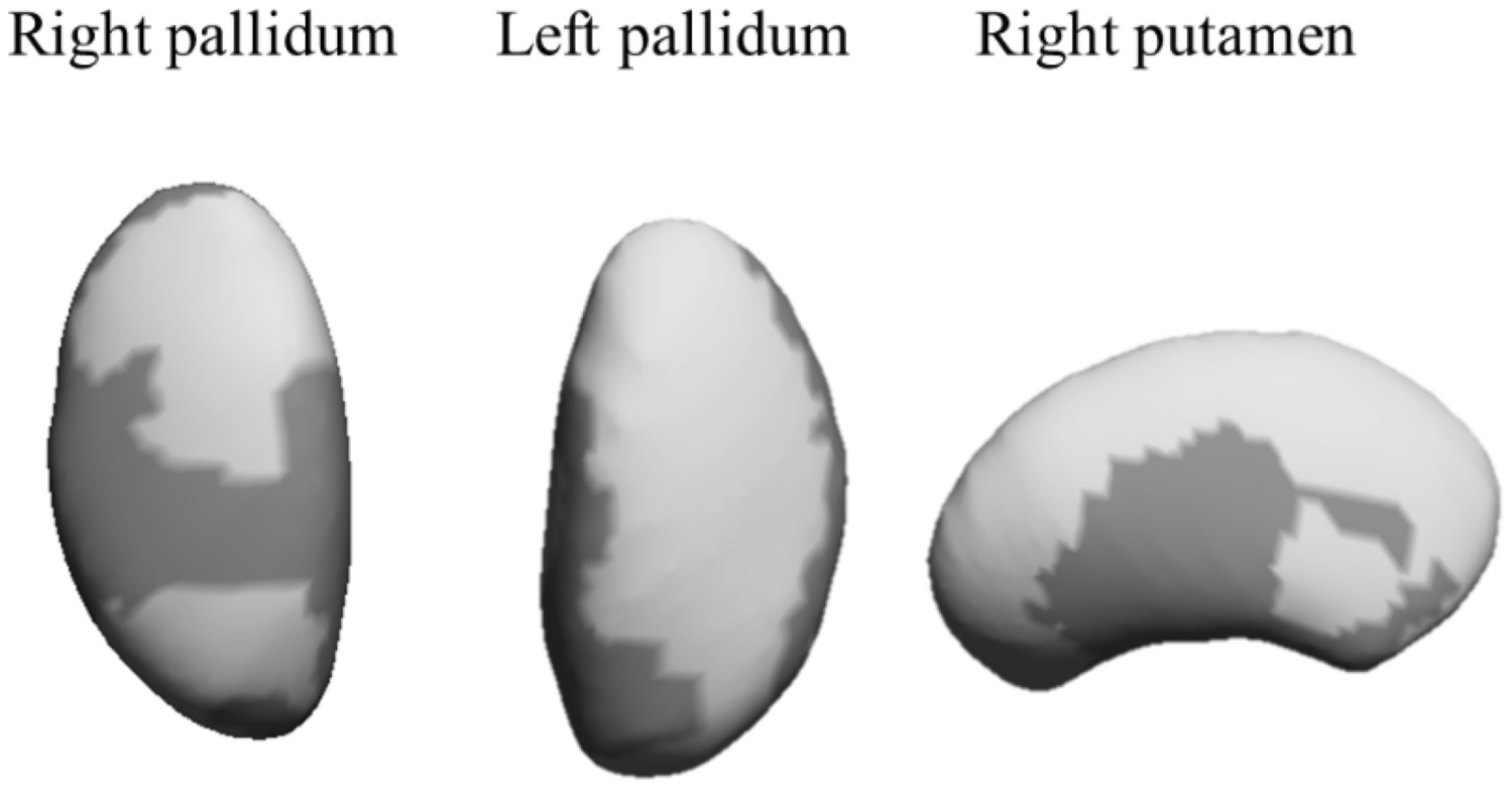
The present findings indicate that more time in bed is related to specific expansions in the right and left pallidum, and the right putamen in children with overweight/obesity. However, no significant correlations were observed between sleep variables and the other subcortical brain structures, i.e., brain stem, thalamus, nucleus accumbens, amygdala, caudate nucleus, and hippocampus.
The present study contributes to the existing literature by studying the relationship between sleep behaviors and the morphology of subcortical brain structures in children. Specifically, the authors observed that for children with overweight/obesity, higher total sleep duration was related to greater expansions in the pallidum and putamen. No previous studies examining these associations in healthy populations or children were found, which hampers the comparisons of present findings with other studies. Replication of these findings in future studies is warranted. To the best of authors’ knowledge, only two studies in adults investigated the association between the shape of subcortical brain structures and sleep in insomnia patients [22, 23]. The first study tested the potential structural alterations of the amygdala in adults with insomnia compared with healthy controls with adequate sleep [22]. The second study examined the associations of the shape of subcortical brain structures with sleep in adults with insomnia [23]. The conclusions from these two studies suggest a potential sleep-related atrophy in the amygdala, putamen, and hippocampus. The authors cannot directly compare their findings with these studies due to different methodological reasons (actigraphy vs. self-reported questionnaire) and characteristics of the study participants (children with overweight/obesity vs. adults with insomnia). Notwithstanding, the results of the previous studies partially agree with present findings, showing a relationship between sleep behaviors and the putamen, yet the present study did not find a sleep-related correlation with amygdala and hippocampus as concluded in the previous studies [22, 23]. Likewise, they did not observe any change in thalamus and caudate nucleus in line with the observations in the present sample [23]. Although the present findings are promising with the potential to decipher the relation between sleep behaviors and brain morphology in children, caution should be paid when interpreting these findings since causal relationships cannot be stated.
When interpreting present findings, it is crucial to understand the role of the basal ganglia (i.e., caudate nucleus, pallidum, putamen, and nucleus accumbens) in sleep regulation and wakefulness. The basal ganglia is a key area involved in motor function, habit formation, reward/addictive behaviors, and executive function [24], all of which depend on wakefulness [25]. Indeed, there are reciprocal connections between the basal ganglia and every part of the sleep–wake circuit: cerebral cortex, brain stem, basal forebrain, thalamus and hypothalamus [25]. Moreover, the role of the pallidum during the sleep cycle may be reflected in the activity of the striatum, which collectively refers to the caudate nucleus, putamen and nucleus accumbens. Data from studies in mice have demonstrated changes in sleep pattern with lesioning or stimulation of the pallidum [26]. Specifically, a lesion in the pallidum decreased sleep behaviors, increased fragmentation and shortened sleep duration. In contrast, stimulation of this region was related to increases in both non-rapid eye movement and rapid eye movement sleep time [26]. Therefore, based on the evidence and confirming the present findings, it seems that the basal ganglia is a key group of structures with an important role in sleep behaviors.
To date, few previous studies suggest a beneficial link between sleep behaviors and gray matter volume in several structures in children and adolescents [3, 27, 28]. For instance, Taki et al. observed that sleep duration during weekdays affected hippocampal gray matter volume in healthy children [27]. Further, Urrila et al. concluded that shorter time in bed during weekdays correlates with smaller brain gray matter volumes in frontal, anterior cingulate, and precuneus cortex regions in adolescents [28]. The present study investigates the week as a whole using weighted averages to account for the correspondent weight of weekdays and weekend days, and thus, providing a clear perspective on the relationship between sleep behaviors and brain shapes rather than assuming different associations dependent upon the day in which sleep occurs [3]. Analyzing sleep behaviors with this approach, a recent study from the same cohort explored the associations of sleep behaviors with gray matter volume in the whole brain, with a particular focus on the hippocampus in children with overweight/obesity [3]. The main finding was that sleep behaviors were associated with gray matter volume in multiple cortical and subcortical brain structures including the hippocampus [3]. The lack of association found in the putamen and pallidum using the whole-brain approach could suggest that an unmasked potential association was detected by the brain shapes analyses, which allows for the detection of locally precise changes in brain morphology of various brain structures.
The study of the sleep-brain connection in this period of the lifespan, when the brain undergoes major developmental changes, is of public health significance. The present study provides important insight into understanding how sleep hygiene is related to brain shapes in children with overweight/obesity. While the current study did not specifically examine the impact of brain volumes on daytime behavior and intelligence, it is noteworthy that a previous study within the same cohort found a positive correlation between expansions in the right pallidum and intelligence [17]. Nevertheless, more research is needed to gain a further understanding of how the various expansions and contractions of certain subcortical structures relate to brain health. Future studies with larger and more diverse samples should aim to replicate these findings, paying particular attention to potential sex and weight status differences, as well as, exploring the broader link between brain shapes and cognition.
The limitations of this study were: i) its cross-sectional design does not allow a causal interpretation of the findings; ii) the inclusion of children with overweight/obesity limits the generalizability of these results, and iii) the use of actigraphy instead of the gold-standard, polysomnography. Moreover, the accelerometer-based estimates of sleep behaviors are an estimation based on movement patterns, and not purely sleep behaviors. As such, these findings should be taken with caution. Still, accelerometers are a non-invasive, objective, and valid technique for assessing sleep behaviors in free-living individuals [16, 29], and they also demonstrate high agreement with polysomnography [30].
Overall, these results indicate that the total time spent in bed is positively associated with further developments in the right and the left pallidum and the right putamen, suggesting a beneficial role of sleep hygiene on brain.

留言 (0)