
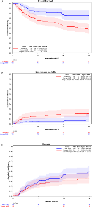
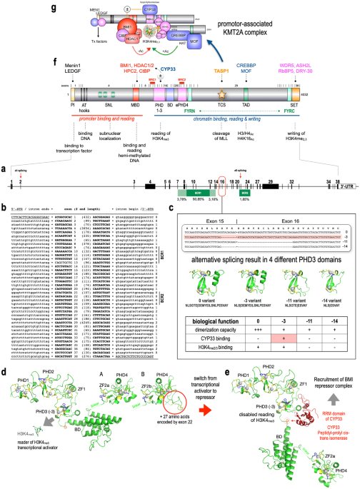
記住我


Upon induced expression of the MLL::ENL as well as MLL::AF4 oncogenes, lymphomyeloid hematopoietic progenitor cells represent the most potent LICs among early hematopoietic stem and progenitor cells (HSPCs) [22,23,24]. Lymphomyeloid multipotent progenitors (LMPPs; Lin− Sca-1+ cKit+ (LSK) Flt3high CD150−) additionally show marked differences in both proteotype and differentiation-associated functions [11] and thus represent highly interesting targets for exploration of the ontogenic effect on their leukemic potential. In humans, the MLL::ENL fusion (resulting from chromosomal translocation (11;19)(q23;p13.3)) is most commonly associated with ALL in infants as well as in adults; however, the mutation almost never gives rise to AML in infant children whereas 22% of adults with the mutation present with myeloid disease [7]. To investigate the lineage potential of normal relative to transformed fetal (embryonic day [E] 14.5) and adult (6–10 week old) LMPPs, we performed an in vitro differentiation assay using a mouse model harboring doxycycline (DOX)-inducible expression of MLL::ENL (iMLL::ENL mice) [23] (Fig. 1A). We observed retained expression of the leukemia stem cell (LSC)-associated marker cKit [25, 26] already at 4 days after induction of MLL::ENL expression and a high production of cKit+ progeny from transformed cells in cultures initiated with fetal LMPPs (Fig. 1B). In contrast, the frequency of cKit+ cells from adult MLL::ENL-expressing LMPPs surpassed the control cells with delay compared to fetal cells, only at day 16 of culture (Fig. 1C).
Fig. 1: LMPPs expressing MLL::ENL show ontogenic differences in lineage output in vitro.
A Workflow for in vitro differentiation of fetal and adult LMPPs derived from iMLL::ENL mice. Expression of the oncogene was induced by addition of doxycycline (DOX) to the cultures. B, C Frequency of cKit+ cells derived from fetal (B) and adult (C) MLL::ENL-induced (+DOX) and non-induced (−DOX) LMPPs. D, E Frequency of cKit+ Flt3+ derived from fetal and adult MLL::ENL-expressing (D) and normal (E) LMPPs. n = 4 for all displayed graphs. Error bars represent SD. ****p < 0.0001, ***p < 0.001, **p < 0.01, and *p < 0.05 and ns not significant. See also Fig. S1.
Myeloid cell output was low from fetal as well as adult MLL::ENL-expressing LMPPs (Fig. S1A), whereas B cell output increased in a near-linear fashion during the culture period (Fig. S1B). Of note, while the frequency of CD11b+ cells in fetal wells was very low at all assayed time points, up to 50% of adult LMPPs intermittently produced high numbers of myeloid progeny (Fig. S1A), indicating an ontogenic difference in the myeloid potential of MLL::ENL-expressing LMPPs, as previously observed for WT LMPPs in a similar culture setting [11] as well as in this experiment (Fig. S1C, D). Interestingly, a population of cKit+ Flt3+ cells emerged in MLL::ENL-induced fetal wells at day 7 of culture (Fig. 1D). Critically, the same population was almost completely absent from adult DOX-treated wells at all investigated time points, and very few, if any, cKit+ Flt3+ cells were produced by fetal and adult LMPPs in the absence of DOX (Fig. 1D, E). Within the hematopoietic system, co-expression of cKit and Flt3 can be found on LMPPs as well as common lymphoid progenitors (CLPs) [27, 28]. In addition, Flt3 expression has been proposed as a hallmark of ALLs harboring MLL rearrangements [29], indicating that fetal, but not adult, MLL::ENL-expressing LMPPs give rise to a lymphoid-like leukemic population in vitro.
Collectively, the results from our in vitro assay show that induction of MLL::ENL in fetal and adult LMPP confers LSC properties to these cells and indicate a stronger lymphoid bias in fetal compared to adult MLL::ENL-expressing LMPPs.
Fetal and adult LMPPs expressing MLL::ENL give rise to aggressive leukemia in vivoNext, we performed an in vivo assay of the leukemic potential of fetal and adult LMPPs via transplantation into WT recipients (Figs. 2A and S2A). To account for the effect of the niche developmental stage in which MLLr leukemia is propagated [20], we included adult as well as neonatal (24–48 h old) recipients. Adult cells engrafted poorly in neonatal recipients, with no peripheral blood (PB) chimerism above 0.2% by 37 weeks post-transplantation (Fig. S2B). Chimerism in neonates transplanted with fetal cells remained above 1% in 3 out of 4 recipients throughout the assayed period, and only fetal cells gave rise to multilineage progeny in mice that were neonatally transplanted (Fig. S2B–E). However, none of the neonatal recipients developed leukemia during the experiment.
Fig. 2: Fetal and adult LMPPs expressing MLL::ENL give rise to aggressive leukemia in vivo.
A Workflow for transplantation of fetal and adult iMLL::ENL LMPPs into WT adult and neonatal recipients. n = 4 per group for neonatal recipients and n = 7 and 5 for adult recipients of fetal and adult iMLL::ENL LMPPs, respectively. For neonatal recipients, the pregnant females were put on a DOX-containing diet at E18.5, and adult recipients were put on a DOX-containing diet 4 days prior to transplantation. B Donor chimerism in peripheral blood of adult recipients transplanted with fetal or adult iMLL::ENL LMPPs. C Myeloid, B- and T cell chimerism in adult recipients transplanted with fetal or adult iMLL::ENL LMPPs. D Survival of adult recipients transplanted with fetal or adult iMLL::ENL LMPPs. E Representative images of spleens harvested from moribund or deceased mice (transplanted with fetal (left) or adult (middle) iMLL::ENL LMPPs) and healthy controls (right). F Distribution of myeloid, B- and T cells in peripheral blood (PB), bone marrow (BM), and spleen of leukemic mice at time of death. Donor sample names are listed on the y-axis. FL fetal liver and ABM adult bone marrow. G Classification of leukemia subtype in adult recipients transplanted with iMLL::ENL fetal or adult LMPPs. Myeloid and B cell chimerism in PB at time of death and/or in the last assayed timepoint before death. Donor sample names are listed. n.a. refer to recipients that died before the onset of detectable morbidity. FL fetal liver, ABM adult bone marrow, my myeloid, B B cell, chim chimerism. See also Fig. S2.
43 and 80% of adult recipients of fetal and adult cells, respectively, succumbed to disease, with recipients receiving adult cells showing a shorter latency (Fig. 2B–E). One fetal and two adult iMLL::ENL LMPP transplanted adult recipients showed a spike in myeloid chimerism, a dramatic drop in B cell chimerism (Fig. 2C), as well as a sharp increase in white blood cell (WBC) count (Fig. S2F) shortly before death, indicating a highly aggressive acute leukemia. Diseased adult cell recipients showed almost exclusively myeloid chimerism at the time of or shortly preceding death (Fig. 2F, G), confirming that adult LMPPs give rise to AML upon induced expression of MLL::ENL in vivo [23, 24]. AML could additionally be confirmed in one recipient of fetal LMPPs (FL5; Fig. 2G). Although the remaining two diseased recipients of fetal cells died before the onset of detectable morbidity, no signs of myeloid disease could be observed in PB collected within three weeks before death, at which point most of the donor-derived cell pool consisted of B cells (Figs. 2G, S2G, H).
Taken together, our results show that both fetal and adult LMPPs can act as potent LICs upon expression of MLL::ENL and give rise primarily to AML in vivo. This contrasts our in vitro findings, hence indicating that the adult microenvironment promotes myeloid programs in cells expressing MLL::ENL, while the neonatal microenvironment supports the fetal counterpart to a greatly elevated capacity to engraft and sustain multilineage hematopoiesis.
Protein expression accurately separates fetal and adult LMPPs on ontogenic and disease stateHaving established the leukemic potential of fetal and adult MLL::ENL-expressing LMPPs, we set out to delineate the molecular features governing the earliest stages of leukemic transformation. Following 4 days of culture in the presence of DOX, a block in differentiation was already evident in fetal and adult iMLL::ENL LMPPs (Fig. 3A, B). We FACS-sorted 40,000 live NK1.1− CD11c− B220− Ly6G− cells per sample from these cultures in four biological replicates and subjected the cells to a quantitative mass spectrometry (MS)-based proteomic workflow adapted for low cell numbers (Fig. 3C) [11, 30,31,32]. We identified 3585 proteins out of which 2769 were quantified across all four conditions with high reproducibility (Fig. S3A, B and Table S1).
Fig. 3: Protein expression accurately separates fetal and adult LMPPs on ontogenic and disease state.
A, B Frequency of immature (CD11b− CD19−), CD11b+, and CD19+ cells (A) and CD11b expression on CD11b+ cells (B) in 4-day cultures of fetal and adult WT and iMLL::ENL LMPPs in the presence and absence of DOX. n = 4 for all conditions. C Workflow for proteomic analysis of early events of MLL::ENL leukemia initiation in fetal and adult LMPPs. LMPPs were derived from WT and iMLL::ENL mice and expression of the oncogene was induced by addition of DOX to the cultures. After 4 days, 40,000 NK1.1− CD11c− B220− Ly6G− cells were FACS-sorted and subjected to quantitative mass spectrometry. n = 4. D PCA based on the top 300 most variably expressed proteins in fetal and adult WT- and iMLL::ENL LMPP-derived 4-day cultures. E Pearson correlation between principal component (PC) 1 and 2 and cellular composition of proteome samples as seen in Fig. 3A (i.e. % Immature, Myeloid, and Lymphoid), as well as % Flt3+, cKit+, and cKit+Flt3+ cells. F, G Radar plots depicting the association of loadings for PC1 (F) and PC2 (G) with known transcriptional profiles of murine hematopoietic cell subsets [33]. In broad terms, PC1 represents the effect of age (PC1low: fetal, PC1high: adult) whereas PC2 represents the effect of oncogene expression (PC2low: WT, PC2high: MLL::ENL). Error bars represent SD. ****p < 0.0001, ***p < 0.001, **p < 0.01, and *p < 0.05 and ns not significant. See also Fig. S3 and Table S1.
Principal component analysis (PCA) of the 300 most variably expressed proteins showed a clear separation of samples on both ontogenic state and oncogene expression (Fig. 3D). PCA indicated greater separation between fetal WT and MLL::ENL-expressing cells than the respective populations in the adult, suggesting more pronounced molecular changes in the fetus compared to the adult upon onset of pre-leukemia, and/or that these molecular changes occur faster in the fetus. Both principal component (PC) 1 and PC2 showed significant correlation with the cellular composition of the assayed samples (Fig. 3E). Intriguingly, PC1, which most clearly separated fetal and adult samples (Fig. 3D), showed a particularly high correlation with the frequency of Flt3+ cells in the samples, in line with the lymphoid bias of fetal pre-leukemic cells observed in vitro (Fig. 1D). We extracted the loadings for PC1 and PC2 and examined which hematopoietic cell types these proteins are associated to [33] (Fig. 3F, G). Samples further left in the PCA plot (“PC1low”; fetal WT and fetal MLL::ENL samples) showed strong association with several different lymphoid cell types, including ProB and T cells (Fig. 3F). PC1high samples (adult WT and adult MLL::ENL) on the other hand showed stronger association with myeloid progenitors. PC2high samples, which include fetal as well as adult MLLr cells, showed stronger association with the most immature hematopoietic progenitors than the WT (PC2low) samples (Fig. 3G).
Collectively, this data shows that protein expression strongly separates healthy and pre-leukemic cells, as well as fetal and adult cells, as early as four days after induction of oncogene expression. In line with the behavior of healthy LMPPs [11], PCA additionally suggests a stronger retainment of lymphoid features in fetal cells compared to adult upon oncogene expression, whereas the opposite is true for myeloid features.
Proteome analysis identifies ontogeny-specific and ontogenically conserved features of MLL::ENL-mediated transformationStatistical analysis identified 31 and 25 proteins as differentially expressed (adjusted p-value < 0.05) between WT and MLL::ENL-expressing cells in fetus and adult, respectively (Figs. 4A–C and S4A). STRING analysis [34] of significantly changed proteins revealed several of these to be interaction partners acting within the same network (Fig. 4D). Among proteins significantly downregulated upon oncogene expression, 5 proteins were shared between fetus and adult: Arid2, Csf1r, Plin2, S100a10, and Prtn3, with several others (e.g. S100a9, Anxa1, Anxa3, and Soat1) showing concordant average expression changes but only reaching statistical significance in one of the two comparisons (Figs. 4C and S4A). Many of these proteins (Arid2, Csf1r, Prtn3, Anxa1, Anxa3, and S100a9) are associated with terminal differentiation [35, 36], and the decrease in their expression is thus in line with the rapid differentiation block induced by MLL::ENL (Fig. 3A, B). Soat1 and Plin2 highlight lipid storage as another shared feature downregulated upon MLL::ENL induction. Shared proteins upregulated in the pre-leukemic samples included Satb1, Tubb2a, Cnn3, Nedd4, and Cand2 (Fig. 4C). Satb1 has previously been shown to be associated with heightened hematopoietic stem cell (HSC) self-renewal [37], whereas the role of Tubb2a, Cnn3, Nedd4, and Cand2 in hematopoiesis and/or leukemia is poorly described. Mapping protein expression to genes previously identified as differentially expressed between normal and pre-leukemic adult pre-granulocytes-monocyte (pGM) progenitor cells [23] showed an overall agreement between adult upregulated protein and mRNA, while Ctse transcript was downregulated and the corresponding protein upregulated (Fig. S4B).
Fig. 4: Proteome analysis identifies ontogeny-specific and ontogenically conserved features of MLL::ENL-mediated leukemic transformation.
A, B Volcano plots of statistical analysis of proteins differentially expressed between MLL::ENL-expressing and WT cells of fetal (A) and adult (B) origin. Significantly changed proteins (adjusted p-value < 0.05) are shown in color. C Heatmap depicting average log2 fold change of proteins significantly differentially expressed between pre-leukemic and WT cells in fetus or adult. D Protein network of significantly differentially expressed proteins between pre-leukemic and WT cells in fetus or adult. Edges represent known and predicted protein–protein interactions from STRING. Node fill color represents up- or downregulated in fetal MLLr. Node border color depicts up- or downregulated in adult MLLr. E, F Gene Set Enrichment Analysis (GSEA) in MLL::ENL vs WT comparisons in fetus (E) and adult (F). Gene sets with positive normalized enrichment scores (NES) are termed ‘activated’ and negative NES are termed ‘suppressed’. Count refers to the number of enriched individual proteins and GeneRatio represent (count of enriched proteins)/(gene set size). The symbol next to the GSEA term shows the GSEA database of the respective gene set. No gene set was found statistically significantly activated in adult MLLr. G log2 fold change MLLr/WT in fetal and adult for proteins belonging to the 40S and 60S ribosomes. H Average log2 fold change of proteins significantly differentially expressed between fetal and adult MLLr cells, and not significantly differentially expressed between fetal and adult WT cells. See also Fig. S4 and Table S2.
Several proteins only showed expression changes in one of the two comparisons, or even opposing changes upon leukemia initiation in fetal versus adult cells. Particularly interesting examples are components of the histone deacetylase (HDAC) signaling pathway – Hdac3 and Rbbp4 – which were upregulated in pre-leukemic relative to control cells in fetal but not in adult cells (Fig. 4C). These proteins may contribute to differential sensitivity of infant and adult leukemia to HDAC inhibitors, which have recently emerged as a promising anti-cancer therapy for MLLr leukemia specifically [38,39,40]. A protein that was strongly upregulated exclusively in adult pre-leukemic cells was Igf2r, which is classified as a growth inhibitor and has also been proposed as a target for tumor control [41]. The opposing expression pattern of the p53 target Trp53i11, with elevated expression in adult cells while reduced in fetal cells upon leukemic transformation, indicates that fetal cells more rapidly suppress apoptotic pathways following mutation acquisition.
Gene set enrichment analysis (GSEA) showed strongly disparate features of leukemic transformation in fetal and adult cells (Fig. 4E, F, and Table S2). Upon expression of MLL::ENL, fetal LMPPs showed an upregulation of proteins associated with translation, whereas proteins downregulated upon transformation in adult cells showed enrichment for mTORC signaling, indicating a decrease in translational activity in adult pre-leukemic relative to healthy cells. The opposing changes in expression of translational proteins upon MLL::ENL expression, including most proteins of the 40S and 60S ribosomes (Fig. 4G), is particularly intriguing considering that a high versus low translation rate is one of the key differences between normal fetal and adult HSPCs [42]. Since ribosomal genes are enriched in primary cells immortalized by MLL::ENL [43], the anticorrelating behavior of associated proteins observed here may indicate that fetal-origin LMPPs take on leukemic features more rapidly than their adult counterpart upon induced expression of MLL::ENL.
Proteins significantly downregulated in pre-leukemic relative to normal fetal cells were enriched mainly for gene sets associated with myeloid differentiation and inflammation (Fig. 4E), again highlighting a broad loss of myeloid features in these cells upon expression of MLL::ENL as previously discussed (Figs. 1D, 3A, B). Critically, this was not the case for adult pre-leukemic cells, which showed downregulation of proteins associated with glycolysis and hypoxia (in addition to mTORC signaling; Fig. 4F), indicating that metabolic rewiring and adaption to changing oxygen levels are features unique to early leukemogenesis in the adult.
Next, we performed statistical analysis of fetal versus adult cells in WT and MLLr samples and overlayed significantly changed proteins (Figs. 4H, S4C, D, and Table S1). 45 proteins that showed differential expression between fetal and adult WT cells maintained similar expression differences upon oncogene expression (Fig. S4C). Interestingly, this set of proteins contained B cell-associated proteins CD79a and Mzb1 that were elevated in fetal cells, and myeloid proteins Mpo and Ctsg that were higher expressed in adult cells. This again highlights that lineage-associated differences that are present in normal fetal and adult cells are retained upon expression of MLL::ENL. The B cell receptor (BCR) component CD79a additionally showed significant downregulation in fetal pre-leukemic cells while remaining unchanged in adult pre-leukemic cells (Fig. 4C). The drop in expression upon induction of MLL::ENL in fetal LMPPs is in line with the differentiation block induced by the fusion oncogene (Fig. 3A, B). Aberrant pre-BCR and BCR signaling play a central role in B cell neoplasia, with enhanced positive signaling of the pre-BCR promoting B-ALL [44], and upregulation of CD79a has been proposed to increase the risk for infiltration of the central nervous system in pediatric B-ALL [45]. Analysis of CD79a surface expression on cultured fetal and adult iMLL::ENL LMPPs showed a higher presence of CD79a+ cells in fetal compared to adult pre-leukemic samples, and a decrease in CD79a surface expression on adult pre-leukemic relative to uninduced cells (Fig. S4E, F). Together with our proteome data, this points towards dysregulation of signaling via this receptor as a feature associated with MLL::ENL-driven transformation, particularly in fetal cells.
Cross-comparison between proteins differentially expressed between fetal and adult, and WT and MLLr, cells revealed a multitude of proteins that were expressed at similar levels between fetal and adult WT cells but showed differential expression when comparing fetal and adult MLLr cells due to changes in their levels upon induction of the oncogene (Fig. 4H). This included several members of the protein folding machinery – specifically, Hspe, Hspd1, and Cebpz. While largely unchanged in adult normal versus pre-leukemic cells, these proteins were upregulated in fetal cells upon induction of MLL::ENL (Table S1), leading to differential expression between fetal and adult MLLr cells (Fig. 4H). Upregulation of heat shock proteins (HSPs), in particular Hspd1, has previously been identified as a poor prognostic factor in AML [46]. Further, we found that fetal relative to adult MLLr cells showed elevated expression of Mthfd2l and Mthfd2, which were previously shown to be the most strongly differentially expressed metabolic enzymes in MLL::AF9 AML samples compared to control cells, and whose knockdown improves survival in a mouse model of the disease [47]. Similarly, the protein Mtap showed differential expression between fetal and adult MLLr cells due to a drop in expression in adult cells upon induction of MLL::ENL. Low expression of this phosphorylase has previously been shown to increase the sensitivity of T-ALL cells to purine synthesis inhibition or methionine starvation [48]. Our data thus suggests ontogenic differences in the sensitivity of MLL::ENL-driven leukemia to inhibition of pathways under the control of Mthfd2 and Mtap, highlighting potential opportunities for age-tailored therapeutic approaches.
The proteomic composition of the extracellular environment of fetal and adult HSPCs displays significant differencesOur in vitro and in vivo assays highlighted that extracellular factors play a role in influencing the behavior of the cells upon leukemic transformation (Figs. 1 and 2). We therefore sought to determine the proteomic composition of the extracellular milieu where fetal and adult LICs reside, i.e. the extracellular fluid (EF) extracted from the FL and adult bones of WT animals (Figs. 5A and S5A). Low between-tissue correlation (Fig. S5B, C) together with a clear separation in principal component space (Fig. 5B) suggest a difference in complexity between these two extracellular compartments. Although the number of identified proteins showed a significant overlap between BMEF and FLEF, with 5276 proteins identified in both compartments, an additional 840 proteins were uniquely identified in FLEF (Fig. 5C), and 2611 proteins showed differential presence in FLEF and BMEF (fold change >2, adjusted p-value < 0.001; Fig. 5D and Table S3). Amongst these, similar to previous EF reports [49], we found 5% (91) FLEF-enriched and 16% (129) BMEF-enriched proteins known to be secreted or annotated as extracellular (Figs. 5E and S5D). The remaining proteins may be secreted through non-canonical secretion pathways such as via extracellular vesicles, and may also represent a certain amount of intracellular leakage.
Fig. 5: The proteomic composition of the extracellular environment of fetal and adult HSPCs displays significant differences.
A Workflow for the extraction and processing of fetal liver and bone marrow extracellular fluid (EF) for proteomic analysis. n = 4. B PCA for the comparison of FLEF and BMEF using all quantified proteins in MSStats. C Venn diagram showing overlap between proteins identified in FLEF and BMEF. D Statistical analysis of proteins differentially expressed between FLEF and BMEF. Significantly changed proteins (adjusted p-value < 0.001, fold change >2) are shown in color and top 10 significant proteins with ‘extracellular’ annotation in FLEF or BMEF are marked. E Proportion of significantly changed proteins (adjusted p-value < 0.001, fold change >2) out of all quantified proteins in FLEF and BMEF, and proportion of proteins annotated as extracellular and with signal peptides (in dark color). F GSEA in FLEF vs BMEF comparisons. Gene sets with positive and negative NES are termed activated in FLEF and BMEF, respectively. Count refers to the number of enriched individual proteins and GeneRatio represent (count of enriched proteins)/(gene set size). The symbol next to the GSEA term shows the GSEA database of the respective gene set. G Expression differences between BMEF and FLEF for proteasome 19S, 20S, 11S, and PA200 subunit components. Significantly changed proteins (adjusted p-value < 0.001, fold change >2) are marked with star. FC fold change. See also Fig. S5 and Tables S3 and S4.
Among the top 10 extracellular proteins at higher levels in FLEF (Figs. 5D and S5E), we found three apolipoproteins (Apob, Apoe, and Apom). Apolipoproteins are major regulators of cholesterol metabolism and the protein constituents of lipoproteins. Apoe has additionally been associated with regulating HSPC proliferation, myeloid cell expansion, and anti-tumor immunity [50, 51]. In addition to lipid metabolism, GSEA showed strong enrichment for various metabolic processes in FLEF (Fig. 5F and Table S4). This included citrate cycle, oxidative phosphorylation, and amino acid degradation, indicating a broad metabolic signaling active in the liver already during embryonic development. On the other hand, cluster-wise enrichment analysis
留言 (0)