We performed a survey among 21 spine surgeons to assess the hypothesis that the intention to intraoperatively reposition pedicle screws differs when spine surgeons evaluate the same screws with 2D or 3D imaging. Radiologic images from eight pedicle screws were shown in a simulated intraoperative setting. Spine surgeons intended to intraoperatively reposition more pedicle screws based on 3D imaging than on 2D imaging.
Our finding that surgeons intend to reposition more pedicle screws based on intraoperative 3D imaging than 2D imaging has been reported previously. In one study among 189 patients, the number of spinal deformity surgeries where surgeons intraoperatively repositioned at least one pedicle screw increased from 13 to 45% [9]. In another study among 810 patients treated for various spinal pathologies, the intraoperative pedicle screw reposition rates almost tripled from 3 to 8% [6].
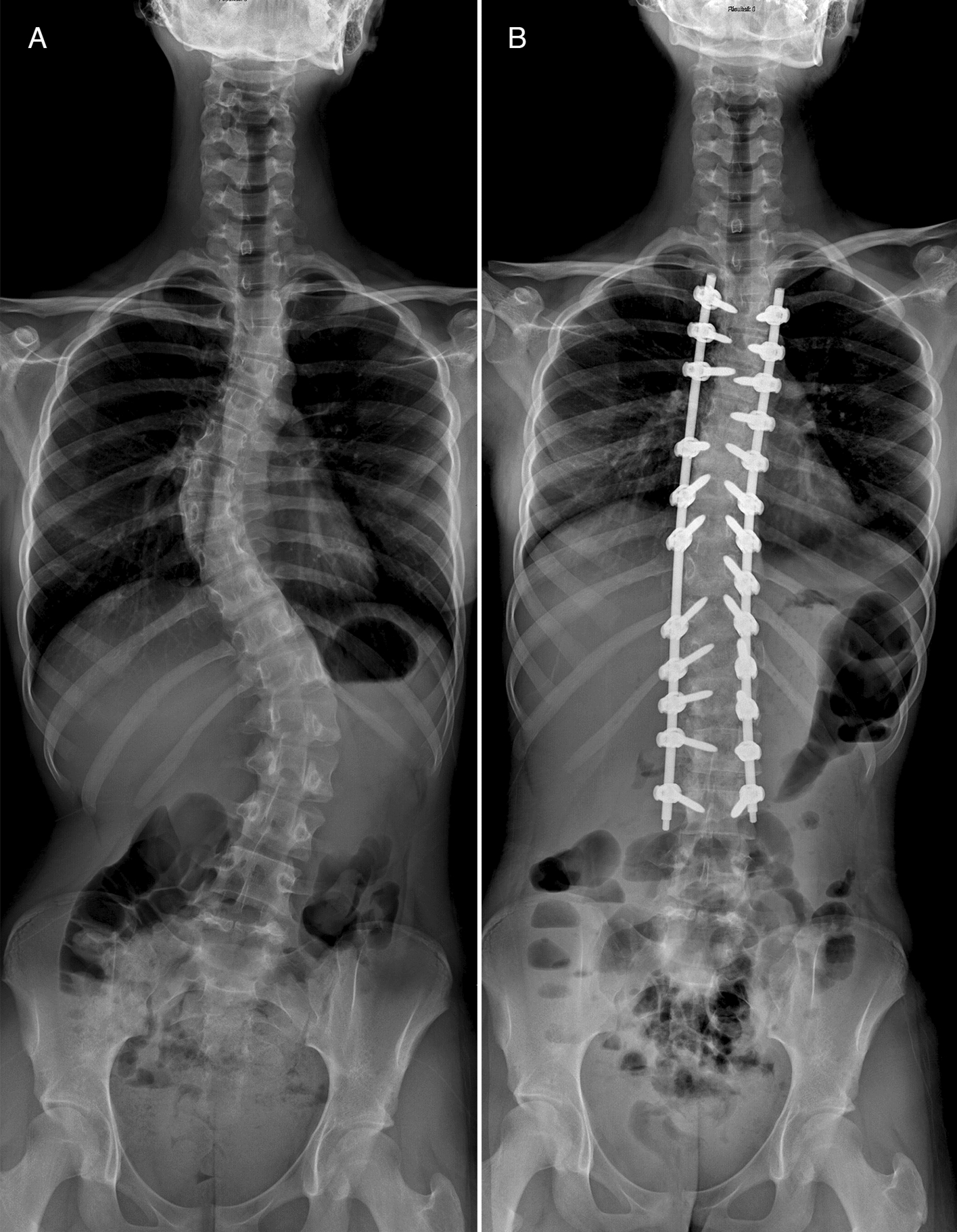
Pedicle screws entirely positioned through the spinal canal often cause clinical symptoms, and immediate repositioning can prevent irreversible (neurologic) damage [5, 10, 11]. The pedicle screws from cases E-L3 and E-T1 were positioned medial to the pedicle (entirely in the spinal canal), and both patients underwent secondary revision surgery due to clinical symptoms related to the misplaced pedicle screws. Based on 2D imaging, 20 of the 21 surgeons accepted the position of at least one of the two pedicle screws. If an intraoperative 3D image had been obtained, then almost all surgeons (20/21) would have repositioned the two pedicle screws immediately, possibly preventing a reoperation and/or irreversible neurological damage. The literature presents different results on whether the number of reoperations for misplaced pedicle screws decreases when an intraoperative 3D image of every placed pedicle screw is obtained compared to a 2D fluoroscopic workflow. One study among 198 patients treated for spinal deformity reported that reoperations due to misplaced pedicle screws decreased from 4.9% to no reoperations in five years [9]. However, another study among 810 patients with various spinal pathologies reported that reoperations due to misplaced pedicle screws did not (yet) decrease in 2.5 years; 0.99% with intraoperative CT available versus 0.99% without intraoperative CT available [6].
Spine surgeons repositioned the pedicle screws from cases B-T8 and C-T7 more often based on 3D imaging. In actual clinics, these two cases did not develop any clinical symptoms related to the breaching pedicle screws. Moreover, based on postoperative CTs and the postoperative clinical status of the patients, the treating spine surgeons did not consider revision surgery necessary. Breaches up to two millimeters are generally considered safe [5, 11,12,13] and breaches of up to four millimeters, when assessed on a postoperative CT, do not, as a rule, lead to clinical symptoms [5, 12, 13]. Therefore, repositioning the pedicle screws from cases B-T8 and C-T7 may be unnecessary.
Our study findings suggest that the additional intraoperative 3D information could increase redundant repositioning of pedicle screws with an acceptable position, a development that has been reported previously [6]. Future studies should specify how to interpret and act on intraoperative 3D information for evaluating pedicle screw positions as its use in spinal practice will only increase. Additionally, future studies should assess when 2D fluoroscopy may become less reliable for intraoperatively evaluating pedicle screw positions due to anatomical factors, such as spine deformity, high body mass index, or overlaying structures such as the pelvis or scapulae [5, 11,12,13].
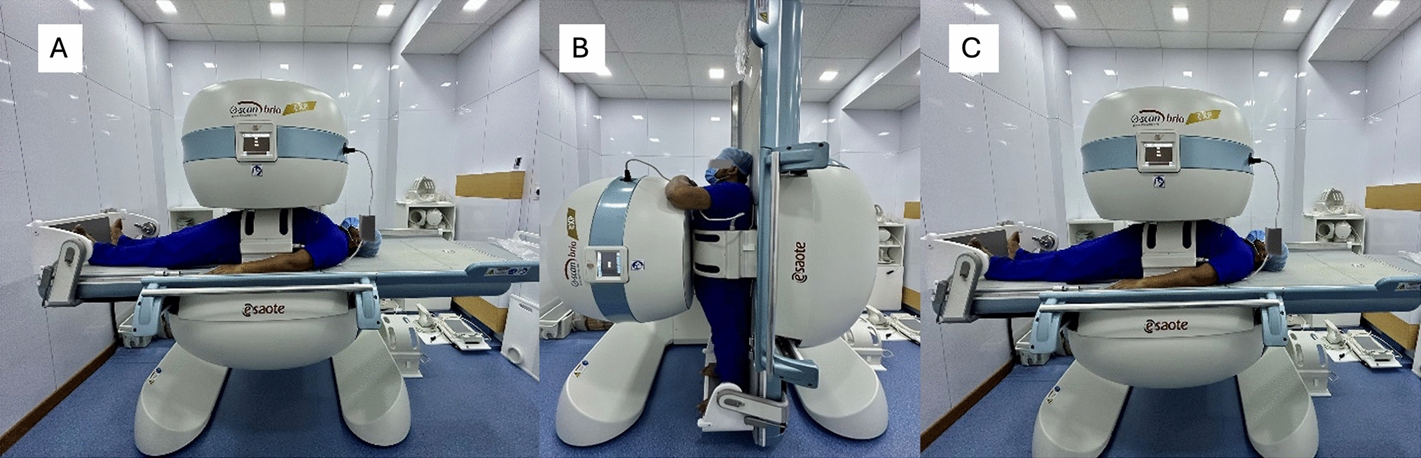
This study has several limitations. First, the survey cases do not represent a real situation in the operating room. During spine surgery, surgeons work with other team members and receive tactile feedback during screw insertion, and if an intraoperative fluoroscopic image is considered insufficient, a new image can be obtained. However, we consider it unlikely that this limitation affected the study findings. Six surgeons made a total of seven comments concerning five of the eight 2D cases, suggesting that, in an actual situation, they would have obtained additional 2D images or would have felt the pedicle walls with an awl first (Supplement 2). Of those five 2D cases, three presented screws without a breach or a breach of < 2 mm. More importantly, regarding the two cases that developed clinical symptoms postoperatively (E-L3 and E-T1), none of the surgeons made a comment on the provided 2D or 3D images, and almost all considered the screws positioned well (enough) based on the 2D images, as did the actual surgical team at that time. Second, spine surgeons assessed pedicle screw positions without knowing the indication for surgery, the function of the screw within the spinal construct, the planned screw trajectory, and the dimensions of the screw or pedicle. For example, spine surgeons can intentionally place thoracic pedicle screws with a lateral breach through the in-out-in technique, limiting the risk of a more critical medial breach. [14] To minimize the impact of specific patient considerations on decision-making, we did not include anatomically deformed pedicles and only included screws with a medial pedicle breach. Third, the survey did not capture individual surgeon thresholds for accepting pedicle screw positions, and our results indicate that those thresholds differ among surgeons. However, almost all spine surgeons intended to reposition more pedicle screws based on the provided 3D imaging than on the provided 2D images. Additionally, the results stratified for years of experience as a spine surgeon and the continent of residency appeared to be similar among the groups, though the number of participants did not allow for a reliable subanalysis. Fourth, we presented postoperative CT scans as intraoperative 3D images. A postoperative CT scan is superior for evaluating soft tissue to intraoperative 3D imaging, such as CBCT. However, for evaluating pedicle screw positions, multiple studies have shown that spine surgeons assess pedicle screw positions with equal accuracy on CT as on CBCT [15,16,17]. Also, some CTs were acquired well after the initial surgery, which, theoretically, may have resulted in late-onset loosening and movement of the pedicle screws. However, none of the selected pedicle screws or attached rods had pulled out or loosened on the used postoperative CTs. In addition, none of the patients had a history of osteoporosis or osteopenia. Therefore, we considered using postoperative CTs justified for our study objectives and unlikely to affect our findings.


留言 (0)