The previous studies on the spinopelvic parameters in TLSV employed the false sacral endplate as the reference line [14, 15, 21, 22], that is, the upper endplate was used as the reference line in sacralization cases and the lower endplate in lumbarization cases. A previous study described two key facts about TLSV [23]. First, sacralization and lumbarization do not affect the upper and lower endplate spinopelvic parameters and LL per se, although they have different numbers of lumbar vertebrae. Second, when measured separately, a statistically significant difference was noted between the spinopelvic parameters and LL of the upper and lower endplates. In the current study, the differences between the PI and LL values for the upper and lower endplates were relatively constant (27° and 14°, respectively). Therefore, it can be reasonably deduced that the selection of the reference sacral plate can affect surgical alignment goals.
Nevertheless, there is no consensus yet on the optimal sacral endplate for patients with TLSV. Zhou et al. calculated the upper and lower endplate spinopelvic parameters and LL of 70 patients with TLSV and spinal pathologies and reported that the lower endplate parameters may imply sagittal malalignment because of the high PT and PI–LL values. The difference between the average values of some of the parameters calculated in our study, and the normative values can be attributed to the positional effect of a CT-based study [20]. In the supine position, PT and LL values decrease by 3°–4°, while SS and PI–LL increased by the same amount [24]. Therefore, in our study, the average PT and PI–LL of lower endplate were approximately 22° and 6° in the standing position, respectively.
When classifying spinal deformities, PT and PI–LL are the chief modifiers of this classification system and should be < 20° and 10°, respectively [25]. On the other hand, the average PT value in healthy subjects was reported to between − 5° and 35° [26]. Thus, there is no optimal PI–LL value, which should be calculated on the basis of individual PI [27,28,29]. According to Le Huec [30], the relationship between PI and LL is not linear, and the formula PI–LL < 10° is valid only for small PI values. Based on this evidence, it can be assumed that, in asymptomatic subjects with TLSV, the upper endplate parameters indicate a low PI alignment, whereas lower endplate parameters indicate a high PI alignment. This observation also implies that both the sets of parameters may not be within the normal ranges in some cases.
Tatara et al. recently proposed a method to standardize the selection of endplates in patients with TLSV. They determined a reference range (mean ± 2 standard deviation [SD]) for PI and PT using values from patients with degenerative spine disease, but without imbalance and TLSV. If either PI, PT, or both endplates were out of the reference range, the other endplate was accepted as the optimum vertebral level. When both the upper and lower endplate PI and PT values came within this mean ± 2 SD range, the situation was interpreted as an “intermediate type” and other parameters such as LL, TK, and C7-plumbline were recruited for decision [17]. Apparently, this method may seem similar to that used in our study, except for some major differences. First, it is more complex and confusing than our current method. Second, they used both PI and PT for the detection of the optimum endplate and determined the upper endplate as the optimum vertebral level in 52% of the cases because the PT value of the lower endplate in their study was frequently out of the normative range and not PI. The main reason for the high PT indicated by the authors was pelvic retroversion to compensate for sagittal balance in patients with degenerative lumbar spine disease. As seen in their example of the upper reference endplate, it is evident that in several cases, the results of Tatara et al.’s method and our method would be different. However, the most important disadvantage of Tatara et al.’s method, in 23% of the cases, was unsatisfactory, and it is unclear how to manage these cases. In addition, the average values of the selected spinopelvic parameters and LL were not reported [17]. In contrast, we observed no statistically significant difference between the spinopelvic parameters and LL of the OPI and control groups, except for SS (difference = 2.5°), which was insignificant at p < 0.01. In the OPI group, for 60% of the cases, the upper endplate parameters were selected, whereas the lower endplate parameters were selected in the remaining 40%. This finding is not surprising because the average PI value of the upper endplate was closer to the normative PI value relative to that of the lower endplate. In terms of sacralization and lumbarization, no statistically significant difference was noted in the frequency distribution of valid endplates between the two groups, although the frequency of valid upper endplates in the sacralization group was higher than that in the lumbarization group (63% vs. 57%). This finding conforms with the result of our previous study wherein the upper and lower endplate parameters were found to be similar between the sacralization and lumbarization groups [23].
Furthermore, nearly similar correlations were observed between the spinopelvic parameters of both the OPI and control groups, expect for the correlation between PI and LL in the former (r = 0.382 versus 0.796); however, this correlation was also statistically significant (r = 0.382, p = 0.007).
The linear regression analysis offered different LL predictive formulas for the OPI and control groups. The two regression lines crossed at 46° PI, and the OPI group appeared as two clusters of patients–one at ~ 41° (average PI of upper endplate selected patients) and the other at ~ 54° (average PI of lower endplate selected patients). The range of PI in the OPI group (32.8°–65.6°) was also narrower than that in the control group (22.3°–72.4°); in this narrow range, the regression lines were very close to each other. In the first cluster, the LL of the OPI group was 2°–3° greater than that of the control group, whereas in the second cluster (PI ~ 54°), the LL of the OPI group was 2°–3° lesser than that of the control group. Therefore, the formula obtained for predicting LL using PI in the control group can be used in patients with TLSV.
Similarly, the PT and PI–LL values of the OPI group appeared as two clusters instead of a normal distribution. These clusters were present at around 6.77° and 14.9° for PT and − 3.5° and 7.27° for PI–LL, respectively; nevertheless, all values were within the normal ranges.
In some previous studies, the authors excluded cases with Castellvi type 2 because they did not have a true bony union between the transverse processes of the TLSV and the sacrum [10]. Moreover, there is a moveable disk between the TLSV and sacrum in this variety. Therefore, it is presumed that TLSV is a part of the pelvis, and hence, the upper endplate should be considered the functional endplate in Castellvi type 3–4 [17]; accordingly, the lower endplate should be used as the functional endplate in Castellvi type 2. However, there is a strong evidence that the joints in Castellvi type 2 restrict movement and absorb the load [31, 32]. Furthermore, disk height is lower than normal at the inferior level of TLSV, implying the presence of a less mobile (and more stable) disk at TLSV [32]. In below the TLSV, there is no option of using the upper endplate as a reference line. As a result, disk degeneration and adjacent segment disease usually occur at the vertebral level above the TLSV; therefore, we included Castellvi type 2 cases in our study. Nevertheless, in cases of disk degeneration below the TLSV, there is no option of using the upper endplate as a reference line.
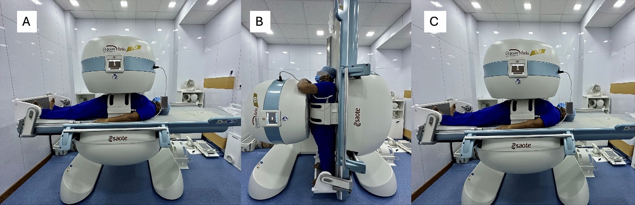
There are some limitations in this study. First, we included only asymptomatic subjects in the analysis. The inclusion of surgical candidates and results in symptomatic patients may have added more value to our results. Second, this was a CT-based study, and the spinopelvic parameters were measured in the supine position. Apart from PI, all other spinopelvic parameters changed with the position, and comparing them with normative values from the standing position did not seem appropriate. On the other hand, using CT scans was a strong point of this study as it is a more reliable imaging modality than plain radiography in the diagnosis and classification of TLSV [33]. CT scan is superior to X-ray for the detection of hypoplastic or short ribs simulating transverse processes on X-ray [34].


留言 (0)