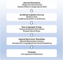
PE is defined differently in different countries, but most follow the criteria given by the International Society for the Study of Hypertension in Pregnancy (ISSHP), which is the one that is most frequently used globally. PE, according to the ISSHP, is defined as the development of new-onset hypertension, proteinuria, or other end-organ damage after 20 weeks of pregnancy and is also associated with the multisystem disorder peculiar to pregnancy (Brown et al., 2018). Addressing PE is critical due to its potential for severe medical consequences for both the mother and fetus. This condition, marked by high blood pressure and organ damage during pregnancy, can escalate to eclampsia, posing life-threatening risks such as seizures. Moreover, it can lead to organ damage, placental abruption, preterm birth, intrauterine growth restriction, and maternal complications like cardiovascular disease. Timely detection through regular prenatal care is essential, enabling interventions such as blood pressure management and close monitoring to mitigate risks. Ultimately, early identification and management of PE are vital for safeguarding maternal and fetal health and ensuring optimal pregnancy outcomes (Fox et al., 2019). Hypertensive disorders during pregnancy, including PE, rank as the second leading cause of maternal mortality globally, accounting for approximately 14% of deaths (with a confidence interval of 11.4–17.4%), resulting in an estimated 62,000–77,000 deaths annually. In pre-eclamptic pregnancies, the risk of fetal death surpasses that of non-pre-eclamptic pregnancies, with an adjusted odds ratio of 3.12 (with a 95% confidence interval of 2.77–3.51), primarily due to fetal growth restriction (FGR) and placental abruption (Abalos et al., 2014). Early-onset PE poses diagnostic challenges but demands early detection to manage risks like fetal growth restriction. In the mid-trimester, typically around 20–34 weeks, diagnosis becomes clearer, focusing on balancing maternal and fetal health through monitoring, medication, and possibly In individuals with delivery. Late-onset cases, occurring after 34 weeks, are generally less severe but still carry potential complications, often warranting delivery as the definitive treatment. Interventions within specific timeframes are key (Wojtowicz et al., 2019). Some women may experience hypertension in pregnancy (HTN-Preg), which can manifest in one of four ways: chronic HTN that precedes pregnancy, PE, chronic HTN with superimposed PE, or non-proteinuric gestational HTN. 5–8% of pregnancies in the US and 8 million pregnancies around the world are impacted by PE (Shah and Khalil, 2015). It is the leading cause of maternal and fetal morbidity and mortality worldwide. This occurs most commonly during the 20th or after the 20th week of the gestational period. Overall it affects 3–7% of all pregnancies. Its hallmark symptoms are Hypertension, Proteinuria, and Endothelial dysfunction causes extensive end-organ damage including Liver, blood, Kidneys, Brain and Placenta (MacDonald et al., 2022). Hemolysis, increased bio liver enzymes, and low platelet count (HELLP) syndrome, which was the first identified by Dr. Louis Weinstein in 1982, may also be a component of PE. Recent research indicates that HELLP syndrome, exhibits a close association with severe PE and eclampsia. Defined by three key characteristics – the destruction of red blood cells (hemolysis), elevated liver enzymes, and a decrease in platelet count – it affects roughly 0.5%-0.9% of all pregnancies. Notably, the incidence rises to 10%-20% amongst pregnancies experiencing severe PE (Sibai, 2004). ISSP reported that PE’s Systolic Blood pressure =>140 mmHg and/or Diastolic Blood Pressure =>90 mmHg measured at least twice at 4-hr intervals and If a woman with Proteinuria is defined as =>300 mg per 24 hour of urine collection (Poon et al., 2021). Currently, there exist various theories, including angiogenesis disorder (Schrey-Petersen and Stepan, 2017), immune dysfunction (Jafri and Ormiston, 2017), inflammasome activity (Socha et al., 2020), and senescence (Scaife et al., 2021), have been put forward to elucidate this phenomenon. Nevertheless, the precise etiology of inadequate placental invasion remains uncertain (Dymara-Konopka et al., 2017). PE is believed to have its origins in the placenta’s dysregulated vascular development. Which then produces a systemic endothelial cell malfunction by broadly dispersing anti-angiogenic substances into the mother’s circulation (Stillman and Karumanchi, 2007).


留言 (0)