
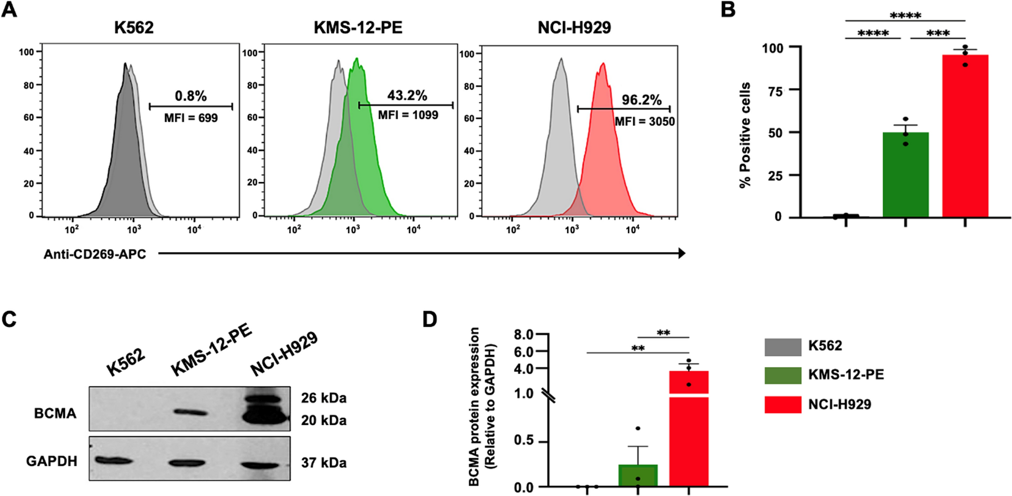
記住我

RNA-seq data were obtained in the TCGA and GTEx databases. By comparing the expression of CSRP1 in normal samples in the GTEX database and corresponding tumor samples in the TCGA database, CSRP1 was found to be significantly overexpressed in 7 cancers, including acute myeloid leukemia (LAML), and underexpressed in 18 cancers (Fig. 1A). For the CSRP gene family, CSRP1 was significantly overexpressed in AML, while CSRP3 was significantly reduced in AML compared to healthy controls (Fig. 1B). Furthermore, by comparing the expression of CSRP1 in 23 normal bone marrow (NBM) and 224 newly diagnosed AML patients in the ZZU cohort, we confirmed that CSRP1 was overexpressed in AML (Fig. 1C).
Fig.1
AML samples showed a higher expression of CSRP1 compared to normal samples. A Expression levels of CSRP1 in paired samples of normal and tumor patients in different cancers. B Comparisons of gene expressions among different CSRP family for normal and AML samples. C AML samples from the ZZU cohort showed a significant increase in CSRP1 expression compared to normal bone marrow samples. *, P < 0.05; **, P < 0.01; ***, P < 0.001
Association between CSRP1 expression and clinical featuresThe main clinical characteristics of AML in the ZZU cohort are shown in Table 1. A total of 224 AML patients were included in the prognostic analysis. The median follow-up time was 529 days. Up to the follow-up time, 138 patients died. The AML patients were divided into the low CSRP1 group (109 cases) and the high CSRP1 group (115 cases) according to the cut-off CSRP1 expression (225%). The high CSRP1 group had higher BM-blasts and more DNMT3A mutations than the low CSRP1 group (P < 0.05, Table 1 and Fig. 5G). We also compared the expression of CSRP1 in AML patients by the other characteristics. The expression levels of CSRP1 showed no significant difference in patients with different gender, age, WBC, cytogenetic risk group, BM-blasts, and PB-blasts (P > 0.05; Fig. 2A–F).
Table 1 Association between CSRP1 expression and clinical features in the ZZU cohortFig.2
Association between CSRP1 expression and clinical features in the ZZU cohort. A–F Comparisons of CSRP1 expression in AML patients based on gender A, age B, cytogenetics risk C, WBC D, BM-blasts E, PB-blasts F. G Association among CSRP1 expression, survival status and common gene mutations in AML. ns: P > 0.05; *, P < 0.05
High CSRP1 impacted the prognosis of AMLPatients with high CSRP1 expression had a significantly worse prognosis than those with low CSRP1 expression (hazard ratio [HR], 2.36 (1.53–3.64); P < 0.001; Fig. 3A) in the TCGA-LAML dataset. The predictive significance of elevated CSRP1 was verified in the Beat-AML dataset (Fig. 3B), the ZZU cohort (Fig. 3C), and the GSE12417 dataset (Fig. 3D–E). The time-dependent ROC curve from the TCGA-LAML dataset demonstrated that CSRP1 was an excellent predictor of AML patient survival.
Fig.3
High expression of CSRP1 was associated with poor OS in AML patients. A–E Kaplan–Meier curves of OS in the TCGA-LAML dataset A, the Beat-AML dataset B, the ZZU cohort C, the GSE12417-GPL96 dataset D, and the GSE12417-GPL570 dataset E and F Time-dependent ROC curve of CSRP1 in TCGA-LAML dataset. OS, overall survival; HR, hazards ratio
Based on the TCGA-LAML dataset, independent prognostic factors for worse OS included CSRP1 (high vs. low, P < 0.001), cytogenetic risk (adverse vs. favorable, P < 0.001; intermediate vs. favorable, P = 0.002), and age (> 60 vs. ≤ 60, P < 0.001) (Table 2 and Fig. 4A). The independent prognostic value of these three factors was further confirmed in the ZZU cohort (Fig. 4B).
Table 2 Univariate analysis and multivariate analysis of the prognostic factors in the TCGA-LAML datasetFig.4
Construction of the nomogram model for AML patients. A–B The forest plots showed that age, cytogenetic risk, and CSRP1 expression are independent factors for poor prognosis in the TCGA-LAML dataset A and the ZZU cohort B. C–D Nomogram for predicting the probability of 1-, 2-, 3-year OS for AML in the C TCGA-LAML dataset and D the ZZU cohort. E–F Calibration plot of the nomogram for predicting the probability of OS at 1, 2, and 3 years for AML in the E TCGA-LAML dataset and F the ZZU cohort
Prognostic model of CSRP1 in AMLA nomogram was developed based on the Cox regression analyses in the TCGA dataset (Fig. 4C) and the ZZU cohort (Fig. 4D) to better predict AML patients' prognoses. Three independent prognostic variables, age, cytogenetic risk, and CSRP1 expression, were included in the model at a statistical significance level of 0.05. A point scale was utilized to allocate points to these factors based on multivariate Cox analysis. Results from the nomogram calibration curve of OS prediction were consistent with observations of all patients in both the TCGA dataset (Fig. 4E) and the ZZU cohort (Fig. 4F).
Identification of DEGs in AML samples with low and high CSRP1 expressionThe gene expression profiles of the high and low CSRP1 groups were analyzed for differences in median mRNA expression. A total of 2758 DEGs from RNA-seq-HTSeq counts were found to be statistically significant between the high and low CSRP1 groups (|log fold change (logFC)|> 0.5, P adj < 0.05). (Fig. 5A). The heat map depicted the top ten up-regulated DEGs and top ten down-regulated DEGs between the high and low CSRP1 groups (Fig. 5B).
Fig.5
GO/KEGG enrichment analysis of DEGs comparing patients with high or low CSRP1 expression in the TCGA-LAML dataset. A Volcano map of the DEGs, including 862 down-regulated genes and 1896 up-regulated genes. B Heat map showing the top ten up-regulated and the top ten down-regulated genes. The samples are shown on the X-axis, while the DEGs are shown on the Y-axis. C–D GO enrichment analysis of the up-regulated DEGs. MF, molecular function. CC, cellular component. BP, biological process. D KEGG enrichment analysis of the up-regulated DEGs. Different categories were shown on the Y-axis, while the X-axis reflected the percentage of DEGs
Functional enrichment analysis of DEGsBiological process (BP) associated with high CSRP1 included neutrophil activation, neutrophil activation involved in immune response, neutrophil degranulation, and neutrophil mediated immunity (Fig. 5C). Cellular components (CC) associated with high CSRP1 included secretory granule membrane, ficolin-1-rich granule, tertiary granule, and cell leading edge (Fig. 5C). Molecular function (MF) associated with high CSRP1 included actin binding, Rho GTPase binding, cytokine binding, and cytokine receptor activity (Fig. 5C). KEGG included phagosome, cytokine-cytokine receptor interaction, lysosome, oxytocin signaling pathway, cell adhesion molecules, rap1 signaling pathway, chemokine signaling pathway, gap junction, HIF-1 signaling pathway, proteoglycans in cancer, Fc gamma R-mediated phagocytosis, natural killer cell mediated cytotoxicity, JAK-STAT signaling pathway, and apoptosis (Fig. 5D).
The prognostic value of the top ten up-regulated DEGs and top ten down-regulated DEGs was further investigated. Six of the top ten DEGs that were up-regulated (ONECUT2, CES1, CD163, KCNN2, THBS1, and B3GALT1) and six of the top ten DEGs that were down-regulated (MYT1L, ASTN1, DLL3, MYO18B, POU4F1, and PCDH10) also had vital prognostic significance in the TCGA AML dataset (Fig. 6).
Fig.6
Prognostic value of DEGs in TCGA-LAML dataset. A–F Six of the top ten up-regulated DEGs (ONECUT2 A, CES1 B, CD163 C, KCNN2 D, THBS1 E, and B3GALT1 F) showed an adverse effect on OS. (G–L). Six of the top ten down-regulated DEGs (MYT1L G ASTN1 H, DLL3 I, MYO18B J, POU4F1 K, and PCDH10 L) had a protective effect on OS
Analysis of immune infiltration in AMLCSRP1 expression in the AML microenvironment was associated with the level of immune cell infiltration as measured by ssGSEA. Mainly, CSRP1 was negatively related to T cells and T helper cells and positively associated with macrophages (Fig.S1C), neutrophils (Fig.S1D), NK CD56 dim cells, NK CD56 bright cells, Tem, and iDC, TFH, Th17 cells, Th1 cells, eosinophils, Treg, and aDC (Fig.S1A). Patients with high CSRP1 had higher enrichment scores of aDC, Eosinophils, iDC, macrophages, neutrophils, NK CD56 bright cells, NK CD56 dim cells, Tem, Th1 cells, Treg, and lower enrichment scores of T helper cells.
PPI enrichment analysis in AML and validation of hub genesThe PPI network was constructed using the String website and the data were generated by Cytoscape (MCODE plug-in). A total of 321 DEGs were imported into the PPI network. Complete module including all genes is presented in Fig.S2A. We obtained 235 nodes and 900 edges. Nineteen genes were present in the most significant module (MCODE score: 7.667; Fig.S2B). The TCGA-LAML dataset was then used for OS analysis on the hub genes. Nine of the 19 hub genes, including CD163, CX3CR1, C5AR1, THSD7A, ADMATS18, IL10, THBS1, ADAMTS15, and LILRB2, were correlated with OS in AML (P < 0.05, Fig.S3).
CSRP1 expression can predict sensitivity to the common chemotherapy agents in AML treatmentWe utilized the “oncoPredict” tool to estimate sensitivity to frequently used chemotherapy agents to better correlate the CSRP1 expression with clinical practice. Accordingly, drug sensitivity of patients in high and low CSRP1 groups to multiple chemotherapy agents including 5-fluorouracil, gemcitabine, rapamycin, cisplatin, and fludarabine was predicted. Based on the findings, the high CSRP1 groups of patients in the TCGA datasets showed higher sensitivity to 5-fluorouracil, gemcitabine, rapamycin, and cisplatin and lower sensitivity to fludarabine (Fig.S4). The relationship of sensitivity to other chemotherapy agents with CSRP1 expression is listed in the supplementary data.
留言 (0)