
記住我


Colorectal cancer (CRC) is the third most common cancer worldwide and the second leading cause of cancer-related deaths (1,2). The current Japanese CRC treatment guidelines identify poor differentiation (PD), submucosal invasion of >1,000 μm, lymphovascular invasion (LVI), and tumor budding grade (BD) 2 or 3 as risk factors of lymph node metastasis (LNM) in patients with CRC (3). Most guidelines and pathology reports, including those in the United States, Europe, and Asia, consider tumor differentiation as a risk factor of LNM in T1 CRC (3–7). Surgical resection with lymph node dissection is recommended for poorly differentiated adenocarcinoma, signet ring cell carcinoma, or mucinous carcinoma identified by histological examination (3–7). Because surgical resection is linked to high morbidity, mortality, and costs and reduced quality of life, compared with endoscopic resection, selecting the appropriate treatment modality is crucial (3–7).
There are 2 classifications of histological differentiation in CRC treatment guidelines: highest grade and predominant (3,4,6). The World Health Organization and International Collaboration on Cancer Reporting classify differentiation according to the highest grade present (6,8). However, the Japanese guidelines and College of American Pathologists Cancer Protocols classify differentiation according to the predominant grade (3,9). The approach to histological differentiation classification varies globally, with some institutions in Europe, South America, and Asia opting for the predominant differentiation classification, establishing that the concept is far from uniform across countries and institutions (10–16). For instance, if a resected tumor is composed of 95% well-differentiated and 5% poorly differentiated tissue, diagnoses of well-differentiated and poorly differentiated tumors would signify using predominant and highest-grade differentiation, respectively.
However, whether predominant or highest-grade differentiation would be a better predictor of LNM in T1 CRC remains to be clarified (17,18). The main indicators of diagnostic accuracy for LNM are sensitivity and specificity (19). While highest-grade differentiation identifies all cases with PD within the tumor through comprehensive examination of serial paraffin-embedded tissue sections, predominant differentiation is based on most of the tumor. Therefore, highest-grade differentiation tends to overestimate malignancy (18), which may elevate the risk estimation of LNM, potentially inducing unnecessary surgeries. To the best of our knowledge, no previous study has systematically reviewed the extent of this overestimation.
In this study, we aimed to assess the diagnostic accuracy of highest-grade or predominant histological differentiation in T1 CRC for determining LNM. We assessed clinical utility through sensitivity and specificity. As an interpretation of these measures, increased sensitivity might reduce the rate of missed metastases, and decreased specificity could lead to unnecessary surgeries (20,21).
METHODSWe adhered to the methodological standards of the Cochrane handbook for systematic review of diagnostic test accuracy and the preferred reporting items for systematic review and meta-analysis of diagnostic test accuracy (see PRISMA-DTA, Supplementary Digital Content 1, https://links.lww.com/CTG/B60) (19,22). Our study protocol was registered in the international prospective register of systematic reviews (PROSPERO, registration number: CRD42023416971) and on the open science framework platform (https://osf.io/TMAUN/) on April 13, 2023.
Study selectionWe included studies that assessed the impact of predominant histological differentiation and/or highest-grade differentiation in T1 CRC. We included prospective and retrospective cohort studies and case-control studies and excluded case reports, case series, reviews, meta-analyses, and duplicate cases. Patients older than 18 years with CRC who had undergone endoscopic or surgical resection were included. The exclusion criteria were familial adenomatous polyposis, Lynch syndrome, inflammatory bowel disease, preoperative chemotherapy, and radiotherapy. The index evaluated in this review was predominant histological differentiation or highest-grade differentiation. The target condition was LNM in patients with T1 CRC. The reference standard was the presence of LNM in surgical specimens. The primary outcome was diagnostic accuracy, including sensitivity and specificity, of LNM prediction in T1 CRC, as determined by predominant histological differentiation or highest-grade differentiation.
Search methodThe Cochrane Central Register of Controlled Trials (CENTRAL), MEDLINE (PubMed), and EMBASE (ProQuest Dialog) databases were searched (see Search Strategy, Supplementary Digital Content 2, https://links.lww.com/CTG/B61). In addition, we searched the World Health Organization International Clinical Trials Platform Search Portal and ClinicalTrials.gov (see Search Strategy, Supplementary Digital Content 2, https://links.lww.com/CTG/B61) for ongoing trials. We did not apply restrictions on the observation period, publication year, language, or country. We considered all studies, including published articles, conference abstracts, and letters. We checked the reference lists of all included studies, international guidelines (3–7), and articles citing eligible studies. We contacted the authors of original studies for additional data.
Data management and assessment of methodological qualityTwo of the 3 independent reviewers (J.W. and S.M. or A.M.) screened the titles and abstracts and subsequently assessed the eligibility of full-text articles. Two of the 3 independent reviewers (J.W. and S.M. or A.M.) performed independent data extraction from the included studies. Disagreements between 2 reviewers were resolved by discussion, and a third reviewer acted as an arbiter if the discussion failed (K.I. or Y.K.). We contacted the original authors if relevant data were missing. Two independent reviewers (J.W. and A.M.) evaluated the risk of bias independently using the quality assessment of diagnostic accuracy studies–2 tool (23). Disagreements between reviewers were discussed, and if agreement could not be reached, a third reviewer (K.I. or Y.K.) arbitrated.
Data synthesis and statistical analysesWe performed a single-group analysis to examine the proportion of positive LNM in T1 CRC. We used the Metaprop command in Stata to calculate the pooled proportions with 95% confidence intervals (CIs), with inverse variance weights obtained from random-effects models (24).
Data for 2 × 2 tables of LNM against the reference standard were extracted from each study. For each of the highest-grade and predominant histological differentiations, we estimated sensitivity and specificity with 95% CIs for LNM per study using forest plots to inspect between‐study variability. In the meta-analysis, we simplified the model to a univariate random-effects logistic regression model to pool sensitivity and specificity with 95% CI separately because we were unable to fit a bivariate model owing to sparse data, few studies, or minimal observed variability in specificity (19,25). The model accounted for both within-study and between-study variability in test performance, including random effects (26). To elucidate the influence of effect modifiers on results, we performed subgroup analysis according to region (Japan vs other countries) for the highest-grade and predominant histological differentiations.
Based on the Cochrane handbook, we did not perform univariate tests for sensitivity and specificity or calculate estimates of the I2 statistic; these methods do not account for heterogeneity attributable to phenomena such as positive threshold effects (19). We performed the following sensitivity analyses to assess whether results were robust enough for the conclusions drawn in the review; only studies with participants who were surgically treated (surgical resection only and/or additional surgical resection after endoscopic resection) and those with participants who met our inclusion/exclusion criteria were included. We did not examine publication bias for the diagnostic accuracy of LNM owing to a lack of appropriate statistical methods (19).
Assuming that there were no biases, including confounding, selection, or misclassification of exposure, we simulated a patient population with T1 CRC considering 2 potential outcomes: positive LNM necessitating surgery and negative LNM not necessitating surgery. With a positive lymph node proportion of 11.2%, according to a recent systematic review (27), the simulation used 1,000 bootstrap resampling, thereby assessing the sensitivity and specificity of the highest-grade and predominant histological differentiations (28,29). This simulation enabled determining the proportion and range of false negatives (missed positive LNM) and false positives (unnecessary surgeries) when comparing the 2 differentiation classifications. We set the sensitivity and specificity of the highest grade of differentiation to the mean values obtained in the meta-analysis. In addition, we calculated the mean and range of the differences in false positives and negatives between the highest-grade and predominant differentiations in Japan and other countries.
We used STATA SE16 (version 16.1; Stata, College Station, TX) with the Metaprop, metandi, and midas packages (24,30,31) and R version 4.1.2 (The R Foundation for Statistical Computing, Vienna, Austria) with the R packages meta version 6.2.1 and boot version 1.3.28 (32,33).
RESULTS Study selection and characteristicsFigure 1 illustrates the study selection process. We identified 4,473 records from 5 electronic databases after removing duplicate records on April 14, 2023. After screening, we ultimately identified 42 studies with 41,290 patients (10–14,16,34–69).
 Figure 1.:
 Flowchart of the study selection process.
Supplementary Table 1 (see Supplementary Digital Content 4, https://links.lww.com/CTG/B63) outlines the characteristics of the studies included in this review. Among the 42 studies, 27 evaluated the diagnostic accuracy of the highest-grade differentiation for LNM in patients with T1 CRC, 14 assessed predominant histological differentiation, and 1 reported outcomes for both classifications. For highest-grade differentiation, 18 studies were from Asia and 10 from Europe and the United States. Regarding predominant differentiation, 12 studies were from Japan and 3 were from Europe and Asia, excluding Japan. In addition, one study from Japan addressed both classifications.
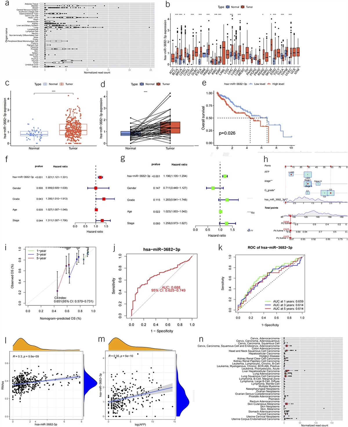
Supplementary Digital Content 3 (https://links.lww.com/CTG/B62) displays the risk of bias assessment of the eligible studies using the quality assessment of diagnostic accuracy studies–2 tool. For the domain of patient selection, we scored 1 study as having an unclear risk of bias and 8 as having high applicability concerns. Regarding the index test, all 42 studies had an unclear risk of bias with low applicability concerns. Regarding the reference standard, 3 studies had a high risk of bias and 8 had high applicability concerns. In the flow and timing domain, 8 studies had a high risk of bias. The pooled proportion of LNM in T1 CRC was 11% (95% CI 10%–12%) (Figure 2).
 Figure 2.:
 Forest plot for the proportion of lymph node metastasis in T1 colorectal cancer. ES, effect size; CI, confidence interval.
Diagnostic accuracy of highest-grade or predominant histological differentiation for LNMFigure 3 shows the analysis of the differentiation classifications. They had pooled sensitivities of 0.18 (95% CI 0.13–0.24) and 0.06 (95% CI 0.04–0.09) (test for subgroup differences, P < 0.0001) and specificities of 0.95 (95% CI 0.93–0.96) and 0.98 (95% CI 0.97–0.99) (test for subgroup differences, P < 0.0001) for the highest-grade and predominant histological differentiations, respectively.
 Figure 3.:
 Forest plot showing the sensitivity and specificity of the highest-grade and predominant histological differentiations for prediction of lymph node metastasis. CI, confidence interval.
In Japan, differentiation classification had pooled sensitivities of 0.28 (95% CI 0.22–0.34) and 0.06 (95% CI 0.04–0.09) (test for subgroup differences, P < 0.0001) and specificities of 0.89 (95% CI 0.85–0.92) and 0.98 (95% CI 0.97–0.99) (test for subgroup differences, P < 0.0001) for the highest-grade and predominant histological differentiations, respectively.
In other countries, differentiation classification had pooled sensitivities of 0.16 (95% CI 0.12–0.23) and 0.09 (95% CI 0.04–0.19) (test for subgroup differences, P = 0.167) and specificities of 0.96 (95% CI 0.94–0.97) and 0.98 (95% CI 0.97–0.99) (test for subgroup differences, P = 0.0029) in the highest-grade and predominant histological differentiations, respectively.
Additional analysesFigure 4 shows the results of subgroup analysis by region (Japan and other countries); highest-grade differentiation had pooled sensitivities of 0.28 (95% CI 0.22–0.34) and 0.16 (95% CI 0.12–0.23) (test for subgroup differences, P = 0.011) and specificities of 0.89 (95% CI 0.85–0.92) and 0.96 (95% CI 0.94–0.97) (test for subgroup differences, P = 0.0003) in Japan and other countries, respectively.
 Figure 4.:
 Forest plot for the subgroup analysis according to region (Japan and other countries) regarding highest-grade differentiation. CI, confidence interval.
Figure 5 shows the results of subgroup analysis by region (Japan and other countries); predominant differentiation had pooled sensitivities of 0.06 (95% CI 0.04–0.08) and 0.09 (95% CI 0.04–0.21) (test for subgroup differences, P = 0.28) and specificities of 0.98 (95% CI 0.97–0.99) and 0.98 (95% CI 0.97–0.99) (test for subgroup differences, P = 0.93) in Japan and other countries, respectively.
 Figure 5.:
 Forest plot for the subgroup analysis according to region (Japan and other countries) regarding predominant differentiation. CI, confidence interval.
The results of sensitivity analyses were consistent with the main results (see Supplementary Table 2, Supplementary Digital Content 5, https://links.lww.com/CTG/B64). In the bootstrap simulation, the difference in sensitivity between the highest-grade and predominant differentiations was 0.12 (95% CI 0.060–0.18), and the difference in specificity was −0.034 (95% CI −0.018 to −0.054). In the simulated cohort of 100 patients with T1 CRC, with an assumed LNM proportion of 11.2%, the differences in false positives and negatives between the highest-grade and predominant differentiations were 3.0 individuals (range 1.6–4.4) and −1.3 individuals (range −2.0 to −0.7), respectively.
In Japan, in the bootstrap estimation, the difference in sensitivity between the highest grade and predominant differentiations was 0.21 (95% CI 0.17–0.27) and that in specificity was −0.091 (95% CI −0.14 to −0.064). In the simulated cohort of 100 patients with T1 CRC, with an assumed LNM proportion of 11.2%, the difference in false positives and negatives between the highest-grade and predominant differentiations was 8.1 individuals (range 5.7–9.8) and −2.3 individuals (range −3.0 to −1.9), respectively.
In other countries, in the bootstrap estimation, the difference in sensitivity between the highest-grade and predominant differentiations was 0.066 (95% CI −0.025 to 0.16), and the difference in specificity was −0.029 (95% CI −0.047 to −0.014). In the simulated cohort of 100 patients with T1 CRC, with an assumed LNM proportion of 11.2%, the differences in false positives and negatives between the highest-grade and predominant differentiations were 2.6 individuals (range 1.3–3.6) and −0.7 individuals (range −2.0 to 0.05), respectively.
DISCUSSIONThe systematic review and meta-analysis of 42 studies involving 41,290 patients investigated the diagnostic accuracy of highest-grade and predominant histological differentiations in T1 CRC for predicting LNM. We found that the sensitivity of highest-grade differentiation was higher than that of predominant histological differentiation, whereas the specificity of the latter was higher than that of the former. In the 100 patients with T1 CRC, using highest-grade differentiation could have prevented oversight of 1 case with LNM, whereas using predominant differentiation could have prevented additional surgeries in 3 patients.
PD was found to have high sensitivity, especially with highest-grade differentiation, and low specificity, especially with predominant differentiation. International guidelines recommend evaluating 4 predictive factors for LNM (PD, depth of submucosal invasion, LVI, and BD) before deciding on additional surgical resection following endoscopic resection. In previous studies, depth of submucosal invasion, LVI, and BD showed sensitivities of 0.95, 0.25, and 0.25 and specificities of 0.40, 0.94, and 0.95, respectively (40,60,70). Compared with other major predictive factors, PD might have the highest specificity but the lowest sensitivity. Given that the specificity of PD is sufficiently high, adopting the highly sensitive high-grade differentiation classification over predominant differentiation as a stand-alone predictor may help prevent misclassification owing to increased sensitivity. Contrarily, previous studies have shown the sensitivity of all 4 predictive factors combined to positive LNM to be 100%, whereas specificity was extremely low (21,71). When considering all 4 factors, adopting the highly specific predominant differentiation classification might lead to increased specificity of the overall predictors.
The choice of highest-grade or predominant differentiation classification hinges on multiple factors, such as patient characteristics and the health care economic circumstances of the country or facility. To illustrate this, selection of the highest-grade differentiation might be more beneficial for patients with certain attributes, including those at low surgical risk (younger patients or those without comorbidities) who wish to survive longer (72,73) because highest-grade differentiation was more effective than predominant differentiation in preventing 1% of missed lymph node–positive patients from undergoing surgery. LNM-positive patients can have potentially fatal outcomes, including metastatic recurrence, if not treated surgically. In addition, the economic situation of the country can significantly influence this choice. In a country with high health care costs or high surgical risk, choosing predominant differentiation could be more cost-effective, providing similar benefits at a lower cost, because additional surgeries could impose a high cost of approximately $8,000 USD, besides being associated with the risk of surgery-related mortality (1.7%) and recurrence (0.7%–1.0%) despite tumor resection (72,74–76). Furthermore, older patients with multiple comorbidities and patients with lower rectal cancer, despite the high risk of LNM, may be candidates for observation rather than for surgery (77). The benefits of surgical resection, especially in older patients and those with severe comorbidities, may be limited owing to its impact on prognosis (78). In addition, for patients with lower rectal tumors, surgery (specifically lymph node dissection) carries the risk of inducing anal, urinary, and sexual dysfunction (79,80). When comparing highest-grade differentiation with predominant differentiation, the choice cannot be made in isolation. Individual patient needs and economic realities need to be weighed in this multifaceted and complex decision, which can have far-reaching implications for patient health outcomes and economic sustainability of the health care system. Hence, an informed decision that balances both considerations is crucial. Thus, selection of highest-grade differentiation or predominant differentiation is not one-size-fits-all, but must be discussed by clinicians, patients, pathologists, and policymakers using the Delphi method according to international guidelines in each country and considering individual and societal factors (81).
We recommend that pathologists assessing cases of T1 CRC use both classification systems and provide information about highest-grade and predominant differentiation. In addition, in cases where a cancerous lesion is associated with multiple coexisting histologic types, it is beneficial to list each histological type in order of area predominance, as outlined in the Japanese guidelines (3). Furthermore, the overall proportion of PD can aid in identifying PD possibly missed in subsequent histological sections. A comprehensive approach incorporating both classifications can provide valuable information for well-informed clinical decisions. This inclusive evaluation ensures better understanding of the differentiation status of the tumor, facilitating the tailoring of treatment strategies and the most appropriate management approach for each patient.
Variations in guideline recommendations for differentiation classification across countries may have contributed to differences in the assessment protocols for histopathological diagnosis and the observed variability in diagnostic accuracy between Japan and other countries in subgroup analysis (3,4,6,8). These disparities could be attributed to differences in the handling and processing of excised specimens, variations in histological evaluation among pathologists with varying levels of expertise and experience, and diverse differentiation classification criteria across countries (3,4,6,8). To ensure consistent and standardized interpretation of diagnostic criteria, it is crucial to establish uniform international diagnostic criteria for differentiation classification in cases of CRC. Such guidelines would facilitate harmonization of practice among pathologists worldwide and improve the reliability and comparability of diagnostic accuracy assessments across different regions.
This study has some limitations. First, considerable variation exists in the analysis and reporting of histological factors, including differentiation classification, the diagnostic concordance rate of histology, and the implementation rate of immunostaining (82) across facilities and countries. In particular, the diagnostic concordance rate between Japanese pathologists and those in Asia, Europe, and the United States is a major problem. However, we investigated specific diagnostic criteria for differentiation and evaluated cross-country differences using subgroup analysis. There is a need to unify tumor grading criteria as a future global initiative. Second, the specific threshold for the proportion of PD that predicts LNM in cases of differentiated cancer with mixed PD components remains unknown. Therefore, further studies should focus on determining this threshold in T1 CRC. Third, the lack of multivariable analysis in our meta-analysis of included studies prevented the determination of potential interactions between different predictive factors for LNM. Caution should be exercised when interpreting results from a meta-analysis of multivariable studies. This is because of inconsistencies in the classification of differentiation for other predictive factors, such as the evaluation of the depth of submucosal invasion or LVI with or without additional staining. Although an earlier meta-analysis of multivariable studies had reported depth of submucosal invasion as an inadequate predictor of LNM (27), one study in the meta-analysis used predominant differentiation and reported depth of submucosal invasion to be associated with positive LNM. A recent multicenter study using predominant differentiation reported depth of submucosal invasion as an independent predictor for LNM (49). The outcomes may differ for these factors because of the varying evaluation methods across the studies. Further studies are needed to evaluate the classification of differentiation, adjusted for other predictive factors, through meta-analysis or multinational and multicenter studies of individual patient data. Last, it is problematic to evaluate the same data for endoscopically treated cases in Japan, where endoscopic submucosal dissection is usually performed as a batch resection, and in Europe and the United States, where segmental resection is more common. However, we evaluated cross-country differences using subgroup analysis.
In conclusion, this systematic review and meta-analysis demonstrated that highest-grade differentiation had greater sensitivity than predominant histological differentiation, while predominant histological differentiation exhibited greater specificity than highest-grade differentiation. Applying highest-grade differentiation could avert missing 1% of LNM, while using predominant differentiation could prevent 3% of unnecessary surgeries. The findings overall implied that clinicians, patients, pathologists, and policymakers should discuss the choice of highest-grade and predominant differentiations, balancing patient health outcomes and economic sustainability of the health care system. Further studies are needed to evaluate differentiation classification adjusted for other predictive factors through individual patient data meta-analyses or multinational and multicenter studies.
CONFLICTS OF INTERESTGuarantor of the article: Jun Watanabe, MD, PhD.
Specific author contributions: J.W., K.I., and Y.K.: contributed to the study concept and design and drafting of the manuscript. J.W.: obtained funding. J.W., K.I., Y.K., S.M., A.M., and K.G.Y.: contributed to the statistical analysis and interpretation of data. K.K. and N.S.: contributed to administrative support and study supervision. J.W., K.I., and Y.K.: developed the software. J.W., K.I., Y.K., S.M., A.M., K.G.Y., F.M.d.J., and I.M.: contributed to data collection and critical revision of the manuscript. S.K., K.K., and N.S.: contributed to critical revision of the manuscript.
Financial support: The study was supported by JSPS KAKENHI (grant number JP21K21121 and 23K16289).
Potential competing interests: None to report.
ACKNOWLEDGMENTSWe thank Hirotoshi Kobayashi from the Department of Surgery, Teikyo University School of Medicine, Mizonokuchi Hospital; Shiro Oka from the Department of Gastroenterology, Hiroshima University Hospital, Kazutomo Togashi from the Department of Coloproctology, Aizu Medical Center, Fukushima Medical University; Hirotoshi Hasegawa from the Department of Surgery, Tokyo Dental College Ichikawa General Hospital; Kazuhiro Yasuda from the Department of Gastroenterological Surgery, Oita Prefectural Hospital; Huann-Sheng Wang from the Department of Surgery, Division of Colorectal Surgery, Taipei Veterans General Hospital; Jung-Wook Huh from the Division of Colorectal Surgery, Department of Surgery, Samsung Medical Center, Sungkyunkwan University School of Medicine; Young-Seok Cho from the Division of Gastroenterology, Department of Internal Medicine, Seoul St. Mary's Hospital, College of Medicine, The Catholic University of Korea; Chang Sil Yu from the Department of Surgery, University of Ulsan College of Medicine & Asan Medical Center; Shinsuke Kazama from the Department of Surgery, Yaizu City Hospital; and Shinji Yoshii from the Department of Gastroenterology and Hepatology, Sapporo Medical University School of Medicine for providing us with the detailed information necessary for this study.
REFERENCES 1. Sung H, Ferlay J, Siegel RL, et al. Global cancer statistics 2020: GLOBOCAN estimates of incidence and mortality worldwide for 36 cancers in 185 countries. CA Cancer J Clin 2021;71(3):209–49. 2. Siegel RL, Miller KD, Fuchs HE, et al. Cancer statistics, 2021. CA Cancer J Clin 2021;71(1):7–33. 3. Hashiguchi Y, Muro K, Saito Y, et al. Japanese Society for Cancer of the Colon and Rectum (JSCCR) guidelines 2019 for the treatment of colorectal cancer. Int J Clin Oncol 2020;25:1–42. 4. Vogel JD, Felder SI, Bhama AR, et al. The American Society of Colon and Rectal Surgeons clinical practice guidelines for the management of colon cancer. Dis Colon Rectum 2022;65(2):148–77. 5. Kim BH, Kim JM, Kang GH, et al. Standardized pathology report for colorectal cancer, 2nd edition. J Pathol Transl Med 2020;54:1–19. 6. Nagtegaal ID, Odze RD, Klimstra D, et al. The 2019 WHO classification of tumours of the digestive system. Histopathology 2020;76(2):182–8. 7. Pimentel-Nunes P, Libânio D, Bastiaansen BAJ, et al. Endoscopic submucosal dissection for superficial gastrointestinal lesions: European Society of Gastrointestinal Endoscopy (ESGE) guideline: Update 2022. Endoscopy 2022;54(6):591–622. 8. Loughrey MB, Arends M, Brown I, et al. Colorectal Cancer Histopathology Reporting Guide. 1st edn. International Collaboration on Cancer Reporting: Sydney, Australia, 2020. (https://www.iccr-cancer.org/datasets/published-datasets/digestive-tract/colorectal/). Accessed June 9, 2023. 9. Washington MK, Berlin J, Branton P, et al. Protocol for the examination of specimens from patients with primary carcinoma of the colon and rectum. Arch Pathol Lab Med 2009;133(10):1539–51. 10. Bae HJ, Ju H, Lee HH, et al. Long-term outcomes after endoscopic versus surgical resection of T1 colorectal carcinoma. Surg Endosc 2023;37(2):1231–41. 11. Yasue C, Chino A, Takamatsu M, et al. Pathological risk factors and predictive endoscopic factors for lymph node metastasis of T1 colorectal cancer: A single-center study of 846 lesions. J Gastroenterol 2019;54(8):708–17. 12. Kim B, Kim EH, Park SJ, et al. The risk of lymph node metastasis makes it unsafe to expand the conventional indications for endoscopic treatment of T1 colorectal cancer: A retrospective study of 428 patients. Medicine 2016;95(37):e4373. 13. Machado I, Valera-Alberni M, Martínez de Juan F, et al. Histological factors predicting loco-regional lymph node metastasis in early invasive colorectal adenocarcinoma pT1. Gastroenterol Hepatol 2016;39:1–8. 14. Yoshii S, Nojima M, Nosho K, et al. Factors associated with risk for colorectal cancer recurrence after endoscopic resection of T1 tumors. Clin Gastroenterol Hepatol 2014;12(2):292–302.e3. 15. Henrique-Filho C, Bromberg SH, Barreto E, et al. Prognostic value of the grade of cellular differentiation, of mucus presence and the growth pattern of the invasive margin in colorectal adenocarcinomas Dukes B. Arq Gastroenterol 2004;41(3):185–9. 16. Tsuruta O, Tsuji Y, Kawano H, et al. Indication for endoscopic resection of submucosal colorectal carcinoma: Special reference to lymph node metastasis. Diagn Ther Endosc 2000;6(3):101–9. 17. Ichimasa K, Kudo SE, Miyachi H, et al. Current problems and perspectives of pathological risk factors for lymph node metastasis in T1 colorectal cancer: Systematic review. Dig Endosc 2022;34(5):901–12. 18. Ichimasa K, Kudo SE, Yeoh KG. Which variable better predicts the risk of lymph node metastasis in T1 colorectal cancer: Highest grade or predominant histological differentiation? Dig Endosc 2022;34(7):1494. 19. Cochrane Screening and Diagnostic Test Methods Group. Cochrane handbook for systematic reviews of diagnostic test accuracy [online]. 2022. (https://training.cochrane.org/handbook-diagnostic-test-accuracy). Accessed June 9, 2023. 20. Kudo SE, Ichimasa K, Villard B, et al. Artificial intelligence system to determine risk of T1 colorectal cancer metastasis to lymph node. Gastroenterology 2021;160(4):1075–84.e2. 21. Ichimasa K, Kudo SE, Mori Y, et al. Artificial intelligence may help in predicting the need for additional surgery after endoscopic resection of T1 colorectal cancer. Endoscopy 2018;50(3):230–40. 22. Salameh JP, Bossuyt PM, McGrath TA, et al. Preferred reporting items for systematic review and meta-analysis of diagnostic test accuracy studies (PRISMA-DTA): Explanation, elaboration, and checklist. BMJ 2020;370:m2632. 23. Whiting PF, Rutjes AWS, Westwood ME, et al. QUADAS-2: A revised tool for the quality assessment of diagnostic accuracy studies. Ann Intern Med 2011;155(8):529–36. 24. Nyaga VN, Arbyn M, Aerts M. Metaprop: A Stata command to perform meta-analysis of binomial data. Arch Public Health 2014;72(1):39. 25. Takwoingi Y, Guo B, Riley RD, et al. Performance of methods for meta-analysis of diagnostic test accuracy with few studies or sparse data. Stat Methods Med Res 2017;26(4):1896–911. 26. Reitsma JB, Glas AS, Rutjes AWS, et al. Bivariate analysis of sensitivity and specificity produces informative summary measures in diagnostic reviews. J Clin Epidemiol 2005;58(10):982–90. 27. Zwager LW, Bastiaansen BAJ, Montazeri NSM, et al. Deep submucosal invasion is not an independent risk factor for lymph node metastasis in T1 colorectal cancer: A meta-analysis. Gastroenterology 2022;163(1):174–89. 28. Efron B, Tibshirani RJ. An Introduction to the Bootstrap. CRC Press: New York, 1994. 29. Zhu W. Making bootstrap statistical inferences: A tutorial. Res Q Exerc Sport 1997;68(1):44–55. 30. Harbord RM, Whiting P. Metandi: meta-analysis of diagnostic accuracy using hierarchical logistic regression. STATA J 2009;9(2):211–29. 31. Dwamena B. Stata module for meta-analytical integration of diagnostic test accuracy studies [online]. 2009. (https://EconPapers.repec.org/RePEc:boc:bocode:s456880). Accessed June 9, 2023. 32. Schwarzer G. meta: An R package for meta-analysis. R News 2007;7:40–5. 33. Canty A, Ripley B. Package “boot.” Bootstrap functions CRAN R Proj [online]. 2017. (https://cran.microsoft.com/snapshot/2016-04-15/web/packages/boot/boot.pdf). Accessed June 9, 2023. 34. Ebbehøj AL, Smith HG, Jørgensen LN, et al. Prognostic factors for lymph node metastases in pT1 colorectal cancer differ according to tumor morphology: A nationwide cohort study. Ann Surg 2023;277(1):127–35. 35. Kajiwara Y, Oka S, Tanaka S, et al. Nomogram as a novel predictive tool for lymph node metastasis in T1 colorectal cancer treated with endoscopic resection: A nationwide, multicenter study. Gastrointest Endosc 2023;97(6):1119–28.e5. 36. Piao Z, Ge R, Wang C.A proposal for grading the risk of lymph node metastasis after endoscopic resection of T1 colorectal cancer. Int J Colorectal Dis 2023;38(1):25. 37. Cho SH, Park BS, Son GM, et al. Differences in factors predicting lymph node metastasis between pT1 rectal cancer and pT1 colon cancer: A retrospective study. Am Surg 2022;2022:31348221111517. 38. Ji X, Kang M, Zhao X, et al. Poorly differentiated cluster grade-a vital predictor for lymph node metastasis and oncological outcomes in patients with T1 colorectal cancer: A retrospective study. BMC Gastroenterol 2022;22(1):409. 39. Kim JK, Rhee YY, Bae JM, et al. Composite scoring system and optimal tumor budding cut-off number for estimating lymph node metastasis in submucosal colorectal cancer. BMC Cancer 2022;22(1):861. 40. Liu Z, Huang C, Tian H, et al. Establishment of a dynamic nomogram for predicting the risk of lymph node metastasis in T1 stage colorectal cancer. Front Surg 2022;9:845666. 41. Morini A, Annicchiarico A, De Giorgi F, et al. Local excision of T1 colorectal cancer: Good differentiation, absence of lymphovascular invasion, and limited tumor radial infiltration (≤4.25 mm) may allow avoiding radical surgery. Int J Colorectal Dis 2022;37(12):2525–33. 42. Ozeki T, Shimura T, Ozeki T, et al. The risk analyses of lymph node metastasis and recurrence for submucosal invasive colorectal cancer: Novel criteria to skip completion surgery. Cancers (Basel) 2022;14(3):822. 43. Rönnow CF, Arthursson V, Toth E, et al. Lymphovascular infiltration, not depth of invasion, is the critical risk factor of metastases in early colorectal cancer: Retrospective population-based cohort study on prospectively collected data, including validation. Ann Surg 2022;275(1):e148–54. 44. Song JH, Hong Y, Kim ER, et al. Utility of artificial intelligence with deep learning of hematoxylin and eosin-stained whole slide images to predict lymph node metastasis in T1 colorectal cancer using endoscopically resected specimens; prediction of lymph node metastasis in T1 colorectal cancer. J Gastroenterol 2022;57(9):654–66. 45. Ahn JH, Kwak MS, Lee HH, et al. Development of a novel prognostic model for predicting lymph node metastasis in early colorectal cancer: Analysis based on the surveillance, epidemiology, and end results database. Front Oncol 2021;11:614398. 46. Lee YJ, Huh JW, Shin JK, et al. Risk factors for lymph node metastasis in early colon cancer. Int J Colorectal Dis 2020;35(8):1607–13. 47. Mochizuki K, Kudo SE, Ichimasa K, et al. Left-sided location is a risk factor for lymph node metastasis of T1 colorectal cancer: A single-center retrospective study. Int J Colorectal Dis 2020;35(10):1911–9. 48. Barel F, Cariou M, Saliou P, et al. Histopathological factors help to predict lymph node metastases more efficiently than extra-nodal recurrences in submucosa invading pT1 colorectal cancer. Sci Rep 2019;9(1):8342. 49. Makimoto S, Takami T, Hatano K, et al. Additional surgery after endoscopic submucosal dissection for colorectal cancer: A review of 53 cases. Int J Colorectal Dis 2019;34(10):1723–9. 50. Zhang Q, Wang L, Huang D, et al. Pathological risk factors for lymph node metastasis in patients with submucosal invasive colorectal carcinoma. Cancer Manag Res 2019;11:1107–14. 51. Han J, Hur H, Min BS, et al. Predictive factors for lymph node metastasis in submucosal invasive colorectal carcinoma: A new proposal of depth of invasion for radical surgery. World J Surg 2018;42(8):2635–41. 52. Belderbos TDG, van Erning FN, de Hingh IHJT, et al. Long-term recurrence-free survival after standard endoscopic resection versus surgical resection of submucosal invasive colorectal cancer: A population-based study. Clin Gastroenterol Hepatol 2017;15(3):403–11.e1. 53. Chen T, Zhang YQ, Chen WF, et al. Efficacy and safety of additional surgery after non-curative endoscopic submucosal dissection for early colorectal cancer. BMC Gastroenterol 2017;17(1):134. 54. Ha RK, Han KS, Sohn DK, et al. Histopathologic risk factors for lymph node metastasis in patients with T1 colorectal cancer. Ann Surg Treat Res 2017;93(5):266–71. 55. Pai RK, Cheng YW, Jakubowski MA, et al. Colorectal carcinomas with submucosal invasion (pT1): Analysis of histopathological and molecular factors predicting lymph node metastasis. Mod Pathol 2017;30(1):113–22. 56. Macias-Garcia F, Celeiro-Muñoz C, Lesquereux-Martinez L, et al. A clinical model for predicting lymph node metastasis in submucosal invasive (T1) colorectal cancer. Int J Colorectal Dis 2015;30(6):761–8. 57. Caputo D, Caricato M, La Vaccara V, et al. T1 colorectal cancer: Poor histological grading is predictive of lymph-node metastases. Int J Surg 2014;12(3):209–12. 58. Nishida T, Egashira Y, Akutagawa H, et al. Predictors of lymph node metastasis in T1 colorectal carcinoma: An immunophenotypic analysis of 265 patients. Dis Colon Rectum 2014;57(8):905–15. 59. Suh JP, Youk EG, Lee EJ, et al. Endoscopic submucosal dissection for nonpedunculated submucosal invasive colorectal cancer: Is it feasible? Eur J Gastroenterol Hepatol 2013;25:1051–9. 60. Wada H, Shiozawa M, Sugano N, et al. Lymphatic invasion identified with D2-40 immunostaining as a risk factor of nodal metastasis in T1 colorectal cancer. Int J Clin Oncol 2013;18(6):1025–31. 61. Kobayashi H, Mochizuki H, Morita T, et al. Characteristics of recurrence after curative resection for T1 colorectal cancer: Japanese multicenter study. J Gastroenterol 2011;46(2):203–11. 62. Ishii H, Ikegami M, Kobayashi H, et al. Risk factors for lymph node metastasis in submucosal invasive colon cancer: Emphasis on comparison between vessel permeation and budding. Tokyo Jikeikai Ika Daigaku Zasshi 2010;125:19–32. 63. Choi PW, Yu CS, Jang SJ, et al. Risk factors for lymph node metastasis in submucosal invasive colorectal cancer. World J Surg 2008;32(9):2089–94. 64. Kazama S, Watanabe T, Ajioka Y, et al. Tumour budding at the deepest invasive margin correlates with lymph node metastasis in submucosal colorectal cancer detected by anticytokeratin antibody CAM5.2. Br J Cancer 2006;94(2):293–8. 65. Wang HS, Liang WY, Lin TC, et al. Curative resection of T1 colorectal carcinoma: Risk of lymph node metastasis and long-term prognosis. Dis Colon Rectum 2005;48(6):1182–92. 66. Watanabe T, Kasamaki S, Kawai K, et al. Clinical significance of tumor surface desmoplastic reaction as a predictor of lymph node metastasis in colorectal cancer with submucosal invasion. Jpn J Gastroenterol Surg 2005;38(11):1675–83. 67. Yamamoto S, Watanabe M, Hasegawa H, et al. The risk of lymph node metastasis in T1 colorectal carcinoma. Hepatogastroenterology 2004;51(58):998–1000. 68. Sakuragi M, Togashi K, Konishi F, et al. Predictive factors for lymph node metastasis in T1 stage colorectal carcinomas. Dis Colon Rectum 2003;46(12):1626–32. 69. Coverlizza S, Risio M, Ferrari A, et al. Colorectal adenomas containing invasive carcinoma. Pathologic assessment of lymph node metastatic potential. Cancer 1989;64(9):1937–47. 70. Kye BH, Jung JH, Kim HJ, et al. Tumor budding as a risk factor of lymph node metastasis in submucosal invasive T1 colorectal carcinoma: A retrospective study. BMC Surg 2012;12:16. 71. Yoshida N, Nakanishi M, Inoue K, et al. Pure well-differentiated adenocarcinoma is a safe factor for lymph node metastasis in T1 and T2 colorectal cancer: A pilot study. Gastroenterol Res Pract 2018;2018:8798405. 72. Dang H, Dekkers N, le Cessie S, et al. Risk and time pattern of recurrences after local endoscopic resection of T1 colorectal cancer: A meta-analysis. Clin Gastroenterol Hepatol 2022;20(2):e298–314. 73. Tian Y, Rong L, Ma Y. Surgical resection after endoscopic resection in patients with T1 colorectal cancer: A meta-analysis. Int J Colorectal Dis 2021;36(3):457–66. 74. Dahan M, Pauliat E, Liva-Yonnet S, et al. What is the cost of endoscopic submucosal dissection (ESD)? A medico-economic study. United European Gastroenterol J 2019;7(1):138–45. 75. Vermeer NCA, Backes Y, Snijders HS, et al. National cohort study on postoperative risks after surgery for submucosal invasive colorectal cancer. BJS Open 2019;3(2):210–7. 76. Kim JB, Lee HS, Lee HJ, et al. Long-term outcomes of endoscopi
留言 (0)